Copyrights © 2016 The Korean Society of Radiology
335
Case Report
pISSN 1738-2637 / eISSN 2288-2928 J Korean Soc Radiol 2016;74(5):335-338 http://dx.doi.org/10.3348/jksr.2016.74.5.335
INTRODUCTION
Juvenile xanthogranuloma (JXG) is a benign, spontaneously regressing lesion that usually occurs during the first year of life, but may also occur in adulthood. This lesion was first described in 1905 by Adamson (1), who termed the entity, “congenital xan- thoma multiplex”. However, the term “juvenile xanthogranulo- ma” has generally been used following the histological findings, by Helwig and Hackney (2) in 1954, of lipid-laden histiocytes and giant cells in the lesion. JXG commonly presents with as- ymptomatic clinical features. The most frequently affected sites are the head and neck, followed by the upper torso and extremi- ties (3). In recent years, various locations and clinical manifes- tations of JXG have been reported. However, there have been only a few reports describing JXG of the external auditory canal and few reports about the radiologic features in the English lit-
erature (4).
We present a case of pathologically proven JXG that occurred in the external auditory canal with a symptomatic clinical pre- sentation.
CASE REPORT
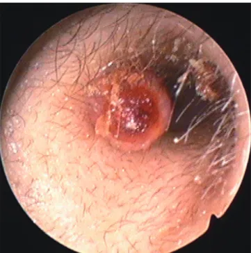
A 30-year-old woman presented to our hospital with otalgia and blood-tinged otorrhea in the left ear, which had been pres- ent for two months after she repeatedly picked her ear. The pa- tient had no hearing difficulty, vertigo, tinnitus, or facial weak- ness. She had no previous history of recurrent otitis media or trauma, and no family history of similar lesions. Otoscopic ex- amination demonstrated a smooth-margined, round, and pink- to-red colored soft tissue mass that was 5 mm in size in the pa- tient’s left external auditory canal (Fig. 1). A tympanic membrane
Adult-Onset Juvenile Xanthogranuloma of the External Auditory Canal: A Case Report
성인에서 발생한 외이도의 소아 황색 육아종: 증례 보고
Joonho Hur, MD
1, Jae Kyun Kim, MD
1,2*, Nara Kim, MD
3, Giyoung Seo, MD
1, Woosun Choi, MD
1, Jun Soo Byun, MD
1, Woong Jae Lee, MD
1, Tae Jin Lee, MD
4Departments of 1Radiology, 4Pathology, Chung-Ang University College of Medicine, Chung-Ang University Hospital, Seoul, Korea
2Department of Radiology, Kangwon National University College of Medicine, Chuncheon, Korea
3Department of Radiology, Samsung Medical Center, Sungkyunkwan University College of Medicine, Seoul, Korea
Juvenile xanthogranuloma (JXG) is a benign, spontaneously regressing lesion that usually occurs during the first year of life, but may also occur in adulthood. Although the most common presentation of JXG is the cutaneous lesion, it can also manifest in various visceral organs. JXG of the external auditory canal is extremely rare, and there have been only a few reports of those cases in the English literature. In this study, we present a case of pathologically proven JXG that occurred in the external auditory canal with a symptomatic clinical presentation.
Index terms
Xanthogranuloma, Juvenile Ear Canal
Tomography, X-Ray Computed
Received June 13, 2014 Revised February 16, 2015 Accepted September 16, 2015
*Corresponding author: Jae Kyun Kim, MD
Department of Radiology, Chung-Ang University College of Medicine, Chung-Ang University Hospital, 102 Heukseok-ro, Dongjak-gu, Seoul 06973, Korea.
Tel. 82-2-6299-2662 Fax. 82-2-6263-1557 E-mail: [email protected]
This is an Open Access article distributed under the terms of the Creative Commons Attribution Non-Commercial License (http://creativecommons.org/licenses/by-nc/3.0) which permits unrestricted non-commercial use, distri- bution, and reproduction in any medium, provided the original work is properly cited.
336
Adult-Onset Juvenile Xanthogranuloma of the External Auditory Canal
jksronline.org
J Korean Soc Radiol 2016;74(5):335-338 with a normal appearance was noted by the otoscopy. There was
no other skin or mucosal lesions. Computed tomography (CT) scans of the temporal bone were performed with a 16-slice CT scanner (LightSpeed Pro 16, GE Medical Systems, Milwaukee, WI, USA). The CT images showed an ovoid soft tissue nodule, which was 5 × 3 mm in size, in the middle of the external audi- tory canal and attached to the antero-superior wall (Fig. 2). There was no evidence of infiltration to the adjacent structures. The
results of the serum chemistry tests were within the normal rang- es, and the other parts of the body were found to be normal upon physical examination.
A complete mass resection was performed with the patient in the supine position and under local anesthesia; the procedure was performed using the transmeatal approach under the sur- gical microscope. The mass was observed in the external audito- ry canal, abutted to the antero-superior wall. The mass was re- moved using micro-scissors, whereas bleeding was controlled by bipolar cauterization. External auditory canal packing was ap- plied, and the specimen was sent to the Department of Pathology.
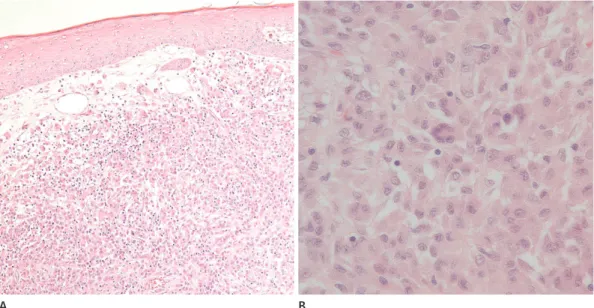
The excised specimen was a soft tissue mass measuring 5 × 5 mm in size. The results of histopathological examinations indi- cated that the mass consisted of foamy histiocytes, Touton giant cells, lymphocytes, and a few eosinophils (Fig. 3). Immunohis- tochemical analysis revealed that this lesion was positive for CD68 and weakly positive for S-100. These histological findings are consistent with those for JXG. After the surgery, the patient had no complications, such as hearing difficulty or facial weakness.
DISCUSSION
JXG is a benign and rare non-Langerhans cell histiocytosis. A bimodal distribution is observed in the onset of JXG. Although it usually occurs during early childhood, about 15% of the cases involve adult patients, with the peak of onset at 20 to 40 years of Fig. 1. An otoscopic image is showing a smooth margined, round soft
tissue mass 5 mm in size in the left external auditory canal.
Fig. 2. Pre-contrast axial (A) and coronal (B) images of temporal bone CT demonstrating an ovoid soft tissue nodule 5 × 3 mm in size, in the middle of the left external auditory canal, attached to the antero-superior wall.
A B
337
Joonho Hur, et al
jksronline.org J Korean Soc Radiol 2016;74(5):335-338 age. Nevertheless, the term “juvenile” is used whether the lesion manifests in adolescents or adults (4). Depending on the age of the patient, the lesion appears different in nature. During early childhood, they tend to occur in multiples and spontaneously re- gress within one year, whereas the adult form tends to be solitary and persistent (4).
The most common presentation of JXG is the cutaneous le- sion, but it can manifest in various visceral organs including the lung, bone, testis, gastrointestinal tract, heart, eye, and oral cav- ity (5, 6). The head, neck, and trunk are the sites most common- ly affected by JXG (3). JXG of the external auditory canal is ex- tremely rare, and it may be difficult to differentiate JXG from other lesions in the external auditory canal (4). Tumor or tumor- like lesions in external auditory canal are exostosis, osteoma, cholesteatoma, squamous cell carcinoma, keratosis obturans, and pleomorphic adenoma. Abnormal osseous proliferation, such as exostosis or osteoma, can be easily seen on CT images.
Cholesteatoma can be distinguishable by bony erosion (7, 8) and squamous cell carcinoma is usually presented as invasive soft tissue mass. Keratosis obturans and pleomorphic adenoma may be distinguishable, but usually diagnosis is established on the ba- sis of a histopathologic finding–not on the CT findings–because of their similarity (9, 10). There are few studies about the MRI finding of JXG, and homogeneous iso-signal intensity on T1- and T2-weighted images have been reported (4).
Histopathology is used to diagnose JXG, based mostly on im-
munohistochemical analysis in the clinical setting. JXG demon- strates a well-circumscribed nodule with severe infiltration of histiocytes. The Touton giant cell is observed in 85% of JXG nod- ules as a multinucleated cell with a peripheral ring of nuclei sur- rounded by a glassy, eosinophilic cytoplasm. Histiocytes and gi- ant cells are of monocyte/macrophage origin. Therefore, JXG is strongly positive for a macrophage marker, CD68, but negative for the S-100 protein, which is a marker used for the diagnosis of Langerhans cell histiocytosis by immunohistochemistry.
Although the pathogenesis of JXG has not been established, it is believed that reactive processes in response to the initiating stimuli, possibly infectious or physical factors, may play a role in the development of JXG (4). In the current case, the patient had a habit of picking her ear. Therefore, we postulate that JXG may have occurred in response to the repeated ear picking. After com- plete excision of the lesion, recurrence is uncommon, although it has been reported.
In summary, we report a rare case of solitary JXG of the exter- nal auditory canal. The lesion was a well-circumscribed soft tis- sue mass 5 mm in size that occurred in the supero-anterior wall of the left external auditory canal, and represented adult-onset JXG. When diagnosing benign soft tissue tumors in the external auditory canal, adult-onset JXG should be considered as an alter- native condition.
Fig. 3. Microscopic images of the specimen showing foamy histiocytes, Touton giant cells, lymphocytes, and a few eosinophils, consistent with the characteristics of xanthogranuloma; hematoxylin and eosin, × 100 (A) and × 400 (B).
A B
338
Adult-Onset Juvenile Xanthogranuloma of the External Auditory Canal
jksronline.org
J Korean Soc Radiol 2016;74(5):335-338
REFERENCES
1. Adamson HG. Society intelligence: The Dermatological So- ciety of London. Br J Dermatol 1905;17:222
2. Helwig EB, Hackney VC. Juvenile xanthogranuloma (nevo- xanthoendothelioma). Am J Pathol 1954;30:625-626 3. Hernandez-Martin A, Baselga E, Drolet BA, Esterly NB. Ju-
venile xanthogranuloma. J Am Acad Dermatol 1997;36(3 Pt 1):355-367; quiz 368-369
4. Yoshihama K, Kato Y, Baba Y. Xanthogranuloma of the ex- ternal auditory canal mimicking a benign tumor: a case re- port. Case Rep Otolaryngol 2012;2012:298089
5. Lesniak MS, Viglione MP, Weingart J. Multicentric paren- chymal xanthogranuloma in a child: case report and review of the literature. Neurosurgery 2002;51:1493-1498; discus- sion 1498
6. Margulis A, Melin-Aldana H, Bauer BS. Juvenile xantho-
granuloma invading the muscles in the head and neck:
clinicopathological case report. Ann Plast Surg 2003;50:
425-428
7. Viswanatha B. Extracanalicular osteoma of the temporal bone. Ear Nose Throat J 2008;87:381-383
8. Woldenberg Y, Nash M, Bodner L. Peripheral osteoma of the maxillofacial region. Diagnosis and management: a study of 14 cases. Med Oral Patol Oral Cir Bucal 2005;10 Suppl 2:E139-E142
9. Heilbrun ME, Salzman KL, Glastonbury CM, Harnsberger HR, Kennedy RJ, Shelton C. External auditory canal cholestea- toma: clinical and imaging spectrum. AJNR Am J Neurora- diol 2003;24:751-756
10. Markou K, Karasmanis I, Vlachtsis K, Petridis D, Nikolaou A, Vital V. Primary pleomorphic adenoma of the external ear canal. Report of a case and literature review. Am J Otolar- yngol 2008;29:142-146
성인에서 발생한 외이도의 소아 황색 육아종: 증례 보고
허준호
1· 김재균
1,2* · 김나라
3· 서기영
1· 최우선
1· 변준수
1· 이웅재
1· 이태진
4소아 황색 육아종은 양성의 자발성 퇴행 병변으로 대개 1세 미만에서 나타나지만 성인에서도 발생하는 것으로 알려져 있 다. 호발 부위로는 피부 병변이 가장 흔하나 다양한 내부 장기에서도 생길 수 있다. 하지만 외이도에 생긴 소아 황색 육아 종은 극히 드물며, 이런 증례는 영문 문헌에서 단지 몇 개만이 보고되었다. 따라서 우리는 임상 증상을 보이던 병리학적으 로 확인된 외이도에 생긴 소아 황색 육아종을 보고하고자 한다.
중앙대학교 의과대학 중앙대학교병원 1영상의학과, 4병리과, 2강원대학교 의과대학 영상의학과,
3성균관대학교 의과대학 삼성서울병원 영상의학과