pISSN : 1229-5418
Implantology 2018; 22(3): 152-160
https://doi.org/10.32542/implantology.20180013
Received: August 27, 2018 Revised: September 28, 2018 Accepted: September 28, 2018
Copyright © 2018. The Korean Academy of Oral &
Maxillofacial Implantology
This is an Open Access article distributed under the terms of the Creative Commons Attribution Non-Commercial License (http://creativecommons.
org/licenses/by-nc/4.0/) which permits unrestricted non-commercial use, distribution, and reproduction in any medium, provided the original work is properly cited.
OPEN ACCESS
Introduction: Patients with oroantral fistula usually have severe alveolar bone defects. Alveolar bone augmentation procedure may be needed when placing implant in atrophic bone. Currently, various alveolar bone augmentation techniques are recognized as promising treatments. The purpose of this study is to evaluate the results of simultaneously performed sinus augmentation, guided bone regeneration (GBR) and block bone graft procedures in patient who have the severe alveolar bone loss with oroantral fistula.
Case: A 58-year-old male patient was referred for implant removal at #15, 16 intruded in the right maxillary sinus. At first, implant fixture was removed and, #14 was extracted. After 7 months, combination technique including sinus augmentation & block bone graft & GBR was carried out.
Sinus augmentation was performed by the lateral approach. The bone block harvested from the mandibular ramus was performed. The bone block is placed on the alveolar bone and fixed with a screw to the alveolar bone with GBR. At 7 months after bone augmentation, Implants were placement at the #14, 16, 17 edentulous site. After implantation 6 months, implant second operation was done. And the prosthetic procedure was completed.
Conclusion: Healing occurs without complications and at 9 months follow-up check, normal clinical and radiological findings were observed.
Keywords: Bone graft, Guided tissue regeneration, Sinus augmentation, Oroantral fistula
Abstract
구강 상악동 누공 동반한 심한 치조골 흡수부위 임플란트 식립: 증례보고
장국원1, 황희성1, 김철훈1, 김복주1*, 김정한1, 김무성1, 김도희2
1동아대학교 의료원 구강악안면외과
2동아대학교 의학대학원
*Corresponding author: Bok-Joo Kim, [email protected]
Implant Placement of Severe Alveolar Bone Resorption Site with Oroantral Fistula: A Case Report
Kuk-Won Jang1, Hee-Sung Hawng1, Chul-Hun Kim1, Bok-Joo Kim1*, Jung-Han Kim1, Moo-Sung Kim1, Do-Hee Kim2
1Department of Oral and Maxillofacial Surgery, College of Medicine, Dong-A University, Busan, Korea
2Graduate School of Medicine, College of Medicine, Dong-A University, Busan, Korea
Ⅰ. 서론
치과영역에서 임플란트 식립술은 현재 보편적으로 행해지는 술식으로 수십 년간 다양한 치료옵션을 제공해왔다. 임플란트는 여러 해부학적 구조물이나, 부족한 치조골 등으로 인하여 식립이 불가능한 경 우가 있으며 이런 경우 여러 가지 골증강술을 이용하여 임플란트를 성공적으로 식립해왔다
1
.상악동 거상술은 함기화된 상악 구치부에서 상악동내의 골이식을 하여 임플란트 식립을 가능하게 하 였다. 상악동 거상술은 상악동에 접근하는 방식에 따라 치조정 접근법과 측방 접근법으로 나눌 수 있다.
위축된 상악골에서의 상악동 거상술은 상당히 예지성있는 술식으로 상악동 거상술을 시행한 부위의 임 플란트 생존율은 90%이상으로 알려져 있다
2
.블록골 이식(Block bone graft)은 여러 가지 골이식재 중 골드 스탠다드인 자가골
3
을 이용하는 방법으 로 상당히 큰 골결손부에 이식이 가능하다는 점에서 심한 치조골 흡수가 있는 환자의 임플란트 식립시 많이 사용하였다. 하지만 골 채취부의 추가적인 수술과 다른 이식재의 발전으로 최근 그 빈도는 줄어들 었지만 여전히 심한 치조골 흡수가 있는 부위의 골증강술시 예지성 있는 치료 옵션 중 하나이다4,5
.구강 상악동 누공(Oroantral Fistula, OAF)은 상악동에서 구강으로의 누공을 의미하는 용어로 심한 치 주질환으로 인한 대구치 발치, 만성 상악동염, 방사선 골괴사증, 종양제거, 임플란트 식립 실패 등이 원 인이다. OAF는 상악동과 구강사이의 경조직, 연조직의 결손을 의미하며, OAF가 발생한 상악 구치부 의 임플란트 식립은 큰 도전이다
6-8
.본 논문에서는 상악 구치부에 임플란트 식립이후 실패로 OAF가 발생한 상악 구치부에 상악동거상 술, 블록골 이식, 골유도재생술을 동시에 시행한 부위에 임플란트 식립 한 증례로 만족할만한 결과를 얻 어 보고하고자 한다.
Ⅱ. 증례
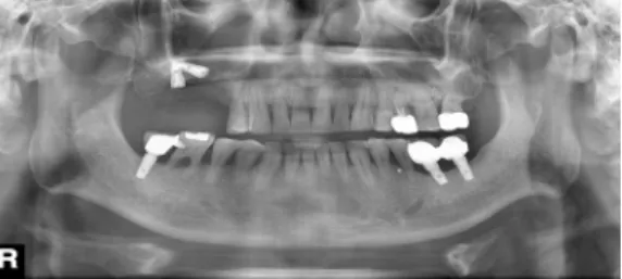
58세 남자환자로 갑상선 기능 저하증 병력이 있으며, 상악동내 임플란트 함입을 주소로 동아대의료 원 구강악안면외과를 내원하였다. 일반치과에서 1년전 골유도재생술(guided bone regeneration, GBR) 을 동반하여 #15,16 임플란트 식립 하였다. 식립 이후 상악동내 임플란트 함입 발생하여 일반치과에서 본원으로 의뢰되었다. 방문시 오른쪽 상악 구치부 #15,16 임플란트는 상악동 내로 함입된 상태이며 치 조골 골절과 OAF가 존재하였다. 추가적으로 #14 치아는 동요도 2도를 보였다. #46 치아는 무치악기간 이 오래되어 정출되어있으며 만성 치주염으로 치근이개부까지 치조골 흡수가 관찰되었다(Fig. 1).
치조골의 심한위축 및 치조골 골절상태를 고려하여 상악동에 대한 측방접근으로 픽스쳐를 제거하기 로 결정 하였다. 측방 골창(lateral bone window) 형성 후 상악동내 석션으로 임플란트를 조심스럽게 제 거하였다. 측방 골창은 골편을 재위치 시켜 봉합 시행하였으며, 이완절개 시행하여 협측피판을 OAF폐 쇄에 이용하였다. 골창편은 제거하는 방법도 있지만 향후 측방접근법의 상악동 거상술시 피판거상을
용이하기 위하여 골창편을 재위치시키는 방법을 선택하였다. 추가적으로 #14 치아를 만성 치주염으로 발치하였다. 이후 경과관찰하며 연조직, 경조직의 회복시켰다.
임플란트 제거 6개월 후 파노라마, CBCT를 촬영하였다(Fig. 2). 방사선 사진을 확인하면 defect로 인 하여 잔존골의 골흡수가 많이 되어있었으며 특히 협측 치조골은 심한 골흡수를 보였다. CBCT상 #16 부위 협측 치조골 흡수, 수직적 치조골 흡수를 확인할 수 있었다. #14,15 부위 골창 형성 시 골삭제 부위 확인 가능하다. 전반적으로 연조직 회복은 되었지만 방사선 사진상 골형성은 거의 없었다.
무치악부 치조골이 많이 위축되어 있어 유치악부 치조골과 높이차이가 큰 상태였다. 상악동 거상술 만을 시행하여 임플란트 식립시 치관의 길이가 비정상적으로 길어져 implant-crown ratio가 상당히 커 질 것이 예상되었다. 이전 임플란트 실패 병력과 심한 치조골 흡수로 본원에서는 수직적인 골증강을 위 하여 골유도재생술, 블록골 이식, 상악동 거상술 3가지 술식을 동시에 진행하는 combination technique 을 계획하였다.
임플란트 제거 7개월 후 골증강술을 실시하였다. 상악동 거상술시 측방접근으로 시행하였다. 본원에 서 임플란트 제거를 위한 측방 골창을 형성했던 부위여서 협측 연조직과 상악동막의 접합으로 피판 거 상시 상악동 막의 천공이 발생할 가능성이 높아 주의 깊게 박리 하였다. OAF가 있었던 자리는 연조직 으로 회복되었지만 경조직 천공이 확인되었으며, 임플란트 제거시 사용했던 골창부위는 얇은 골로 회
Fig. 1. First visit Panorama. #15,16 implants are into the maxillary sinus. The #14 natural teeth observated bone resorption on the distal side and the #46 is excessive erupted.
Kuk-Won Jang et al. : Implant Placement of Severe Alveolar Bone Resorption Site with Oroantral Fistula: A Case Report. Implantology 2018
Fig. 2. Implant removal 6 month. (A) Panorama after implant fixture removal 6 month; Severe alveolar bone defect in the right maxillary posterior, (B) #15 site Conebeam CT after implant fixture removal 6 month; CBCT findings in #15 region of maxillary alveolar bone. It is possible to confirm the formation site of bone window, (C) #16 site Conebeam CT after implant fixture removal 6 month; CBCT findings in #16 region of maxillary alveolar bone. Buccal alveolar bone severe resorption. yellow arrow is OAF.
Kuk-Won Jang et al. : Implant Placement of Severe Alveolar Bone Resorption Site with Oroantral Fistula: A Case Report. Implantology 2018
A B C
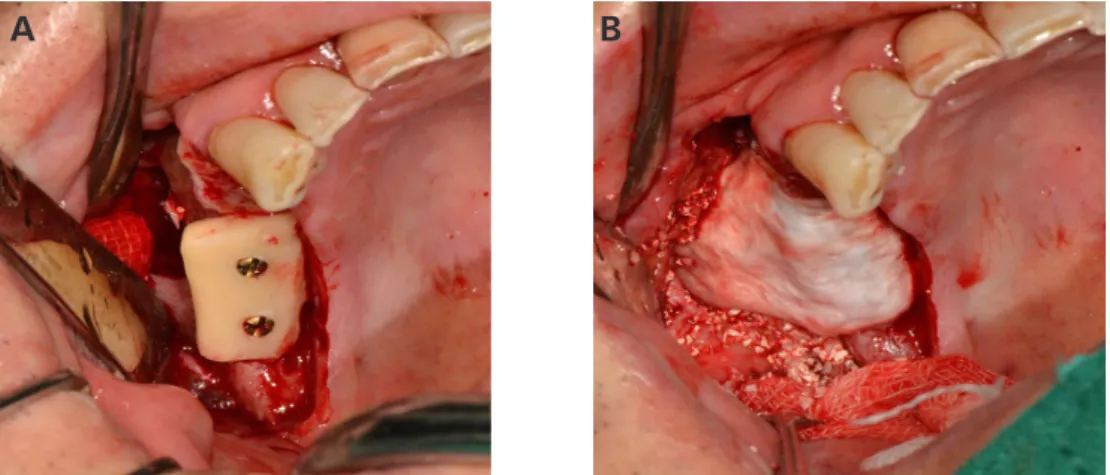
복된 것을 확인 하였다. 이후 측방 골창을 형성 및 상악동막 거상 시행하였다. 상악동내로 동종골 Ora- graft(LifeNetHealth, USA) 1.5 g, 이종골 Ovis XENO(Oscotec, Korea) 1.5 g을 섞어서 이식하였으며 오 른쪽 하악지에서 약 20 × 10 mm 블록 골을 채취하였고 치조골정 수여부에 screw 2개로 고정을 하였 다. 흡수성막 Ossguide (SKbioland, Korea)와 동종골 oragraft 0.5 g, 이종골 Ovis XENO 0.5 g를 사용하 여 추가적인 GBR을 실시하였다(Fig. 3). 수직적인 골증강을 시행한 후 협측 피판에 대한 이완절개를 시 행하여 연조직을 장력없이 봉합하였다. 625 mg Augmentin TID, Tiroxin 250 mg TID, Clanza S 100 mg BID 2주 약물 처방, Dicknol 90 mg, Pazeron 0.5 g, Dexametasone 5 mg 주사처방하였다. 수술 다음날 과 3일후 dressing 실시하였다. 골이식 일주일후 발사를 시행하였다. 발사 이후에도 1주 간격으로 주기 적인 체크 시행하였으며 공여부 신경손상, 연조직 천공, 감염, 상악동내 골이식재 침범 등 부작용은 없 었다(Fig. 4).
골이식 5개월 후 CBCT 확인결과 임플란트 식립에 충분한 골생성을 확인할 수 있었다(Fig. 5). 이후 골이식 7개월째 #14,16,17 임플란트 식립을 계획하였다. block bone을 고정하는 screw제거 후 임플란 트 식립하였다. 피판거상시 자가골이 골융합이 일어난 것을 확인 할 수 있었다. #16,17 부위는 폭 4.8 mm,
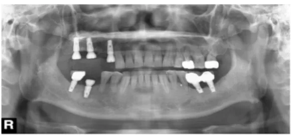
Fig. 4. Panorama Immediately after bone augmentaion. Immediately after bone augmentation. The bone graft in the maxillary sinus can be identified and the block bone can be identified.
Kuk-Won Jang et al. : Implant Placement of Severe Alveolar Bone Resorption Site with Oroantral Fistula: A Case Report. Implantology 2018
Fig. 3. During operation. (A) lateral approach sinus lift & block bone graft; Lateral approach Maxillary sinus elevation - lateral window can be identified. Fixed the block bone with a screw, (B) adiitional GBR performed; Photo after GBR. Block bone graft and insufficient area, add a bone graft. The membrane was covered with the lateral window and the GBR area.
Kuk-Won Jang et al. : Implant Placement of Severe Alveolar Bone Resorption Site with Oroantral Fistula: A Case Report. Implantology 2018
A B
높이 10 mm 임플란트를 식립하였고 #14 부위는 폭 4.3 mm, 높이 10 mm 임플란트(Dentis, South korea) 를 식립하였다 식립 후 cover screw를 채결하였다. 추가적으로 치주가 좋지 않는 #46 치아 발치 후 즉시 식립을 폭 4.3 mm, 높이 10 mm 임플란트로 식립하였다(Fig. 6).
이후 계속적인 경과관찰 후 임플란트 식립 6개월 후 2차 수술을 시행하여 임플란트 지대주를 연조직 상방으로 노출하였다(Fig. 7). 이후 최종보철 과정 진행하여 #14=16,17 4 unit PFM bridge, #46 PFM crown으로 보철치료 완료하였다.
이후 6개월, 9개월 경과관찰을 진행하였다. 6개월 경과관찰 시 #16 근심부위 약간의 골소실이 보이지
Fig. 5. Bone augmentation. (A) #16 site CBCT after bone augmentation 5months; Confirmation of sufficient bone formation in maxillary sinus. confirmation of block bone osseointegration, (B) Panorama after bone augmentaion 5months; Confirmation of sufficient bone formation in maxillary sinus. confirmation of block bone osseointegration.
Kuk-Won Jang et al. : Implant Placement of Severe Alveolar Bone Resorption Site with Oroantral Fistula: A Case Report. Implantology 2018
A B
Fig. 6. Implant first surgery. (A) Before incision; Oral photos before implant placement. It can be confirmed that there is shortage of buccal attached gingiva, (B) Before removal screw, (C) implant installation, (D) Panorama after implant first surgery; A panorama after reimplantation of the implant.
and #46 Additional extraction was performed and immediate implantation.
Kuk-Won Jang et al. : Implant Placement of Severe Alveolar Bone Resorption Site with Oroantral Fistula: A Case Report. Implantology 2018
A B C
D
만 임상적 증상은 없었다. 9개월 경과관찰 시 골소실 부위 증가하지 않았고, 방사선학적, 임상적으로 양 호한 상태를 보이고 있다(Figs. 8, 9).
Ⅲ. 총괄 및 고찰
치아 결손부의 임플란트 수복 시 장기적인 생존, 심미적 수복을 위해서는 식립 부위에 충분한 치조골 이 필요하다. 하지만 환자에 따라 골 흡수가 일어나 치조골의 양을 증가시키는 것이 필요할 수 있다. 치 조골의 양을 증가시키는 술식은 보통 치조골 증강술 (alveolar bone augmentation) 또는 골증강술(bone augmentation) 이라고 한다. 이러한 골증강술은 1. 골유도 재생술 2. 상악동 거상술 3. 블록골 이식술 4.
골신장술 5. 치조골 분할술 이 흔히 쓰이고 있다. 이중 본 증례에서는 골유도 재생술, 상악동 거상술, 블 록골 이식술을 동시에 시행하였다
9
.일반적인 상악 구치부 치조골 소실일 경우 상악동 거상술만을 시행한다. 하지만 본 증례에서는 유치
Fig. 7. Panorama after implant second surgery.
Kuk-Won Jang et al. : Implant Placement of Severe Alveolar Bone Resorption Site with Oroantral Fistula: A Case Report. Implantology 2018
Fig. 8. 6month panorama after prosthesis delivery.
Kuk-Won Jang et al. : Implant Placement of Severe Alveolar Bone Resorption Site with Oroantral Fistula: A Case Report. Implantology 2018
Fig. 9. 9month panorama after Prosthesis delivery.
Kuk-Won Jang et al. : Implant Placement of Severe Alveolar Bone Resorption Site with Oroantral Fistula: A Case Report. Implantology 2018
악부와 무치악부의 심각한 골 높이 차이, OAF 존재로 단순한 상악동 거상술시 implant-crown ratio의 악화, OAF로 인한 상악동내 골이식재의 소실, 감염 등이 예상된다. implant-crown ratio가 불리하면 음 식물 저류, 주위 연조직 염증 및 그로 인한 하방 치조골 흡수, 임플란트의 보철적 문제로 임플란트 장기 생존률에 영향이 있을 수 있다
10
. 추가적인 수직골 증가가 필요하여 이에 대하여 본원에서는 골유도 재 생술, 블록골 이식, 상악동 거상술를 선택하였다.OAF(oroantral fistula) 발생시 폐쇄방법은 기본적으로 누공의 크기에 따라 달라진다. 5 mm이하의 구 강 상악동 누공은 상악동의 감염이 없다면 특별한 처치 없이 코풀기 금지, 유동식 섭취등의 주의사항을 잘 지키면 이차성 치유로 회복될 수 있다. 누공의 크기가 5 mm이상일 경우 결손부 위치, 크기에 따라 Sliding bridge flap, Rehrmann's buccal advancement flap, Palatal flap을 이용할 수 있다
11
. 위와 같은 방 법은 연조직의 폐쇄방법으로서 임플란트 식립이 계획된 경우는 경조직에 대한 처치가 필요할 수 있다.OAF 부위의 경조직 회복에 대하여 proctor등은 자가장골의 해면골 조각으로 OAF 부위를 수복하는 술 식을 보고 하였고
12
, Hass등은 원형의 피질골편을 이용하여 OAF 부위에 끼워 넣는 방법을 보고 하였다7
. 본 증례에서는 임플란트 직경 정도의 크기인 5 mm 크기였다. 크기만 보면 특별한 처치 없이 2차 회복 을 기다려도 되지만, 치조골 골절이 있고, 함입된 임플란트 제거 위하여 협측 피판 형성이 필요하여 협 측 피판을 이용한 누공의 폐쇄를 하였다. 임플란트 식립을 위해서는 경조직의 회복도 필요하였지만, 누 공의 크기가 크지 않고, 누공 주변의 자가골이 충분하다는 점, 상악동내 감염증상이 없다는 점에서 연조 직으로 OAF의 폐쇄 이후 골증강술시 누공 부위에 골이식제를 이식하는 방법을 선택하였다.본 증례에서 함입된 임플란트 제거를 위한 골창 형성과 상악동 거상술시 골창 형성으로 2번의 상악동 측방 골창을 형성해야 한다. 첫번째 골창 형성시는 일반적인 증례와 같이 시행하면 되지만 2번째 골창 형성시 첫번째 수술로 인한 협측 치은과 상악동막의 접합으로 피판 형성시 상악동막 천공이 발생할 수 있다. 그래서 본 증례에서는 임플란트 제거시 골창편을 재위치시켜 상악동막의 접합부위가 최소화를 유도하였다. 이로 인해 상악동 거상술시 상악동막 천공 없이 피판거상이 가능하였다.
3가지 술식의 동시시행은 치료기간의 단축과 수술 횟수 감소로 인한 불편감 감소, 연조직의 양호한 치 유 등 장점이 있지만 수술부위 회복지연, 긴 수술시간으로 인한 감염 가능성 등이 존재하며 특히 차폐막 노출, 상악동 감염 등 처치가 필요한 부작용 발생시 모든 술식이 같이 실패할 수 있는 위험성을 가지고 있다. 이에 대하여 본 증례에서는 부작용 발생을 줄이기 위해 골증대술 이후 확실한 정기체크를 시행하 여 부작용발생시 즉각적인 조치가 가능하게 하였다. 감염가능성에 대하여 환자분께 금주, 금연, 주의사 항 준수에 대하여 상당히 강조하여 설명하였으며 수술직후 2주간 약물처방 실시하였으며 추가적인 주 사처방을 실시하였다.
수직적인 골증강술시 창상열개 방지를 위한 무장력 폐쇄(tension free closure)가 수술성공에 제일 중 요한 요인일 것이다. 많은 연구에서 알려진대로 강한 장력이 있는 경우 봉합부는 혈류의 감소가 일어나 며 이로 인하여 산소분압이 낮아져 피판의 부분적 괴사가 일어나 창상열개가 일어날 수 있다
13-16
. 이를예방하기 위하여 본 증례에서는 이완절개(releasing incision)을 사용하였으며 측방접근 상악동 거상술 을 동시에 시행하여 보다 넓은 피판을 형성하여 통상적인 이완절개보다 넓게 시행하여 무장력 폐쇄를 이루었다.
임플란트 식립전 임상사진상 협측 부착치은의 양이 거의 없는 것을 확인할 수 있다(Fig. 6). 이는 일반 치과의 임플란트 수술을 포함한 여러번의 수술, OAF 폐쇄을 위한 피판 형성, 수직골 증강술시 이완절 개 등으로 인하여 발생하였다. 이러한 부착치은의 부족은 잇솔질 중 통증 및 구강위생관리에 문제가 발 생할 수 있다. 장기적인 임플란트 유지를 위해서는 구강위생관리가 중요하다
17,18
. 이러한 이유로 임플 란트 2차수술시 근단 변위 판막술(apical positioned flap)시행하여 임플란트 협측에 2-3 mm 부착치은 을 형성해주었다.상악골은 골질이 D3, D4로 분류되는 경우가 많다
19
. 더욱이 본 증례에서는 매식된 임플란트가 함입될 정도로 골질이 안 좋은 것을 확인할 수 있다. 하지만 하악의 피질골을 이식 해놓은 상태여서 하악의 피 질골에 식립하는 것과 같이 초기고정은 충분히 나왔다.Ⅳ. 결론
임플란트 실패로 구강 상악동 누공이 발생한 환자에서 상악동 거상술, 블록골 이식술, GBR 동시 시 행은 구강 상악동 누공을 폐쇄하고 수직적 골증강을 획득할 수 있는 예지성 있는 치료로 판단된다.
References
1. Nyman S. Bone regeneration using the principle of guided tissue regeneration. J Clin Periodontol.
1991; 18: 494-498.
2. Ki SI, Yu MG, Kim YJ, et al. A Retrospective Evaluation of Implant Installation with Maxillary Sinus Augmentation by Lateral Window Technique. Maxillofac Plast Reconstr Surg. 2008; 30: 457-464.
3. Kim JW, Cho MH, Kim SJ, et al. Alveolar distraction osteogenesis versus autogenous onlay bone graft for vertical augmentation of severely atrophied alveolar ridges after 12 years of long-term fol- low-up. Oral Surg Oral Med Oral Pathol Oral Radiol. 2013; 116: 540-549.
4. Donos N, Mardas N, Chadha V. Clinical outcomes of implants following lateral bone augmentation:
systematic assessment of available options (barrier membranes, bone grafts, split osteotomy). J Clin Periodontol. 2008; 35: 173-202.
5. Rocchietta I, Fontana F, Simion M. Clinical outcomes of vertical bone augmentation to enable dental implant placement: a systematic review. J Clin Periodontol. 2008; 35: 203-215.
6. Kraut RA, Smith RV. Team approach for closure of oroantral and oronasal fistulae. Atlas Oral Maxil- lofac Surg Clin North Am. 2000; 8: 55-75.
7. Haas R, Watzak G, Baron M, et al. A preliminary study of monocortical bone grafts for oroantral fistu- la closure. Oral Surg Oral Med Oral Pathol Oral Radiol. 2003; 96: 263-266.
8. Raghoebar GM, Timmenga NM, Reintsema H, et al. Maxillary bone grafting for insertion of endosse-
ous implants: results after 12–124 months. Clin Oral Implants Res. 2001; 12: 279-286.
9. Aghaloo TL, Moy PK. Which hard tissue augmentation techniques are the most successful in fur- nishing bony support for implant placement? Int J Oral Maxillofac Implants. 2007; 22: 49-70.
10. Urdaneta RA, Rodriguez S, McNeil DC, et al. The effect of increased crown-to-implant ratio on sin- gle-tooth locking-taper implants. Int J Oral Maxillofac Implants. 2010; 25: 729-743.
11. Güven O. A clinical study on oroantral fistulae. J Craniomaxillofac Surg. 1998; 26: 267-271.
12. Proctor B. Bone graft closure of large or persistent oromaxillary fistula. Laryngoscope. 1969; 79: 822- 826.
13. Larrabee WF Jr, Holloway GA Jr, Sutton D. Wound tension and blood flow in skin flaps. Ann Otol Rhinol Laryngol. 1984; 93: 112-115.
14. Shapiro AL, Hochman M, Thomas JR, et al. Effects of intraoperative tissue expansion and skin flaps on wound closing tensions. Arch Otolaryngol Head Neck Surg. 1996; 122: 1107-1111.
15. Carranza FA, Jr., Carraro JJ, Dotto CA, et al. Effect of periosteal fenestration in gingival extension operations. J Periodontol. 1966; 37: 335-340.
16. Burkhardt R, Preiss A, Joss A, et al. Influence of suture tension to the tearing characteristics of the soft tissues: an in vitro experiment. Clin Oral Implants Res. 2008; 19: 314-319.
17. Salvi GE, Lang NP. Diagnostic parameters for monitoring peri-implant conditions. Int J Oral Maxillo- fac Implants. 2004; 19: 116-127.
18. Warrer K, Buser D, Lang N, et al. Plaque-induced peri-implantitis in the presence or absence of kera- tinized mucosa. An experimental study in monkeys. Clin Oral Implants Res. 1995; 6: 131-138.
19. Truhlar RS, Orenstein IH, Morris HF, et al. Distribution of bone quality in patients receiving endosse- ous dental implants. J Oral Maxillofac Surg. 1997; 55: 38-45.