www.jpis.org
pISSN 2093-2278 eISSN 2093-2286 Copyright © 2010 Korean Academy of PeriodontologyThis is an Open Access article distributed under the terms of the Creative Commons Attribution Non-Commercial License (http://creativecommons.org/licenses/by-nc/3.0/).
Incomplete bone formation after sinus augmentation: A case report on radiological findings by computerized tomography at follow-up
Kyung-Shil Lee1, Young-Hyuk Kwon1,2, Yeek Herr1,2, Seung-Il Shin1, Ji-Yeon Lee1, Jong-Hyuk Chung1,2*
1Department of Periodontology, Kyung Hee University School of Dentistry, Seoul, Korea
2Institute of Oral Biology, Kyung Hee University School of Dentistry, Seoul, Korea
Purpose: The aim of this case report is to present a case of incomplete bone formation after sinus augmentation.
Methods: A patient having alveolar bone resorption of the maxillary posterior edentulous region and advanced pneumatiza- tion of the maxillary sinus was treated with sinus elevation using deproteinized bovine bone in the Department of Periodon- tology, Kyung Hee University School of Dentistry and re-evaluated with computed tomography (CT) follow-up.
Results: Even though there were no significant findings or abnormal radiolucency on the panoramic radiograph, incomplete bone formation in the central portion of the augmented sinus was found fortuitously in the CT scan. The CT scan revealed peri-implant radiolucency in the apical portion of the implant placed in the augmented maxillary sinus. Nevertheless, the den- tal implants placed in the grafted sinus still functioned well at over 15 months follow-up.
Conclusions: The result of this case suggests that patients who received maxillary sinus augmentation may experience incom- plete bone formation. It is possible that 1) osteoconductive graft material with poor osteogenic potential, 2) overpacking of graft material that restricts the blood supply, and 3) bone microbial contamination may cause the appearance of incomplete bone formation after sinus augmentation. Further studies are needed to elucidate the mechanism of this unexpected result and care must be taken to prevent it.
Keywords: Bone formation, Computed tomography, Maxillary sinus.
INTRODUCTION
Alveolar bone resorption in maxillary posterior edentulous region and advanced pneumatization of the maxillary sinus can result in a lack of bone support for dental implants, leav- ing only a thin wall of bone between the maxillary sinus and oral cavity. When this area does not offer adequate conditions for anchoring, long-term maintenance of dental implants is hard to achieve and a bone graft is required.
The placement and integration of dental implants in such
patients requires augmentation of the maxillary sinus. The classic procedure for maxillary sinus augmentation was first introduced by Boyne and James [1]. The periosteal membrane of the sinus mucosa that adheres to the maxillary sinus floor has few elastic fibers. Thus, separation of this sinus membrane is relatively simple and has become a standard method with good results [2,3]. Numerous clinical studies have reported the clinical outcomes of placing implants in the augmented maxillary sinus. The insertion of dental implants in combi- nation with maxillary sinus floor elevation is a predictable
Received: Jul. 23, 2010; Accepted: Oct. 12, 2010
*Correspondence: Jong-Hyuk Chung
Department of Periodontology & Institute of Oral Biology, Kyung Hee University School of Dentistry, 1 Hoegi-dong, Dongdaemun-gu, Seoul 130-701, Korea E-mail: [email protected], Tel: +82-2-958-9380, Fax: +82-2-958-9387
low incidences of surgical complications [4]. However, it sometimes leads to complications due to anatomical struc- ture, infections, iatrogenic factors, or other unknown factors.
Therefore, pre- and post-operative evaluation of the maxil- lary sinus is very important. However, there has been limited availability of data on this procedure based on computed to- mography (CT).
This case report describes a 53-year-old male patient who received maxillary sinus augmentation and implant installa- tion with a staged approach. Although the patient had no sub- jective symptoms and there was no abnormal radiolucency on the panoramic radiograph, incomplete bone formation in the central portion of the augmented sinus was found fortu- itously in the CT scan.
CASE DESCRIPTION
A 53-year-old male patient who was in good systemic con- dition visited the Department of Periodontology, Kyung Hee University School of Dentistry with the chief complaints of pain and gingival bleeding in the upper right 1st molar (#16) area. The upper right 2nd molar (#17) area was in an edentu-
gical/surgical periodontal therapy, extraction of #16 was in- evitable. The residual bone height was between 2 to 4 mm (Fig. 2). Augmentation of the maxillary sinus was scheduled to be conducted with a diagnostic stent 3 months after the extraction of #16, followed by placement of two dental im- plants 6 months after the augmentation of the maxillary si- nus (Fig. 3). The maxillary sinus was clinically healthy. The si- nus membrane was elevated from a lateral approach and the sinus was grafted with deproteinized bovine bone (DBB; Bio- Oss, Geistlich Pharma AG, Wolhusen, Switzerland) (Fig. 4).
Prophylactic antibiotics (amoxicillin 500 mg, Chong Kun Dang Pharm., Seoul, Korea) were prescribed three times a day for 14 days and 0.12% chlorhexidine solution (Hexame- dine, Bukwang Pharm., Seoul, Korea) was also prescribed twice a day for the first 2 weeks to prevent infection of the surgical wound. Healing was uneventful and there was no infection or other post-surgical complications during the healing period.
After a healing period of 6 months, a two stage implant sur- gery was planned (Fig. 5). Full thickness flap reflection under local anesthesia was followed by placement of two dental im- plants (Replace Select tapered HA, Nobel Biocare, Göteborg,
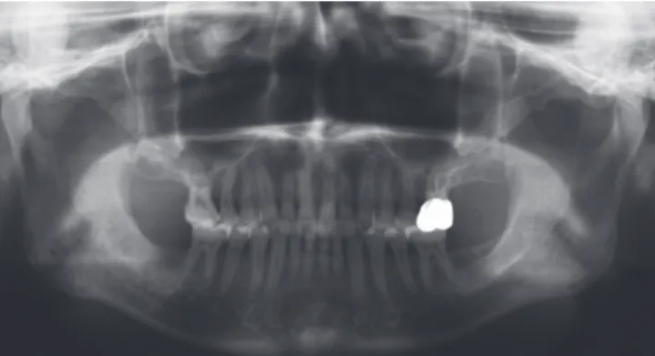
Figure 1. Panoramic radiograph at first visit. Figure 3. Diagnostic stent for the implant placement at #16i and 17i.
Figure 2. After extraction of #16, marked alveolar bone resorption and pneumatization of the maxillary sinus is visible.
Figure 4. Maxillary sinus augmentation was performed.
the site of #16i and #17i (Fig. 6). Both antibiotics and analge- sics were administered for seven days, and mouth rinse with 0.12% chlorhexidine was also recommended for the follow- ing two weeks. No complications occurred after implant in- stallation.
The second implant surgery was performed 6 months later (Fig. 7) and the implants were first loaded 2 months after the second surgery. Final prosthetic loading was initiated 13 months after the first implant surgery (Fig. 8).
The implants placed in the augmented sinus were clinically
functioning successfully at 17 months after initial loading.
Unexpectedly, the patient visited the dental clinic with the chief complaints of pain on biting in the upper right 2nd pre- molar (#15) since he had eaten hard food 3 days earlier. The
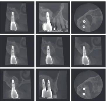
#15 tooth was diagnosed as cracked and endodontic therapy was required. During endodontic therapy, a CT scan was tak- en to locate the buccal canal of the tooth. Peri-implant radio- lucency in the apical portion of the implant placed in the aug- mented maxillary sinus was found by accident in the CT scan although a conventional (panoramic) radiograph revealed no signs of peri-implant radiolucency (Fig. 9). This was after a healing period of 32 months since sinus augmentation. The fortuitously discovered radiolucent portion can be described as incomplete bone formation or bone cavity in the augment- ed maxillary sinus. Nevertheless, the dental implants that
Figure 5. Radiopacity of the augmented sinus had increased at 7 months after the operation.
Figure 7. Second implant surgery on #16i and 17i was performed 6 months after the first implant surgery.
Figure 6. First implant surgery on #16i and 17i was performed.
Figure 8. Final prosthetic loading was initiated 13 months after the first implant surgery.
Figure 9. Bone cavity in #16i and 17i area was found in the comput- ed tomography scan (32 months after the sinus augmentation).
were placed in the grafted sinus had been functioning well after prosthetic loading for more than 60 months and no en- largement of the bone cavity was found in follow-up radio- graphic views (Fig. 10). The patient has had no subjective symptoms such as discomfort or pain in the #16i and 17i area and has been receiving follow-up care on a regular basis.
DISCUSSION
This study presents the fortuitous discovery of incomplete bone formation (a bone cavity) in the central portion of the maxillary sinus after sinus augmentation using DBB.
Although sinus augmentation is very predictable [5] and complications caused by sinus graft are very rare, clinicians have observed various types of complications, such as perfo- ration of the sinus membrane, excessive bleeding, infection of the grafted sinus, and failure of bone formation during and after sinus augmentation procedures [6-9]. Among these complications, incomplete bone formation of the grafted si- nus is not common and the reason for this phenomenon has not yet been determined.
Several causes of incomplete bone formation can be sug- gested. First, the graft material used in augmentation was solely DBB, which is a cell-free grafting material with osteo- conductive properties [10-13]. Cells possessing osteogenic po- tential are rich in residual host bone and elevated sinus mem- brane, whereas osteogenic potential of the graft is poor [1,14].
Therefore, new bone originates from the maxillary bone and progresses towards the augmented area [15,16]. The area of newly mineralized bone on the sinus floor is expected to be larger in the vicinity of the residual host bone [17]. Busenlech- ner et al. [16] reported the relative portion of newly formed bone after sinus augmentation with DBB in a minipig model study. In that study, with increasing distance from the host bone, the relative portion of newly formed bone declined from 38±13.3% at a 0-1 mm distance to 6.6±7% at a 4-5 mm dis- tance. Fuerst et al. [14] and Roldan et al. [15] have also described
within the augmented sinus in analysis by focusing on select- ed regions. The addition of autogenous bone, which contains bone-derived progenitor cells and osteoblasts, to bone sub- stitutes is thought to enhance new bone formation [14]. In several animal model studies, e.g. in sheep [18] and in mon- keys [19-21], the percentage of newly formed bone was in- creased by adding autogenous bone to bone substitutes. Ex- perimental studies have shown that culture expanded autog- enous bone-derived cells (ABC) added to cell-free grafting materials also enhanced the percentage of bone newly formed in critical-size defects of the rodent calvaria, and the dog and sheep mandible [22-25]. ABC possess an osteogenic potential and therefore increase bone formation in regions with a low number of bone-forming cells [23]. It can be speculated that supplementation of bovine bone mineral (BBM) with ABC may be recommended in cases where bony consolidation is complicated by the large volume of the grafting material or the poor regenerative potential of the host bone. If we had added autogenous bone to cell-free DBB during the sinus graft, more prominent bone formation could have been achieved.
Second, overpacking of graft material could restrict the blood supply. Rosenberg [26] and Garrett et al. [27] suggested pack- ing the bone graft loosely. Loose packing of the graft can cre- ate better interparticulate spacing, which facilitates more rapid vascularization and more abundant bone formation. In con- trast, tightly packed particles allow cellular and vascular access only to the outside layer of particles [28].
Third, Verdugo et al. [29] reported that bone contamination by specific pathogens could impair osteogenesis and induce greater bone loss in regenerative procedures. Similarly, Choukroun et al. [30] reported the accidental discovery of bubble-like lacunae in the grafted maxillary sinus when a si- nus lift was performed with graft material without a 0.5%
metronidazole solution. In that study, the formation of bub- ble-like lacunae within the graft was hypothesized to have resulted from anaerobic contamination after sinus grafting:
the “septic theory.” This “septic theory” suggests that grafted bone contamination by anaerobic bacteria could possibly in- duce problems with healing. They suggested the local use of a very small quantity of metronidazole (10 mg) as a sterile so- lution incorporated into the sinus bone graft.
Fourth, whether the healing period was long enough should also be considered. Hanisch et al. [31] reported that newly formed bone after sinus augmentation procedures using an allogenic-xenogenic bone graft in the grafted area at 12 months (20.7±8.3%) was significantly higher than at 6 months (8.1±3.0%), but it still remained lower than the volume of re- sidual bone. In other words, the mineralization process of an Figure 10. Panoramic radiograph of the most recent visit.
According to Nkenke and Stelzle [32], healing periods after simultaneous implant placement ranged from 2 to 10 months.
In staged approaches, healing periods for the graft material from 3 to 13 months were chosen. After implant placement, additional healing periods of up to 10 months were reported.
In the present study, the healing period between sinus eleva- tion and implant placement was 6 months, and the implant was loaded 8 months after the implant placement. Therefore, the healing period of the present case was long enough.
As the placement of endosseous implants has become the treatment of choice for restoring function and reconstruct- ing edentulous areas, the number of patients having surgical complications is also on the rise. In this case report, we de- scribed one of these complications: the incomplete forma- tion of maxillary sinus bone after sinus augmentation. As we have discussed above, it is possible that 1) osteoconductive graft material with poor osteogenic potential, 2) overpacking of graft material that restricts the blood supply, or 3) bone microbial contamination may cause the appearance of in- complete bone formation after sinus augmentation. Although the irregular appearance of the augmented maxillary sinus does not preclude implant placement and the success rate of implants placed in the subsinus area is very similar to that of implants placed in other regions [5,30,33], there is still lack of sound scientific data about whether the heterogeneity can be considered as a suitable condition for implant placement and survival. Further studies are needed to elucidate the mecha- nism of this unexpected result and care has to be taken to prevent it.
CONFLICT OF INTEREST
No potential conflict of interest relevant to this article was reported.
REFERENCES
1. Boyne PJ, James RA. Grafting of the maxillary sinus floor with autogenous marrow and bone. J Oral Surg 1980;38:
613-6.
2. Peleg M, Chaushu G, Mazor Z, Ardekian L, Bakoon M. Ra- diological findings of the post-sinus lift maxillary sinus: a computerized tomography follow-up. J Periodontol 1999;
70:1564-73.
3. Murakami K, Itoh T, Watanabe S, Naito T, Yokota M. Peri- odontal and computer tomography scanning evaluation of endosseous implants in conjunction with sinus lift pro- cedure. A 6-case series. J Periodontol 1999;70:1254-9.
4. Pjetursson BE, Tan WC, Zwahlen M, Lang NP. A systemat-
al of implants inserted in combination with sinus floor el- evation. J Clin Periodontol 2008;35:216-40.
5. Wallace SS, Froum SJ. Effect of maxillary sinus augmenta- tion on the survival of endosseous dental implants. A sys- tematic review. Ann Periodontol 2003;8:328-43.
6. Mardinger O, Nissan J, Chaushu G. Sinus floor augmenta- tion with simultaneous implant placement in the severely atrophic maxilla: technical problems and complications. J Periodontol 2007;78:1872-7.
7. Barone A, Santini S, Sbordone L, Crespi R, Covani U. A clinical study of the outcomes and complications associ- ated with maxillary sinus augmentation. Int J Oral Maxil- lofac Implants 2006;21:81-5.
8. Schwartz-Arad D, Herzberg R, Dolev E. The prevalence of surgical complications of the sinus graft procedure and their impact on implant survival. J Periodontol 2004;75:
511-6.
9. Zijderveld SA, van den Bergh JP, Schulten EA, ten Brug- genkate CM. Anatomical and surgical findings and com- plications in 100 consecutive maxillary sinus floor eleva- tion procedures. J Oral Maxillofac Surg 2008;66:1426-38.
10. Hallman M, Sennerby L, Zetterqvist L, Lundgren S. A 3-year prospective follow-up study of implant-supported fixed prostheses in patients subjected to maxillary sinus floor augmentation with a 80:20 mixture of deproteinized bovine bone and autogenous bone clinical, radiographic and resonance frequency analysis. Int J Oral Maxillofac Surg 2005;34:273-80.
11. Hallman M, Cederlund A, Lindskog S, Lundgren S, Senne- rby L. A clinical histologic study of bovine hydroxyapatite in combination with autogenous bone and fibrin glue for maxillary sinus floor augmentation: results after 6 to 8 months of healing. Clin Oral Implants Res 2001;12:135-43.
12. Skoglund A, Hising P, Young C. A clinical and histologic examination in humans of the osseous response to im- planted natural bone mineral. Int J Oral Maxillofac Im- plants 1997;12:194-9.
13. Yildirim M, Spiekermann H, Biesterfeld S, Edelhoff D.
Maxillary sinus augmentation using xenogenic bone sub- stitute material Bio-Oss in combination with venous blood: a histologic and histomorphometric study in hu- mans. Clin Oral Implants Res 2000;11:217-29.
14. Fuerst G, Tangl S, Gruber R, Gahleitner A, Sanroman F, Watzek G. Bone formation following sinus grafting with autogenous bone-derived cells and bovine bone mineral in minipigs: preliminary findings. Clin Oral Implants Res 2004;15:733-40.
15. Roldan JC, Jepsen S, Schmidt C, Knuppel H, Rueger DC, Acil Y, et al. Sinus floor augmentation with simultaneous
rich plasma or recombinant human bone morphogenetic protein-7. Clin Oral Implants Res 2004;15:716-23.
16. Busenlechner D, Huber CD, Vasak C, Dobsak A, Gruber R, Watzek G. Sinus augmentation analysis revised: the gra- dient of graft consolidation. Clin Oral Implants Res 2009;
20:1078-83.
17. Quinones CR, Hurzeler MB, Schupbach P, Kirsch A, Blum P, Caffesse RG, et al. Maxillary sinus augmentation using different grafting materials and osseointegrated dental implants in monkeys. Part II. Evaluation of porous hydroxy- apatite as a grafting material. Clin Oral Implants Res 1997;
8:487-96.
18. Haas R, Donath K, Fodinger M, Watzek G. Bovine hydroxy- apatite for maxillary sinus grafting: comparative histomor- phometric findings in sheep. Clin Oral Implants Res 1998;
9:107-16.
19. Margolin MD, Cogan AG, Taylor M, Buck D, McAllister TN, Toth C, et al. Maxillary sinus augmentation in the non- human primate: a comparative radiographic and histo- logic study between recombinant human osteogenic pro- tein-1 and natural bone mineral. J Periodontol 1998;69:
911-9.
20. Hurzeler MB, Quinones CR, Kirsch A, Gloker C, Schup- bach P, Strub JR, et al. Maxillary sinus augmentation using different grafting materials and dental implants in mon- keys. Part I. Evaluation of anorganic bovine-derived bone matrix. Clin Oral Implants Res 1997;8:476-86.
21. Hurzeler MB, Quinones CR, Kirsch A, Schupbach P, Krausse A, Strub JR, et al. Maxillary sinus augmentation using dif- ferent grafting materials and dental implants in monkeys.
Part III. Evaluation of autogenous bone combined with porous hydroxyapatite. Clin Oral Implants Res 1997;8:401- 11.
22. Bruder SP, Kurth AA, Shea M, Hayes WC, Jaiswal N, Kadi- yala S. Bone regeneration by implantation of purified, culture-expanded human mesenchymal stem cells. J Or- thop Res 1998;16:155-62.
23. Schliephake H, Knebel JW, Aufderheide M, Tauscher M.
Use of cultivated osteoprogenitor cells to increase bone formation in segmental mandibular defects: an experi- mental pilot study in sheep. Int J Oral Maxillofac Surg
24. Kon E, Muraglia A, Corsi A, Bianco P, Marcacci M, Martin I, et al. Autologous bone marrow stromal cells loaded onto porous hydroxyapatite ceramic accelerate bone repair in critical-size defects of sheep long bones. J Biomed Mater Res 2000;49:328-37.
25. De Kok IJ, Peter SJ, Archambault M, van den Bos C, Kadi- yala S, Aukhil I, et al. Investigation of allogeneic mesen- chymal stem cell-based alveolar bone formation: prelimi- nary findings. Clin Oral Implants Res 2003;14:481-9.
26. Rosenberg MM. Free osseous tissue autografts as a pre- dictable procedure. J Periodontol 1971;42:195-209.
27. Garrett S, Loos B, Chamberlain D, Egelberg J. Treatment of intraosseous periodontal defects with a combined ad- junctive therapy of citric acid conditioning, bone grafting, and placement of collagenous membranes. J Clin Peri- odontol 1988;15:383-9.
28. Vastardis S, Yukna RA, Mayer ET, Atkinson BL. Periodon- tal regeneration with peptide-enhanced anorganic bone matrix in particulate and putty form in dogs. J Periodontol 2005;76:1690-6.
29. Verdugo F, Castillo A, Moragues MD, Ponton J. Bone mi- crobial contamination influences autogenous grafting in sinus augmentation. J Periodontol 2009;80:1355-64.
30. Choukroun J, Simonpieri A, Del Corso M, Mazor Z, Sam- martino G, Dohan Ehrenfest DM. Controlling systematic perioperative anaerobic contamination during sinus-lift procedures by using metronidazole: an innovative ap- proach. Implant Dent 2008;17:257-70.
31. Hanisch O, Lozada JL, Holmes RE, Calhoun CJ, Kan JY, Spiekermann H. Maxillary sinus augmentation prior to placement of endosseous implants: a histomorphometric analysis. Int J Oral Maxillofac Implants 1999;14:329-36.
32. Nkenke E, Stelzle F. Clinical outcomes of sinus floor aug- mentation for implant placement using autogenous bone or bone substitutes: a systematic review. Clin Oral Implants Res 2009;20 Suppl 4:124-33.
33. Raghoebar GM, Timmenga NM, Reintsema H, Stegenga B, Vissink A. Maxillary bone grafting for insertion of en- dosseous implants: results after 12-124 months. Clin Oral Implants Res 2001;12:279-86.