1 2
1)국민건강보험공단 일산병원 치주과, 2)연세대학교 치과대학 치주과학교실 이성배1), 박유선1), 김병헌1), 이보아1), 최성호2), 김영택1)
심미적 영역에서 즉시 임플란트 식립 및 임시 수복을 통한 단일치의 수복에 대한 증례 보고
Immediate implant placement and provisionalization of single implant in the esthetic region : Cases report
1)Department of Periodontology, National Health Insurance Service Ilsan Hospital
2)Department of Periodontology, College of Dentistry, Yonsei University
Song-Bea Lee1), Yu-Seon Park1), Byoung-Heon Kim1), Bo-Ah Lee1), Seong-Ho Choi2), Young-Taek Kim1)*
In the anterior maxillary area, dental implants for tooth replacement are challenging due to the need to satisfy high esthetic level as well as functionality. Immediate implant placement and provisionalization can dramatically reduce the edentulous period, and then fulfill patient’s demand for esthetics. The aim of present case report is to demonstrate two cases that suc- cessfully restored single tooth with immediate implant placement and provisionalization in the anterior maxillary area.
A 47 years old female was scheduled to replace her maxillary right central incisor due to crown-root fracture by trauma.
Another 54-year-old female was planned to place dental implant following tooth extraction of maxillary right lateral inci- sor owing to continuous pus discharge despite repetitive treatments including apicoectomy. In these two cases, surgical and prosthetic procedures progressed in a similar way. After minimal flap elevation, atraumatic tooth extraction was performed.
Implant was placed in proper 3-dimensional position and angulation with primary stability. Bone graft or guided bone regeneration for peri-implant bone defect was conducted simultaneously. Provisionalization without occlusal loading was carried out at the same day.
Each definitive crown was delivered at 7 and 5 months after the surgery. Two cases have been followed uneventfully for 2 to 5 years of loading time.
In conclusion, Immediate implant placement and provisionalization could lead to esthetic outcome for single tooth re- placement with dental implant under proper case selection.
Key words : Immediate implant placement, Provisionalization, Esthetic zone, Single implant Corresponding Author
Young-Taek Kim, DDS, PhD
Department of Periodontology, National Health Insurance Service Ilsan Hospital E-mail : [email protected]
ABSTRACT
I. 서론
일반적으로 임플란트는 높은 성공률을 가지는 예지성 있는 술식으로 받아들여지지만 상악 전치부에서 임플 란트 식립은 기능은 물론이고 높은 심미성이 요구된다 는 점에서 임상가들에게 매우 도전적인 술식이다. 심미 적인 결과를 얻기 어렵게 만드는 요소로는 일반적으로 좁은 치조제의 폭경, 환자의 높은 심미적 욕구, 치간 유 두 유지의 어려움, 연조직 및 경조직의 결함 등이 있다1) . 임플란트 식립은 발치 후 식립까지 경과된 시기에 따 라 즉시 식립(Type I, 발치 당일 식립), 연조직 피개 후 조 기 식립(Type II, 발치 후 4-8주), 일부 경조직 치유를 동 반한 조기 식립(Type III, 발치 후 12-16주), 지연 식립 (Type IV, 발치 후 6개월 이상)과 같이 4가지로 분류된 다2). 전통적으로 조기 식립(Type III)이나 지연 식립이 권장되어 왔으나, 전치부의 경우 장기간의 치아상실을 유발하므로 환자에게 큰 심미적 불편감을 초래할 수 있 다. 반면에 즉시 식립도 역시 임플란트를 성공적으로 수 복할 수 있음이 보고되었고, 이러한 접근은 환자의 치아 상실 기간을 획기적으로 줄여 심미적 불편감을 감소시 키는 큰 장점을 가진다.
즉시 식립 임플란트는 이상적인 자연치의 위치에 식 립이 가능하고, 수술 시 술자가 식립 위치를 결정하기에 용이하다. 또한 치료기간과 수술 횟수의 단축이 가능하 고 치조골과 치은 조직의 소실을 방지할 수 있다. 반면에 일차 연조직 봉합을 달성하거나 적절한 초기고정을 얻 기 어려울 수 있어 술자에 따라 기술적 민감성이 크다3).
즉시 임플란트 식립과 동시에 임시 보철물을 장착할 경우 심미적인 면에서 큰 도움이 된다. 임시 보철물을 장 착함으로써 환자의 치아상실 기간을 줄일 수 있고, 임플 란트 주변부 연조직의 형태변화를 유도할 수 있어 심미 성이 크게 요구되는 부위에 적용하기 유리하다4).
발치 후 즉시 임플란트 식립 및 임시 수복을 시행한 두 증례를 보고하고자 한다.
II. 증례보고
CASE 1
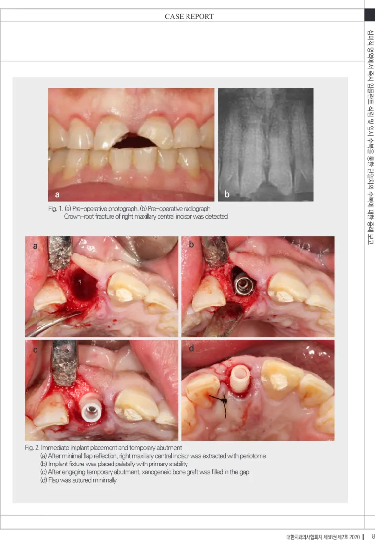
47세 여성 환자가 외상 후 상악 전치부 치아 파절을 주소로 내원하였다. 특별한 의과적 병력은 없었으며, 상 악 우측 중절치에 치관치근파절과 상악 좌측 중절치에 3급치관파절이 있는 상태였다(Fig. 1). 상악 좌측 중절치 는 근관치료 후 레진 수복으로, 상악 우측 중절치는 발치 후 임플란트로 수복하기로 계획하였다. 본 증례는 해당 치아의 협측 치조골판과 인접 치간골이 건전하고, 치은 이 thick biotype이며, 환자가 비흡연자였기 때문에 즉 시 식립 임플란트의 적응증으로 고려되어 계획되었다.
상악 우측 중절치에 열구내절개와 원심측 치간유두 에 절개를 시행하여 최소한으로 전층판막을 거상하였 다. 페리오톰을 사용하여 최소한의 외상으로 상악 우측 중절치를 발치하였다(Fig. 2a). 임플란트 드릴을 사용하 여 발치와의 구개측을 향하도록 임플란트 식립 부위를 형성하였다. 이후 인접치의 백악법랑경계 하방 3mm 에 위치하도록 임플란트(Implantium®, Dentium, Seoul, Korea; Φ4.3x14mm)를 식립하였다(Fig. 2b).
20Ncm의 초기 고정력을 얻었고, 준비한 임시지대주(Φ 5.5mm)를 체결하였다. 남은 발치와의 결손부는 이종골 (Bio-Oss®, Geistlich, Wolhusen, Switzerland; 0.25g) 로 충전하고(Fig.2c) 천연콜라겐막(CollaTape®, Zim- mer Dental, Carlsbad, CA, USA)을 트리밍하여 협측 만 덮은 후 봉합하였다(Fig. 2d).
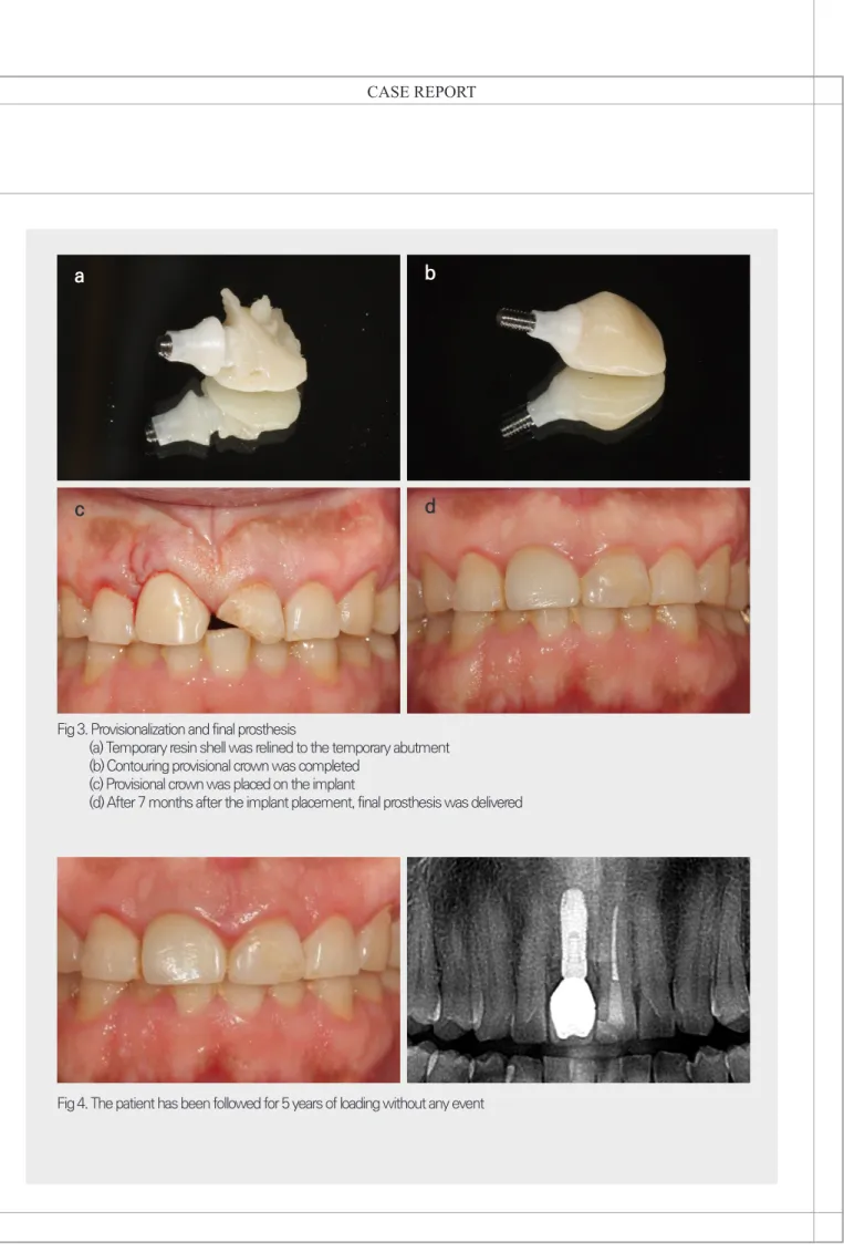
미리 제작한 temporary shell을 이용해 식립한 임플 란트에 임시보철물을 제작하였다(Fig. 3a). 조직압박을
Fig. 1. (a) Pre-operative photograph, (b) Pre-operative radiograph Crown-root fracture of right maxillary central incisor was detected
Fig. 2. Immediate implant placement and temporary abutment
(a) After minimal flap reflection, right maxillary central incisor was extracted with periotome (b) Implant fixture was placed palatally with primary stability
(c) After engaging temporary abutment, xenogeneic bone graft was filled in the gap (d) Flap was sutured minimally
a b
a
c
b
d
a
c
b
d
Fig 3. Provisionalization and final prosthesis
(a) Temporary resin shell was relined to the temporary abutment (b) Contouring provisional crown was completed
(c) Provisional crown was placed on the implant
(d) After 7 months after the implant placement, final prosthesis was delivered
Fig 4. The patient has been followed for 5 years of loading without any event
태로 형성하였다(Fig. 3b, 3c). 대합치와의 교합은 형성 하지 않았으며 이후 점진적인 임시보철물의 형태 수정 을 통해 적절한 치은 형성을 도모하였다. 수술 후 5개월 째에 최종 인상을 채득하였고, 7개월째에 최종 보철물을 장착하였다(Fig. 3d).
이후 5년의 하중기간동안 임플란트 주변의 연조직과 경조직에 변화는 관찰되지 않으며 특별한 수술적 및 보 철적 합병증 없이 잘 유지되고 있다(Fig. 4).
CASE 2
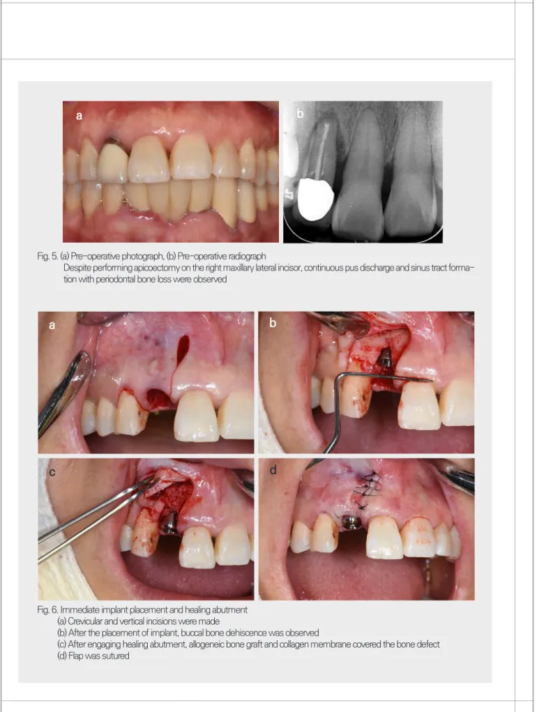
54세 여성 환자가 상악 우측 측절치의 동요도를 주소 로 내원하였다. 이미 근관치료가 되어 있던 치아로 치근 단절제술을 시행하였으나 이후에도 동요도는 소실되지 않고 지속적인 누공 형성과 배농이 있어 발치 후 임플란 트 수복을 계획하였다(Fig. 5). 치근이 짧고, 골파괴가 광 범위하지 않아 고정을 얻을 수직골이 충분하고, 인접한 치간골의 높이가 양호하여 발치 후 즉시 임플란트 식립 을 계획하였다. thin biotype으로 인하여 식립 후 치은 퇴축이 발생할 경우 연조직이식의 시행 가능성을 고지 하였다.
최소한의 외상으로 치아를 발치한 후 협측골의 일 부 소실을 확인하였다. 협측 판막 거상과 치간유두 보 존을 목적으로 열구내절개와 수직절개를 시행하고 전 층판막을 거상하였다(Fig. 6a). 임플란트 드릴을 사용하 여 발치와의 구개측을 향하도록 임플란트 식립 부위를 형성한 후, 인접치의 백악법랑경계 하방 3mm에 위치 하도록 임플란트를 식립하였다 (Implantium®, Den- tium, Seoul, Korea; Φ3.8x10mm). 40Ncm의 초기 고정력으로 식립하였고, 협측골의 열개형 결손부가 관 찰되었다(Fig. 6b). 치유지대주 체결 후 골결손부는 동 종골(ICB®, Rocky Mountain Tissue Bank, Aurora, CO, USA; 0.25g)과 흡수성 콜라겐막(Collagen mem- brane®, Genoss, Suwon, Korea; 10 x 20mm)으로 골
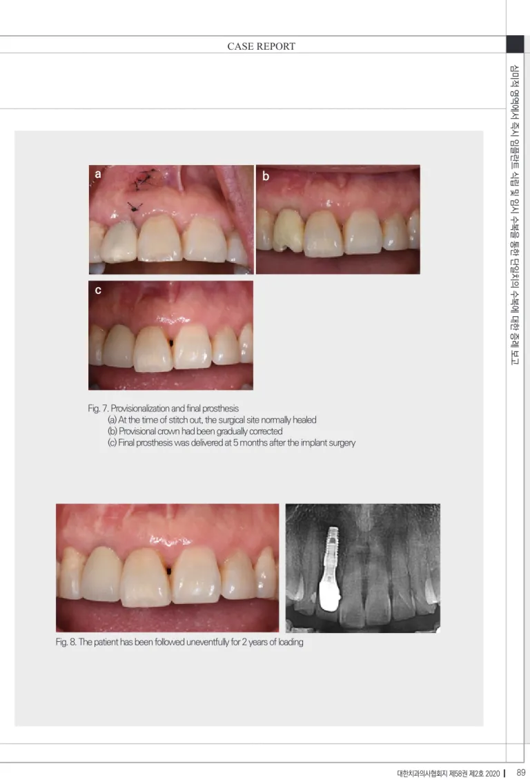
유도재생술을 시행한 뒤 봉합하였다(Fig. 6c, 6d) 수술 당일에 식립한 임플란트 위로 임시 보철물을 제작하였다(Fig. 7a). 보철물은 대합치와 교합되지 않 게 형성하였다. 이후 점진적으로 임시 보철물의 형태 를 수정하였으며(Fig. 7b), 4개월 후 최종 인상을 채득 하고 5개월째에 최종 보철물을 장착하였다(Fig. 7c).
수술 직후보다 미약한 협측 치은연의 퇴축이 관찰되지 만 환자와 상의하여 임플란트 주위 점막의 높이는 양 호한 것으로 판단하여 추가적인 연조직이식은 시행하 지 않기로 하였다.
이후 2년의 하중 기간동안 특별한 합병증없이 잘 유 지되고 있으며, 특히 협측 연조직의 위치는 변화가 없 었다(Fig. 8).
III. 고찰
이번 증례보고에서는 발치 후 임플란트 식립이 계획 된 상악 전치부에 즉시 식립 임플란트와 즉시 임시 보철 물을 통하여 심미성을 유지하면서 수복한 증례들을 다 루었다.
즉시 임플란트 식립은 기본적으로 발치와에 식립한 다는 점에 있어서 시간에 따른 발치와의 형태 변화에 대 한 고려가 필요하다. 발치 후 치조제는 위축이 진행되는 데, 발치 후 첫 3개월에 그 변화가 가장 크고5), 평균적으 로 폭이 3.87mm, 순측 높이가 1.53mm 감소하는 것으 로 나타났다.6) 즉시 임플란트 식립에서는 그 변화량이 감소하는 것으로 보이지만 변화는 불가피해 보인다7,8) . 따라서 발치와의 변화를 고려한 3차원적인 임플란트의 식립 위치가 중요하며, 이는 임플란트를 통한 심미수복 의 시작이 된다.
임플란트 식립에 있어 심미적으로 예지성 있는 결과 를 얻기 위해서는 발치와의 연조직과 경조직이 충분한
Fig. 5. (a) Pre-operative photograph, (b) Pre-operative radiograph
Despite performing apicoectomy on the right maxillary lateral incisor, continuous pus discharge and sinus tract forma- tion with periodontal bone loss were observed
Fig. 6. Immediate implant placement and healing abutment (a) Crevicular and vertical incisions were made
(b) After the placement of implant, buccal bone dehiscence was observed
(c) After engaging healing abutment, allogeneic bone graft and collagen membrane covered the bone defect (d) Flap was sutured
a b
a
c d
b
Fig. 7. Provisionalization and final prosthesis
(a) At the time of stitch out, the surgical site normally healed (b) Provisional crown had been gradually corrected
(c) Final prosthesis was delivered at 5 months after the implant surgery
Fig. 8. The patient has been followed uneventfully for 2 years of loading
a
c
b
상태여야 한다. 이를 평가하기 위한 요소로서 연조직의 양과 질, biotype, 치조돌기의 높이, 치근단 하방의 가용 골의 양, 순측 치조골판의 높이와 두께, 발치와의 병소, 치간골의 높이, 인접치 사이의 거리, 발치와의 순구개측 경사도 등이 제시되었다. 부적절한 요소가 있을 경우, 골 유도재생술이나 연조직이식술을 동반하거나 단계적 접 근법으로 임플란트를 식립하는 것이 추천된다9).
치간유두의 존재는 심미적인 치료 결과에 필수적인 요소이다. 단일치의 임플란트 수복에서는 인접치의 존 재로 인해 치간골을 유지하기 용이하다. 한 연구에서는 임플란트와 자연치 사이의 치간 유두 높이는 임플란트 보다는 자연치쪽의 골 높이가 결정짓는다고 하였다.10) 따라서 본 두 증례에서는 추후 보철물의 형태만 잘 조 절된다면 치간유두의 형성은 어렵지 않을 것으로 사료 된다.
또한 임플란트의 심미성을 평가하는 방법 중 가장 빈 번히 사용되는 지표로 정중 협측 임플란트 주위 점막 높 이가 있다11). 임플란트 주위 점막의 안정성 저하로 점막 퇴축이 나타날 수 있다. 상악 전치부에서 즉시와 조기 임 플란트 식립 후 심미적 결과를 평가한 한 체계적 문헌고 찰에서 즉시 식립 임플란트 후 1년에서 3년동안 9%에 서 41%의 부위에서 1mm 이상의 임플란트 주위 점막 의 퇴축이 나타나는 것으로 조사되었다12). 이것의 위험 요소로서 얇은 순측 골판, 온전한 순측 골판의 결손, 임 플란트의 협측 위치, 연조직의 thin biotype이 보고되 고 있다13).
임플란트 주위 점막의 퇴축을 최소화하기 위해서는 우선 연조직 하방의 골이 온전해야 한다. 얇은 순측골판 또는 순측골판의 결손이 있는 경우 골유도재생술을 고 려할 수 있다. 다양한 문헌에서 즉시 식립 임플란트에서 골폭을 증강시키기 위해서 또는 골결손부를 수복하기 위해서 골유도재생술을 동반하는 것을 추천한다1,14,15) .
재생술을 동반한 즉시 식립 임플란트로 수복하여 심미 적으로 안정적인 결과를 보였음을 보고하였다.16,17) 하지 만 술후 7-9개월동안 관찰한 단기간의 결과라는 한계가 있어 장기적인 추적관찰이 필요하다.
첫번째 증례에서는 발치 후 순측골은 온전하였으나 골의 형태를 최대한 보존하기 위하여 Bio-col tech- nique을 응용하였다. 이 술식은 발치와에 Bio-Oss®를 이식한 후 흡수성의 콜라겐 재료로 상부를 덮는 방식으 로 이루어지는데, 즉시 식립 임플란트에서도 잔존한 발 치와에 사용할 수 있다. Sclar(2004)는 본 술식을 소개 하면서 원래 자연치에서 경조직과 연조직의 형태를 최 대한 보존하여 심미적인 결과를 얻을 수 있다고 하였다
18). 즉시 임시보철을 동반할 경우, 보철물이 콜라겐 재료 의 유지에 도움을 줄 수 있는 부가적인 장점도 있다. 두 번째 증례의 경우, 협측골의 열개형 결손이 존재하기 때 문에 차폐막을 사용한 골유도재생술을 시행하기로 계획 하였고, 보다 빠른 신생골의 형성을 위해 동종골을 선택 하였다. 사용한 ICB®는 빠르게 신생골로 치환되고, 안정 적인 신생골 형성능력을 보여 자가골과 가장 비슷하다 고 생각되고 있다.
두 증례는 임플란트 식립 부위에서 경조직의 상태에 차이가 있다. 첫번째 증례의 경우 치아파절만 발생한 상 태로 온전한 경조직을 유지하고 있으나, 두번째 증례는 지속적인 염증으로 인해 순측골의 일부가 소실되었으 며, 임플란트 식립 시에도 열개형 결손이 관찰되었다. 이 로 인해 결과적으로 순측 점막의 안정성에 차이가 있을 것으로 예상되며, 실제로도 첫번째 증례는 점막 높이를 유지하는데 반해 두번째 증례는 수술 시와 비교하여 미 약한 점막 퇴축이 관찰되었다. 그럼에도 치유 초기에 점 막 퇴축이 발생한 이후로는 수술 시 동반했던 골유도재 생술의 효과로 인해 순측 점막 높이는 잘 유지되는 것으 로 보인다.
에 결합조직이식술을 동반할 경우, 임플란트 주위 점막 의 퇴축을 방지하는데 도움이 될 수 있다19,20). 이는 임플 란트 주변의 연조직을 thin biotype에서 thick biotype 으로 전환시킴으로써 점막의 퇴축에 좀더 저항성을 띄 는 것으로 생각된다. 따라서 thin biotype으로 점막퇴 축이 우려되는 경우, 결합조직이식을 고려해 볼 수 있다.
즉시 임플란트 식립과 동시에 임시 수복물을 장착하 면 환자에게 높은 심미적 만족감을 줄 수 있으나 보철물 에 작용하는 측방력으로 임플란트의 초기 안정성이 저 하되면서 임플란트의 실패 가능성을 증가시킬 수 있다.
상악전치부에서는 임시 보철물에 교합력이 가해지면 측 방력으로서 작용할 수 있다. Grutter(2009) 등은 심미적 인 부위에서 즉시 식립 임플란트 후 임시보철물을 장착 하였을 때, 교합의 형성 유무에 따라 임플란트 생존률이 각각 92.8%와 97.1%로 교합력이 작용하는 군에서 생존 률이 저하된다고 보고한 바 있다21). 이러한 연구의 결과 로 2014년 ITI consensus에서는 심미적 영역에서 즉시 부하보다는 조기 부하를 일상적으로 적용할 것을 권장 하였다13).따라서 즉시 임시 수복시에 중심위와 편심위 에서 임시 보철물이 대합치와 접촉하지 않도록 형성하 는 것이 중요하다.
최종 인상 채득의 시기는 여러 문헌들마다 수술 후 3-6개월로 다양하게 나타난다. 한 연구에서는 수술 후
협측 점막의 퇴축은 초기 3개월동안에 대부분이 발생하 므로 심미성이 요구되는 부위에서는 최종 인상 채득을 적어도 술 후 3개월이 지나고 진행할 것을 권유하였다
22). 또한 Grunder(2000) 등은 최종 보철물을 장착하기 전에 임시 수복물로 최소 6개월을 지켜볼 것을 조언하 였다10). 따라서 최종 인상 채득은 술 후 3-6개월의 범위 에서 임플란트 주변의 연조직 변연이 안정되었다고 판 단되는 시기에 시행할 수 있다. 본 증례에서도 앞서 기 술한대로 최종 인상은 각각 술 후 5개월과 4개월에 채 득하였고, 1-2달의 관찰기간을 더 가진 후 최종 보철물 을 장착하였다.
IV. 결론
본 보고에서는 심미성이 요구되는 상악 전치부에서 발치 후 즉시 임플란트 식립과 임시 보철물을 통해 성공 적으로 수복한 증례에 대해 다루었다. 즉시 식립 임플란 트와 즉시 임시 보철물은 환자의 상태가 양호한 경우 환 자의 전치부 상실기간을 없애고 심미적 수복을 이룰 수 있는 술식이다. 환자의 상태와 술자의 숙련도에 따라 일 부 제한점들을 극복한다면 보다 더 많은 환자에게 빠르 고 심미적인 임플란트 수복이 가능할 것이다.
1. Buser D, Martin W, Belser UC. Optimizing esthetics for implant res- torations in the anterior maxilla: anatomic and surgical considerations.
Int J Oral Maxillofac Implants 2004;19 Suppl:43-61
2. Chen ST, Buser D. Clinical and esthetic outcomes of implants placed in postextraction sites. Int J Oral Maxillofac Implants 2009;24 Sup- pl:186-217
3. Bhola M, Neely AL, Kolhatkar S. Immediate implant placement:
clinical decisions, advantages, and disadvantages. J Prosthodont 2008;17(7):576-581
4. Saito H, Chu SJ, Reynolds MA, Tarnow DP. Provisional Restorations Used in Immediate Implant Placement Provide a Platform to Pro- mote Peri-implant Soft Tissue Healing: A Pilot Study. Int J Periodon- tics Restorative Dent 2016;36(1):47-52
5. Schropp L, Wenzel A, Kostopoulos L, Karring T. Bone healing and soft tissue contour changes following single-tooth extraction: a clini- cal and radiographic 12-month prospective study. Int J Periodontics Restorative Dent 2003;23(4):313-323
6. Van der Weijden F, Dell'Acqua F, Slot DE. Alveolar bone dimensional changes of post-extraction sockets in humans: a systematic review.
J Clin Periodontol 2009;36(12):1048-1058
7. Araujo MG, Sukekava F, Wennstrom JL, Lindhe J. Ridge alterations following implant placement in fresh extraction sockets: an experi- mental study in the dog. J Clin Periodontol 2005;32(6):645-652 8. Lee CT, Chiu TS, Chuang SK, Tarnow D, Stoupel J. Alterations of
the bone dimension following immediate implant placement into extraction socket: systematic review and meta-analysis. J Clin Peri- odontol 2014;41(9):914-926
9. Juodzbalys G, Sakavicius D, Wang HL. Classification of extraction sockets based upon soft and hard tissue components. J Periodontol 2008;79(3):413-424
10. Grunder U. Stability of the mucosal topography around single-tooth implants and adjacent teeth: 1-year results. Int J Periodontics Re- storative Dent 2000;20(1):11-17
11. Benic GI, Wolleb K, Sancho-Puchades M, Hammerle CH. Sys- tematic review of parameters and methods for the professional assessment of aesthetics in dental implant research. J Clin Peri- odontol 2012;39 Suppl 12:160-192
12. Chen ST, Buser D. Esthetic outcomes following immediate and early implant placement in the anterior maxilla - a systematic re- view. Int J Oral Maxillofac Implants 2014;29 Suppl:186-215
13. Morton D, Chen ST, Martin WC, Levine RA, Buser D. Consensus statements and recommended clinical procedures regarding opti- mizing esthetic outcomes in implant dentistry. Int J Oral Maxillofac Implants 2014;29 Suppl:216-220
14. van Steenberghe D, Callens A, Geers L, Jacobs R. The clinical use of deproteinized bovine bone mineral on bone regeneration in con- junction with immediate implant installation. Clin Oral Implants Res 2000;11(3):210-216
15. Funato A, Salama MA, Ishikawa T, Garber DA, Salama H. Timing, positioning, and sequential staging in esthetic implant therapy: a four-dimensional perspective. Int J Periodontics Restorative Dent 2007;27(4):313-323
16. Waki T, Kan JY. Immediate placement and provisionalization of maxillary anterior single implant with guided bone regeneration, connective tissue graft, and coronally positioned flap procedures. Int J Esthet Dent 2016;11(2):174-185
17. Sarnachiaro GO, Chu SJ, Sarnachiaro E, Gotta SL, Tarnow DP. Im- mediate Implant Placement into Extraction Sockets with Labial Plate Dehiscence Defects: A Clinical Case Series. Clin Implant Dent Relat Res 2016;18(4):821-829
18. Sclar AG. Strategies for management of single-tooth extraction sites in aesthetic implant therapy. J Oral Maxillofac Surg 2004;62(9 Suppl 2):90-105
19. Yoshino S, Kan JY, Rungcharassaeng K, Roe P, Lozada JL. Effects of connective tissue grafting on the facial gingival level following single immediate implant placement and provisionalization in the esthetic zone: a 1-year randomized controlled prospective study.
Int J Oral Maxillofac Implants 2014;29(2):432-440
20. Zuiderveld EG, Meijer HJA, den Hartog L, Vissink A, Raghoe- bar GM. Effect of connective tissue grafting on peri-implant tis- sue in single immediate implant sites: A RCT. J Clin Periodontol 2018;45(2):253-264
21. Grutter L, Belser UC. Implant loading protocols for the partially edentulous esthetic zone. Int J Oral Maxillofac Implants 2009;24 Suppl:169-179
22. Small PN, Tarnow DP. Gingival recession around implants: a 1-year longitudinal prospective study. Int J Oral Maxillofac Implants 2000;15(4):527-532
참 고 문 헌