ISSN 2288-8101(Print) ISSN 2288-8586(Online)
Case Report
RECEIVED August 7, 2014, REVISED August 28, 2014, ACCEPTED September 22, 2014 Correspondence to Kyoung-Won Kim
Department of Oral and Maxillofacial Surgery, Medical Research Institute, Chungbuk National University College of Medicine 52 Naesudong-ro, Seowon-gu, Cheongju 362-763, Korea
Tel: 82-43-269-6383, Fax: 82-43-269-6387, E-mail: [email protected]
Copyright © 2014 by The Korean Association of Maxillofacial Plastic and Reconstructive Surgeons. All rights reserved.
CC
This is an open access article distributed under the terms of the Creative Commons Attribution Non-Commercial License (http://creativecommons.org/licenses/
by-nc/3.0) which permits unrestricted non-commercial use, distribution, and reproduction in any medium, provided the original work is properly cited.
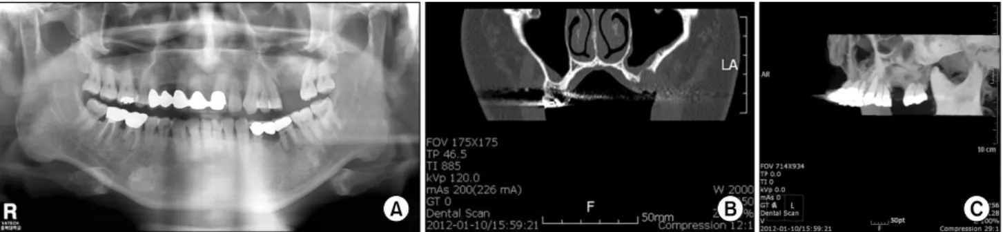
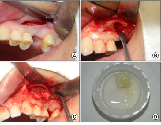
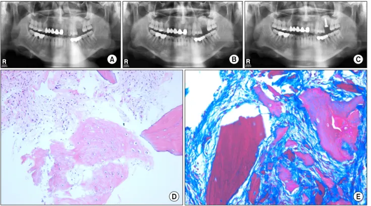
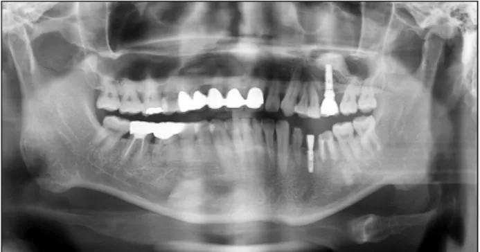
Vertical Augmentation of Maxillary Posterior Alveolar Ridge Using Allogenic Block Bone Graft and
Simultaneous Maxillary Sinus Graft
Eun-Young Lee, Eun-Suk Kim 1 , Kyoung-Won Kim
Department of Oral and Maxillofacial Surgery, Medical Research Institute, College of Medicine, Chungbuk National University,
1