Case Report
원고 접수일 2010년 11월 23일, 게재 확정일 2011년 2월 24일 책임저자 김수관
(501-759) 광주시 동구 서석동 375번지, 조선대학교 치의학전문대학원 구강악안면 외과학교실
Tel: 062-220-3815, Fax: 062-228-7316, E-mail: [email protected]
RECEIVED November 23, 2010, ACCEPTED February 24, 2011 Correspondence to Su-Gwan Kim
Department of Oral and Maxillofacial Surgery, School of Dentistry, Chosun University
375, Seosuk-dong, Dong-gu, Gwangju 501-759, Korea
Tel: 82-62-220-3815, Fax: 82-62-228-7316, E-mail: [email protected]
CC This is an open access article distributed under the terms of the Creative Commons Attribution Non-Commercial License (http://creativecommons.org/licenses/
by-nc/3.0) which permits unrestricted non-commercial use, distribution, and reproduction in any medium, provided the original work is properly cited.
자가치아골이식재를 이용한 치조능 수직 및 수평증대술: 증례보고
김영균1ㆍ김수관2ㆍ엄인웅3
1분당서울대학교병원 치과 구강악안면외과, 2조선대학교 치의학전문대학원 구강악안면외과학교실, 3한국치아은행
Abstract
Vertical and Horizontal Ridge Augmentation Using Autogenous Tooth Bone Graft Materials: Case Report
Young Kyun Kim 1 , Su-Gwan Kim 2 , In-Woong Um 3
Department of Oral and Maxillofacial Surgery,
1Section of Dentistry, Seoul National University Bundang Hospital,
2
School of Dentistry, Chosun University,
3Korea Tooth Bank
Horizontal and vertical ridge augmentation was performed using autogenous tooth bone graft block and powder in 44-year old male patient. Excellent bony healing was obtained 2∼4 months after ridge augmentation. Implant treatment was performed successfully.
Key words: Autogenous tooth bone, Ridge augmentation
서 론
골결손량이 큰 부위의 재건이나 치조능증대술과 같이 많은 양의 골증대량이 필요한 경우엔 자가골 이식이 가장 좋은 결과를 보인다. 자가골은 장골, 늑골, 경골과 같은 연골내골(endochon- dral bone)과 두개골, 안면골과 같은 막내골에서 채취할 수 있다.
치조능증대술은 수직적, 수평적으로 골량이 부족한 경우 치조능 의 상방 혹은 협측에 입자형 혹은 블록형 골이식을 시행하여 치조능의 높이 혹은 폭경을 증가시키는 술식을 말하며 동시에
수직 수평 증대술을 시행할 수도 있지만 개별적으로 시행하기도 한다. 일종의 온레이 이식이기 때문에 이식 후 골흡수가 많이 발생되고 상부 연조직 열개가 일어날 가능성이 높다[1].
한편 자가골이식은 공여부 합병증이 있고 시술시간이 많이
소요되며 채취량의 제한 등 여러 가지 문제점이 있어 환자 및
임상의들이 기피하는 경우가 많다. 자가골을 대체하기 위해 동종
골, 이종골, 합성골 등의 골이식재료들이 개발되었지만 수직 및
수평적으로 골조직을 증대시키는 술식에서는 단독 사용이 추천되
지 않는다[1,2].
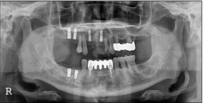
Fig. 1. Initial panoramic radiography. There were generalized al-
veolar bone resorption and multiple teeth mobility.Fig. 2. Autogenous tooth block was adapted and stabilized with
titanium screws.Fig. 3. Vertical ridge augmentation was performed using autoge-
nous tooth block at right maxillary posterior area. Block was stabi- lized with titanium screw. Surrounding defect was restored using allograft. Simultaneous sinus bone graft was performed with au- togenous tooth bone powder and allograft.Fig. 4. Vertical ridge augmentation was performed with autoge-
nous tooth bone graft block and powder at right mandibular pos- terior area.저자 등은 최근 개발된 입자 및 블록형 자가치아골이식재를 사용하여 성공적인 치조능증대술을 시행할 수 있었으며 증례와 함께 보고하는 바이다.
증례보고
44세 남자 환자가 다수치아 유동성 및 소실 부위에 대한 임프란 트 수복을 위해 내원하였다. 임상 및 방사선 검사 결과 전반적인 치조골 퇴축 및 하악 전치부 치아 상실 소견을 보였으며 진행성 만성 치주염 진단 하에 다수 치아들을 발치하고 치조능 증대술 및 상악동골이식과 임프란트 식립을 계획하였다(Fig. 1).
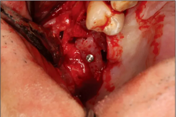
2009년 10월 20일 #16, 15, 12, 11, 21을 발치한 후 #15, 16은 블록, 나머지는 분말로 처리하여 보관하였다. 2009년 11월 19일 #45, 46을 발치한 후 1개는 블록 다른 1개는 분말로 처리하 였으며 당일 #12-21 부위 치조능 증대술을 시행하였다. 피판을 거상한 후 자가치아골이식재 블록을 잘라서 순측에 적합시킨 후 티타늄 나사로 고정하고 주변에 자가치아골이식재 분말을 이식하 였다(Fig. 2). 흡수성 콜라겐막(Ossix plus)을 피개한 후 창상을 봉합하였다. 상악 우측 구치부 치조정 절개를 시행한 후 피판을
거상하였고 측방접근법을 통한 상악동골이식술을 시행하였다.
골이식재는 자가치아골이식재 분말과 동종골(Orthoblast II)을 혼합하여 사용하였다. 동시에 치조능 수직증대술을 위해 자가치 아골이식재 블록을 잘라서 이식하였고 주변에는 동종골(AlloBT) 을 이식하고 흡수성 콜라겐막을 피개한 후 창상을 봉합하였다 (Fig. 3). 2009년 12월 15일 하악 우측 구치부 피판을 거상한 후 자가치아골이식재 블록과 분말을 이용하여 치조능증대술을 시행하였다. 블록은 #45 부위에 위치시켰으며 흡수성 콜라겐막을 피개한 후 창상을 봉합하였다(Fig. 4). 2010년 2월 23일(골이식 2개월 후) 하악 우측 구치부 임프란트 식립술이 시행되었다 (Osstem GS III, #45:4D/8.5L, #46:5D/8.5L). 블록형 골이식재 는 잘 생착되었으며 주변의 골이식재 치유가 매우 우수한 양상을 보였다(Fig. 5). 오스텔 멘토로 측정한 임프란트 초기고정 값은
#45:63, #46:62 ISQ였다. 2010년 3월 16일(골이식 4개월 후)
Fig. 5. Right mandibular posterior area was exposed for implant
placement 2 months after ridge augmentation. Excellent bony healing was observed.Fig. 6. Maxillary anterior ridge was exposed for implant place-
ment 4 months after ridge augmentation. Excellent bony healing was observed.Fig. 7. Maxillary right posterior area was exposed for implant
placement 4 months after ridge augmentation. Excellent bony healing was observed.Fig. 8. Panoramic radiography after implant placement.
상악 전치부 및 우측 구치부 임프란트 식립술(Osstem TS III SA, #15:4.5D/11.5L, #15:5D/11.5L, GS III, #12:3.5D/11.5L,
#21:4D/11.5L)이 시행되었다. 블록형 골이식재는 잘 생착되었고 주변 골이식재 치유가 매우 우수한 양상을 보였다(Fig. 6, 7).
임프란트 초기고정 값은 #15:75, #16:70, #12:63, #21:73 ISQ였 다(Fig. 8). 2010년 4월 20일 #45, 46 이차수술을 시행하였으며 이차고정 값은 #45:83, #46:70 ISQ였다. 2010년 6월 1일 상악 임프란트 이차수술이 시행되었고 이차고정 값은 #12:70, #21:74,
#15:76, #16:79 ISQ였다. 2010년 6월 25일 #15, 16, 45, 46 임프란트 상부 보철물이 장착되었고 2010년 8월 27일 #12-21 부위 상부 보철물이 장착되었다(Fig. 9).
고 찰
자가골의 단점을 보완하기 위해 개발된 동종골, 이종골, 합성골
등은 생활력 있는 골조직을 재생시켜야 하는 경우 즉 골벽이 없는 상태에서 수직 혹은 수평적으로 치조골을 증대시킬 경우엔 단독으로 사용하는 것은 추천되지 않는다.
자가 블록골은 강도가 세면서 형태 유지 효과가 좋고 막내골을 사용할 경우에는 흡수가 적기 때문에 치조능 수직 혹은 수평 치조능증대술을 시행할 때 유용한 효과를 발휘할 수 있다[3,4].
Raghoebar 등[5]은 상악전치부의 국소적인 치조골 결손부의 골증대를 위해 자가골이식을 시행한 증례들을 분석하였다. 1군은 하악골 정중부와 상행지에서 채취한 블록골을 사용하였고 2군은 상악결절에서 채취한 해면골을 사용하였다. 3개월의 치유기간을 부여한 후 임프란트를 식립하였으며 6개월 후 상부 보철물을 장착 하였다. 모든 부위에서 충분한 골증대 효과를 얻었고 보철 하중 24∼68개월 후에 모든 임프란트들이 잘 기능하고 있었다고 보고 하였다.
Ahn 등[6]은 자가 블록골을 이용한 수평 치조능증대술과 임프
란트 식립의 증례들을 분석한 결과 97.6%의 높은 임프란트 생존
율을 보였고 임프란트 주변 치조정골은 보철 기능 후 평균 17개월
의 관찰기 중에 약 0.9 mm의 흡수를 나타내어 비교적 안정적인
결과를 보였다. 그러나 술 후 합병증은 7명의 환자들에서 일시적
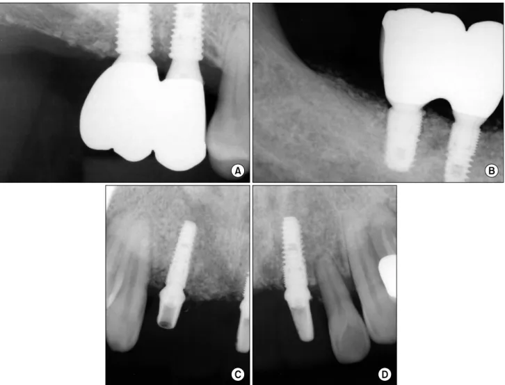
Fig. 9. Radiography and intraoral photography after final prosthetic delivery. (A) Right maxillary posterior area periapical radiography.
(B) Right mandibular posterior area periapical radiography. (C) Periapical radiography of maxillary right lateral incisor area. (D) Periapical radiography of maxillary left central incisor area.
인 지각이상이 발생한 경우가 가장 많았고 술 후 감염, 피하출혈 및 창상열개 등의 순이었지만 적절한 술 후 관리를 통해 심각한 문제점없이 잘 치유될 수 있었다고 보고하였다. 한편 Kim과 Im[7]
은 입자형골이식재를 이용한 수평 치조능증대술이 블록골 이식술 과 임상성적에 있어서 큰 차이를 보이지 않았다고 보고하였다.
최근 환자들에서 발치된 치아들을 최첨단 의료 공법으로 처리 하여 자가치아골이식재로 제조한 후 동일 환자의 골이식술에 이용 하는 방법이 개발되었다. 자가치아골이식재(AutoBT, Korea Tooth Bank, Seoul, Korea)는 이식 후 골유도 및 골전도에 의한 치유를 보이며 생체적합성이 매우 우수한 것으로 판명되었다 [8].
치아와 골의 화학적 조성은 매우 유사하다. 즉 enamel의 성분 은 총 무기성분 95%, 유기성분 0.6%, 물 4%이고 dentin은 무기 성분 70∼75%, 유기성분 20%, 물 10%로 구성되어 있다. 치조골 의 성분은 무기성분 65%, 유기성분 25%, 물 10%로 알려져 있다 [9]. Bhaskar[10]이 출판한 Orban’s Oral histology and embry-
ology에서는 enamel의 성분은 무기성분 96%, 유기성분과 물 4%, dentin은 무기성분 65%, 유기성분과 물 35%, cementum은 무기성분 45∼50%, 유기성분과 물 50∼55%, alveolar bone은 무기성분 65%, 유기성분 35%로 기술되어 있다.
자가치아골이식재 분말과 탈회시킨 블록은 점착성이 우수하고 결손부에 잘 적합되는 특성을 가지고 있으며 조작이 매우 편리하 다. 골유도 및 골전도에 의해 매우 우수하고 빠른 골치유를 보이며 창상이 일부 벌어지면서 노출되더라도 감염에 대한 저항성이 우수 하고 이차치유가 잘 이루어지는 장점이 있다[11]. 따라서 치조능 수직 및 수평 증대술에서 자가치아골이식재는 자가골 이식의 대체 수단이 될 수 있으며 양이 모자랄 경우엔 타 골이식재와 혼합하여 사용한다면 임상에서 매우 유용하게 사용할 수 있다고 사료된다.
본 증례에서는 상악 전치부 순측 증대술을 위해 블록을 나사로
고정하고 주변에 분말형 자가치아골이식재를 이식하였다. 또한
우측 구치부 치조능 수직증대술을 위해 블록과 분말형 이식재를
함께 사용하여 임상적으로 양호한 결과를 얻었으며 이차 수술
시 피판을 거상하였을 때 블록형 이식재가 잘 생착되어 있는 것이 확인되었다.
References