Ⅰ.
서 론
임플란트 치료 시 골열개 또는 골천공이 발생하는 경우 가 많으며 골이식재를 이용한 골유도재생술은 보편적인 술식이 되었다. 골유도재생술을 위한 가장 이상적인 재료 는 자가골이다. 그러나 자가골이식은 채취량이 제한되어 있고 공여부 합병증이 크며 이식 후 흡수가 많이 발생하는 문제점이 있다. 따라서 동종골, 이종골, 합성골과 같은 골 대체재료가 개발되어 임상에서 사용하고 있으나 각각의 장점을 공유하기 위해 자가골과 다른 골이식재를 혼합하 여 사용하는 경우가 많다. 한편 골유도재생술의 범위가 적 을 경우엔 다른 골대체재료를 사용해도 무방하다. 그러나 생활력 있는 골조직의 재생이 필요한 경우, 즉 광범위한 치 조능증대술, 결손부의 골벽이 거의 존재하지 않는 경우에 는 반드시 자가골이식을 선택해야 할 것이다. 동종골과 이
종골은 감염전파 등에 대한 막연한 불안감으로 인해 임상 의 및 환자들이 사용을 간혹 기피하는 경향을 보이기도 한 다. 합성골은 가격이 저렴하면서 질병전염의 위험성은 전 혀 없지만 골형성과 골유도 능력이 전혀 없어 생활력 있는 골조직 형성을 목적으로 할 경우엔 사용에 제한을 받고 있 다1.
저자 등은 1993년부터 자가골과 유사한 골재생 능력을 가지면서 동종골, 이종골, 합성골의 단점을 극복할 수 있는 새로운 골이식재 개발에 착수하였으며, 최근 발치한 자가 치아를 이용한 골이식재를 개발하였다. 본 논문에서는 임 플란트 식립 시 자가치아골이식재(AutoBT, Korea Tooth Bank Co., Seoul, Korea)분말을 이용하여 골유도재생술을 병행한 증례들을 소개하고 조직학적 소견과 함께 자가치 아를 이용한 골이식재의 우수한 골치유 능력을 언급하고 자 한다.
Ⅱ.
증례보고
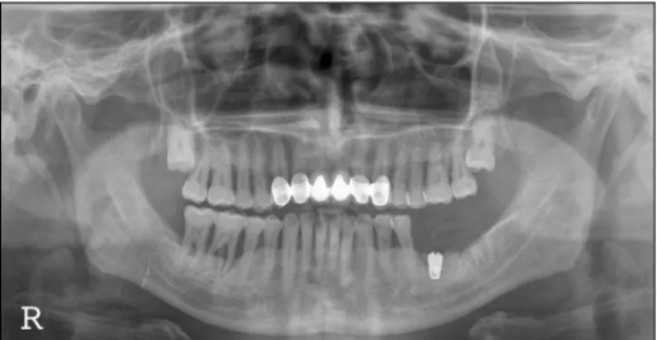
44세 남자환자가 다수 치아 유동성 및 통증을 주소로 내 원하였다. 임상 및 방사선검사에서 전반적인 치조골 소실 및 치근단 농양소견이 관찰되었으며 치주치료 및 다수 치 아들을 발치(#12, 22, 25, 27, 28, 35, 36, 37, 47)한 후 임플란 트치료를 계획하였다. 발치한 치아들은 자가치아골이식재 엄 인 웅
110-847 서울특별시 종로구 평창동229-4 한국자가치아뼈은행 기술개발연구소 In-Woong Um
Research & Development Center, Korea Auto-teeth & Bank 229-4, Pyeongchang-dong, Jongno-gu, Seoul, 110-847, Korea TEL: +82-31-787-7541 FAX: +82-31-787-4068
E-mail: [email protected]
자가치아골이식재를 이용한 골유도재생술: 증례보고
김영균1∙이효정2∙김경욱3∙김수관4∙엄인웅5 분당서울대학교병원 치과1구강악안면외과, 2치주과,
3단국대학교 치과대학 구강악안면외과, 4조선대학교 치의학전문대학원 구강악안면외과, 5한국자가치아뼈은행 기술개발연구소
Guide bone regeneration using autogenous teeth: case reports
Young-Kyun Kim1, Hyo-Jung Lee2, Kyung-Wook Kim3, Su-Gwan Kim4, In-Woong Um5
Departments of 1Oral and Maxillofacial Surgery, 2Periodontology, Section of Dentistry, Seoul National University Bundang Hospital, Seongnam,
3Department of Oral and Maxillofacial Surgery, College of Dentistry, Dankook University, Cheonan,
4Department of Oral and Maxillofacial Surgery, School of Dentistry, Chosun University, Gwangju,
5Research & Development Center, Korea Auto-teeth & Bank, Seoul, Korea
The authors installed implants combined with guided bony regeneration (GBR) using autogenous tooth bone graft material in the patients. In one patient, GBR and simultaneous implant placement were performed. In two patients, GBR was performed and the implants were placed after 6 months.
All patients achieved favorable clinical outcomes. Excellent osteoconductive bony healing was observed in the 6 month histology examination after the bone graft.
Key words:Autogenous tooth bone graft material, Osteoconductive bony healing
[paper submitted 2010. 11. 18 / revised 2011. 2. 9 / accepted 2011. 3. 26]
Abstract (J Korean Assoc Oral Maxillofac Surg 2011;37:142-7)
로 처리하여 치조능증대술 및 골유도재생술에 사용하기로 하였다.(Fig. 1) 2009년 1월 6일 하악 좌측 제1, 2대구치를 발치한 후 이종골(BioCera, Oscotec Inc., Cheonan, Korea)과 collagen sponge (Teruplug, Terumo Co., Ltd., Tokyo, Japan) 를 사용하여 치조능보존술(ridge preservation)을 시행하였 다. 하악 제2소구치는 수일 전에 저절로 탈락되었다.(Fig. 2) 2009년 3월 23일 하악 좌측 구치부 임플란트(Osstem GS III,
Osstem Implant Co., Ltd., Busan, Korea), 하악 좌측 제2소구 치(직경 4 mm, 길이 11.5 mm), 하악 좌측 제1, 2대구치(직경 5 mm, 길이 11.5 mm)를 식립하였다. Osstell Mentor (Integration Diagnostics AB, S¨avedalen, Sweden)로 측정한 초기 안정성은 하악 좌측 제2소구치 57, 제1대구치 74, 제2 대구치 78 implant stability quotient (ISQ) 값을 보였으며 식 립 후 주변 골열개 부위에는 자가치아골이식재를 이식하 고 흡수성 콜라겐막을 피개하고 창상을 1차 봉합하였 다.(Fig. 3) 육 개월 후 2차 수술을 시행하였으며 이전의 자 가치아골이식재의 골치유는 매우 우수한 결과를 보였 다.(Figs. 4, 5) #15 surgical blade를 사용하여 골이식 부위에 서 조직시편을 채취하였다.(Fig. 6) Osstell Mentor로 측정한 2차 안정성은 하악 좌측 제2소구치 86, 제1대구치 88, 제2 대구치 86 ISQ 값을 보였으며 치유지대주를 연결하고 창상 을 봉합하였다. 상부 보철물이 장착되었으며 6개월 경과관 찰 시점에 임플란트 주변의 연조직 상태가 매우 우수하고 치조정골이 안정적으로 유지되고 있었다.(Fig. 7)
그 외 2명의 환자들에서는 발치한 자가치아골이식재를
Fig. 2. Preoperative intraoral view. Teeth were extracted 2 months ago.
Fig. 3. Implants were placed and dehiscence defects were covered autogenous tooth bone graft material.
Fig. 4.Periapical radiography 6 months after implant placement. Fig. 5. Secondary surgery was performed and flap was elevated. Excellent bony healing was observed.
Fig. 1.Initial panoramic radiography.
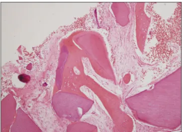
이용하여 골유도재생술을 시행하고 6개월 후에 임플란트 를 식립하였으며 성공적인 보철치료가 이루어졌다. 골이식 6개월 후 채취한 조직시편들에서는 자가치아골이식재들과
신생골들이 직접 유합하는 소견이 관찰되었고 신생골에서 는 골개조가 이루어지면서 새롭게 골수를 형성하는 자가골 의 이식과 유사한 치유과정이 관찰되었다.(Figs. 8-20)
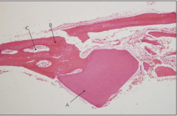
Fig. 6. The remodeling of new bones formed in the vicinity of graft materials is observed. A was graft materials, B was newly-formed bones, C was bone marrow.(H&E staining, original magnification x100)
Fig. 7.Periapical radiography 6 months after final prosthetic delivery.
Fig. 8. Guided bony regeneration (GBR) was performed at right 1st molar area of 49-year old female patient.
Autogenous tooth bone graft material and collagen mem- brane were used.
Fig. 9.Periapical radiography 3 weeks after bone graft.
Fig. 10. Periapical radiography 6 months after bone graft.
Alveolar crestal level was stable.
Fig. 11. Implant was installed 6 months after bone graft.
Bony quality was type I.
Fig. 12.Periapical radiography after final prosthetic delivery. Fig. 13. Microphotograph 6 months after AutoBT transplan- tation. Higher magnification demonstrated new bone forma- tion (arrows) around the implant chips (asterisks).(H&E staining, original magnification x100)
Fig. 14.Periapical radiography of 50-year old male patient 2 months after extraction of mandibular left 1st molar.
Fig. 15. Periapical radiography 2 weeks after autogenous tooth bone graft.
Fig. 16. Periapical radiography 5 months after autogenous tooth bone graft. Alveolar crestal bone level was stable.
Fig. 17. Implant was placed 6 months after bone graft.
Adjacent 2nd molar was extracted.
Ⅲ.
고 찰
저자 등은 1993년도부터 사람의 치아를 이용한 생체재료 개발과 실험연구를 진행하였으며 국내 특허를 획득하였다2. 또한 동물의 치아를 이용한 골이식재를 개발하여 미국 특 허를 취득하였다3. 치아를 이용한 골이식재의 무기질은 주 로 수산화인회석으로 구성되어 있으며 골대체재료로 사용 할 수 있는 가능성이 지금까지의 다양한 실험연구에서 입
증되었다4-7. 기존의 연구들을 바탕으로 환자들에서 발치되
는 치아들을 최첨단 의료공법으로 처리하여 자가이식재로 제조한 후 동일 환자의 골이식술에 이용하는 방법을 개발 하였다8.
치아와 치조골의 화학적 조성은 매우 유사하다. 즉 법랑 질의 성분은 총 무기성분 95%, 유기성분 0.6%, 물 4%이고 상아질은 무기성분 70-75%, 유기성분 20%, 물 10%로 구성 되어 있다. 치조골의 성분은 무기성분 65%, 유기성분 25%,
물 10%로 알려져 있다9. Orban’s Oral histology and embryol- ogy에서는 법랑질의 성분은 무기성분 96%, 유기성분과 물 4%, 상아질은 무기성분 65%, 유기성분과 물 35%, 백악질 은 무기성분 45-50%, 유기성분과 물 50-55%, 치조골은 무 기성분 65%, 유기성분 35%로 기술되어 있다10. 상아질에 존재하는 유기성분의 약 90%는 콜라겐이며 석회화에 중요 한 역할을 담당한다. 콜라겐은 주로 type I으로 구성되어 있 고 그 외의 유기성분은 비콜라겐성 단백질(non-collagenous proteins), 탄수화물(carbohydrate), 지질(lipid), citrate, lactate 등으로 구성되어 있다9. 단백질에는 bone morphogenetic protein (BMP)를 포함한 다양한 골성장 요소들이 존재하는 것으로 알려져 있으며 Choung11,12은 최초로 발치된 치아로 부터 치아단백질을 추출하는 방법과 그것의 이용에 관한 특허를 취득하였다.
자가치아를 이용한 골이식재는 치조골과 매우 유사한 무기질과 유기질 성분을 모두 포함하고 있기 때문에 골이 식 후 우수한 골유도 및 골전도 기능에 의한 골치유가 예 상된다8,9. 본 증례들에서 자가치아골이식재 이식 부위의 6 개월 후 조직학적 치유과정을 살펴볼 때 이식재가 서서히 흡수되면서 신생골로 대체되고, 신생골은 잔존 자가치아 골이식재들과 직접적인 유합을 이루고 있었다. 매우 우수 한 골전도에 의한 치유과정이 전 시편들에서 관찰되었고 골개조가 양호하게 이루어지는 것을 확인할 수 있었다. 임 플란트 상부 보철물이 완성된 후 경과관찰 기간 중에 임상 및 방사선학적으로 매우 안정적인 상태가 유지되었다. 따 라서 자가치아골이식재 분말은 골전도에 의한 골치유가 이루어지는 생체적 합성이 있는 재료로 임플란트 주변 골 유도재생술에 이용할 경우 좋은 결과를 보일 것으로 생각 한다.
Fig. 18. Second surgery was performed at left 1st molar area. Additional implant was placed at left 2nd molar area.
Fig. 19.Periapical radiography after final prosthetic delivery.
Fig. 20. Microphotograph 6 months after AutoBT transplan- tation. Higher magnification demonstrated new bone forma- tion around the implant chips.(H&E staining, original mag- nification x200)
References
1. Kim MJ, Kim YK, Kim SG. A variety of biomaterial used in dental surgery. Seoul: Narae Publishing Co.; 2004.
2. Kim YK, inventor; Tooth plaster and manufacturing method thereof. Korean patent 1019980008980. 1998 Mar 17.
3. Kim SG, Kim YK, inventors; Restorative and grafting material for hard tissue defects prepared from animal teeth. US patent 20030717801. 2003 Nov 19.
4. Kim YK, Yeo HH, Ryu CH, Lee HB, Byun UR, Cho JO. An ex- perimental study on the tissue reaction of toothash implanted in mandible body of the mature dog. J Korean Assoc Maxillofac Plast Reconstr Surg 1993;15:129-36.
5. Kim SG, Yeo HH, Kim YK. Grafting of large defects of the jaws with a particulate dentin-plaster of paris combination. Oral Surg Oral Med Oral Pathol Oral Radiol Endod 1999;88:22-5.
6. Kim SG, Chung CH, Kim YK, Park JC, Lim SC. Use of particu- late dentin-plaster of Paris combination with/without platelet-rich
plasma in the treatment of bone defects around implants. Int J Oral Maxillofac Implants 2002;17:86-94.
7. Kim SY, Kim SG, Lim SC, Bae CS. Effects on bone formation in ovariectomized rats after implantation of tooth ash and plaster of Paris mixture. J Oral Maxillofac Surg 2004;62:852-7.
8. Kim YK, Kim SG, Byeon JH, Lee HJ, Um IU, Lim SC, Development of a novel bone grafting material using autogenous teeth. Oral Surg Oral Med Oral Pathol Oral Radiol Endod 2010;109:496-503.
9. Min BM, ed. Oral biochemistry. Seoul: Daehan Narae Publishing, Inc.; 2007.
10. Bhaskar SN, ed. Orban’s Oral histology and embryology. 9th ed.
St. Louis: Mosby Co.; 1980.
11. Choung PH, inventor; Method for extracting tooth protein from extracted tooth. Korean patent 1020020008789. 2002 Feb 19.
12. Choung PH, inventor; Tooth protein extracted from extracted tooth and method for using the same. Korean patent 1020040051812. 2004 Jul 3.