서론
Brånemark이 완전 무치악 환자를 대상으로 골유착 임플 란트에 의한 수복 치료를 소개한 이후 단일 무치악, 부분 무 치악 또는 완전 무치악 부위에서 양호한 결과들을 나타내고 있다1). 상악 구치부는 하악이나 상악 전치부에 비하여 피질 골이 얇고 대부분이 망상골로 구성된 약한 골질로 임플란트
식립시 초기 고정이 어려운 경우가 많다. 또한 치아 상실 후 진행되는 치조골의 빠른 흡수와 호흡으로 인한 상악동 내의 공기압의 상승으로 함기화(pneumatization) 현상이 동반된 다2). 결국 상악동저와 치조제의 거리가 가까워져 충분한 길 이의 임플란트 식립이 어려워지기도 한다. 이러한 어려움을 극복하기 위하여 1980년 boyne과 James3)는 Caldwell-luc 수술법을 이용한 상악동 거상술을 발표하였다. 1986년 Tatum4)은 여러 가지 기구, bur, curette을 이용한 치조정접 근법을 소개하였고 이후 1994년 Summers5)는 Osteotome을 이용하여 골이식과 함께 임플란트를 식립하는 개선된 치조 정접근법을 발표하였다. 초기 측방 접근법에서 골이식재로 자가골이 사용되었고 1987년 Misch6)는 tricalcium phos- phate, 탈회골, 혈액 등을 이용하여 이식 후 98%의 성공률
상악동 거상술을 동반한 임플란트 식립 후 생존율에 대한 후향적 연구
유정아
1,2, 이상민
2, 유미경
2, 정의원
1, 김창성
1, 최성호
1, 박필규
2, 조규성
1*1. 연세대학교 치과대학 치주과학교실, 치주조직재생연구소 2. 서울 보훈병원 치과부 치주과
The retrospective study of survival rate of implants with maxillary sinus floor elevation Jeoung-A Yu
1,2, Sang-Min Lee
2, Mi-Kyung Yoo
2, Ui-Won Jung
1, Chang-Sung Kim
1, Seong-Ho Choi
1, Pil-Kyoo Park
2, Kyoo-Sung Cho
1*1. Department of Periodontology, Research Institute for Periodontal Regeneration, College of Dentistry, Yonsei University
2. Department of Periodontology, Dental Medical Center, Seoul Veterans Hospital
ABSTRACT
Purpose: The purpose of this study is to show the total survival rate of implants with maxillary sinus floor elevation and the effects that reach the survival rate by classifying types of graft materials, implant surface, operation method, bone height.
Methods: In a total of 131 patients, 251 implants with sinus floor elevation were installed simultaneously or after regular healing. Various bone grafts (autograft, xonograft, allograft, alloplast) and implant surface (MTX-HA implant, chemical etching implant, Titanium oxide surface implant, resorbable blasting media implant, resorbable blast texturing implant, HA-coated implant) were used. All implants were investigated clinically and radiographically, being with 1 to 5 years fol- low-up period after installation.
Results: The survival rate of 251 implants with maxillary sinus floor elevation was 94%. The types of implant, surface, graft material, bone height have no statistically signi-ficant differencies.
Conclusions: It can be suggested that maxillary sinus floor elevation may have predictable result with various bone graft materials and implant surface. (J Korean Acad Periodontol 2009;39:293-301)
KEY WORDS: maxillary sinus; dental implants; survival rate.
Correspondence: Dr. Kyoo-Sung Cho
Department of Periodontology, Research Institute for Periodontal Regeneration, College of Dentistry, Yonsei University, 250 Seongsanno, Seodaemun-gu, Seoul, 120-752, Korea
E-mail: kscho@yuhs.ac, TEL: 02-2228-3188, Fax: 02-392-0398 Received: Apr 27, 2009; Accepted: Jun 15, 2009
본 연구의 일부는 연세대학교 치과대학 치주조직재생연구소 연구비에 의해 이루어졌음.
을 보고하였고, Smiler와 Holmes7)는 이식재로 hydrox- yapatite를 사용하였다. 1988년 Wood와 Moore8)는 이식재 의 공여부위로 하악지와 오휘돌기를 사용하여 좋은 결과를 얻었다고 하였다. 1996년 Academy of OsseoIntegration Sinus Consensus Conference9)에서는 자가골이 상악동 이 식재로 유용하고 더 나아가 동종골, 이종골, 합성골 등의 다 른 이식재들의 유용성에 관해 긍정적인 발표를 하였고 더 많은 연구가 필요하다고 보고하였다. 이후 Hising10)은 Bio-ossⓇ와 같은 이종골의 단독 혹은 자가골과 혼합 이식 시에 비슷한 성공률을 발표하였고 Froum 등11)은 mineral- ized cancellous bone graft(Puros™)의 우수한 골형성력을 증명하였다. 또한 Lee 등12)은 MBCP™를 상악동 거상술에 사용시 예견성 있는 결과를 보고하기도 하였다. Kim 등13)은 Panoramic 방사선 사진에서 상악동 골이식 후의 이식재의 변화율을 측정하였는데 자가골의 혼합 배율을 낮추거나 또 는 자가골을 혼합하지 않은 골이식재를 사용하였을 때 더욱 안정적으로 유지된다고 하였다. 이처럼 이식재에 대해 다양 하게 임상적, 조직학적인 연구가 보고되고 있다. 상악동 거 상술에 사용된 이식재, 임플란트의 표면처리, 임플란트의 직경과 길이의 선택, 잔존골에 따른 수술 방법의 적절한 선 택은 임플란트의 생존율에 중요한 영향을 미친다. 이번 연 구의 목적은 상악동 거상술 후 식립된 임플란트의 전체 생 존율을 구하고 임플란트의 직경, 길이, 식립 위치, 이식재 종류, 임플란트 표면처리, 잔존골 고경, 수술 방법들을 분류 하여 생존율에 미치는 영향을 알아보고자 한다.
재료 및 방법
1. 연구 대상
1) 연구 환자
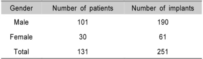
본 연구는 서울 보훈병원 치과에서 2002년 5월에서 2006 년 12월까지 상악동 거상술을 동반한 임플란트 수술을 받은 환자를 대상으로 하였으며 환자군은 남성 101명(58.6±8.1 세), 여성 30명(52.8±8.8세)으로 구성되어 있다(Table 1).
연령별로 분류하였을 때 51~60세의 환자가 43%로 가장 많 았다. 식립된 임플란트는 총 251개였으며 보철치료가 완료 된 환자들을 선별하였고 평균 관찰기간은 24개월이었다. 식 립 부위별 분류는 제 1소구치가 11개, 제 2소구치가 38개,
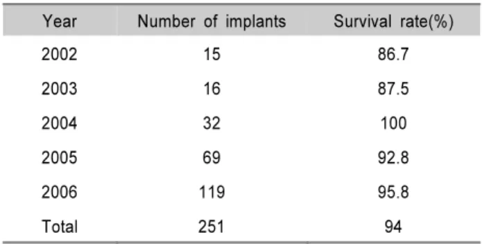
제 1대구치가 119개, 제 2대구치가 83개이고 이 중 제 1대 구치가 119개로 가장 많이 식립되었다. 임플란트 식립 연도 별 환자의 분포는 2002년부터 2006년까지 각각 15, 16, 32, 69, 119개의 임플란트가 식립되어 상악동 거상술이 점차적 으로 증가하는 것을 볼 수 있었다(Table 1, 2, 3).
Table 1. Patient Distrubution According to Gender Gender Number of patients Number of implants
Male 101 190
Female 30 61
Total 131 251
Table 2. Patient Distribution According to Age
Age Number of implants %
31~40 5 2
41~50 62 24
51~60 107 43
61~70 74 29
71~ 3 1
Total 251 94
Table 3. Implant Distribution According to Year Placed
Year Number of implants %
2002 15 6
2003 16 7
2004 32 13
2005 69 27
2006 119 47
Total 251 251
2) 연구 재료
(1) 이식재의 종류
자가골(autogenous bone), 이종골 Bio-ossⓇ(Geistlig- Pharma, Wolhusen, Switzerland), 동종골 DFDB(Demineralized Freeze Dried Allogenic bone), ICBⓇ(Rochy mount tissue bank, Denvor, CO), Puros™(Zimmer Dental, Carlbad, Calif), 합성골 MBCP™(Biomatlante Sarl, France), Hydroxyapatite 등을 단독 이식 및 혼합 이식하였다.
(2) 임플란트의 종류
총 6가지로 분류되며 MTX-HA implant(Taper Screw VentⓇ, Zimmerdental, Carlsbad, CA), Chemical etching im- plant(CamlogⓇ, Altatec, Wimsheim, Germany), Titanium- oxide implant(Tioblast™, Astra Tech, Mlndal, Sweden), Resorbable blasting media implant(GS Ц, Osstem, Busan, Korea), Resorbable blast texturing implant(BiohorozonⓇ, Biohorozons Implant System, Birmingham, AL), Tiunite (ReplaceⓇ Select, Nobel Biocare, Goteborg, Sweden) 등이 다. MTX-HA Implant는 가공 전의 순수 crystalline을 97%까 지 증가시킨 MP-1 HA(dual transition surface) 코팅과 생체 호환성 용매로 Grit-blasting하여 acid-washing한 것이다.
Tioblast™는 TiO2로 grit blasting하여 골 치유 과정을 촉진시키고 RBM은 HA(수산화인회석 Ca10(PO4)6(OH2)) 가 루를 이용한 표면처리의 특징을 가진다. RBT는 pure tita- nium oxide surface 위에 blast process에서 사용되는 calcium phosphate가 제조과정에서 용해되도록 하는 특징 을 가지고 있다. Tiunite는 산화막의 두께가 치근단 부위쪽 으로 두꺼워진다. 형태적 특성을 보면 Tioblast™와 RBM은 microthread, RBT는 사각형 나사 형태이고 다른 4개의 임 플란트는 아래가 좁아지는 치근형이다.
2. 연구 방법
1) 상악동 거상술의 수술 방법
(1) 측방 접근법(lateral approach)
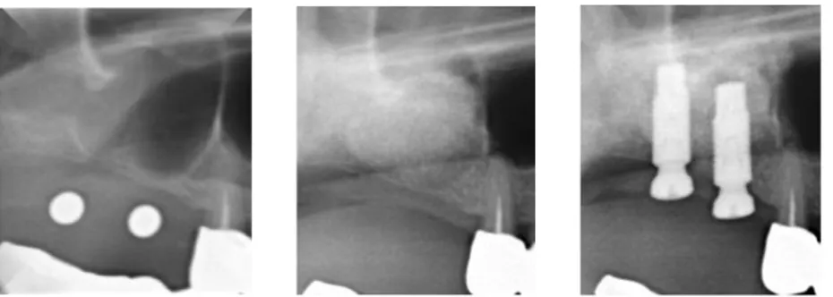
측방 접근법을 통한 상악동 거상술은 Kent와 Block 등이 제안한 변형된 Caldwell-Luc술식에 따라 시행되었다. 치조 정 수평 절개와 충분한 수직 절개를 가한 후에 전층 판막을 조심스럽게 형성하고 상악동 측벽을 고속 라운드 버로 상악 동 기저부에서 2~3mm 상방 위로 원형 골절제술을 시행하 여 상악동막을 거상하였다. 그 후 임플란트의 초기 고정이 가능한 경우 이식재와 함께 임플란트를 함께 동시 식립하였 고 초기 고정이 불가능한 경우는 임플란트를 식립하지 않고 골이식만 시행하였다(Fig. 1). 술 후 판막을 재위치시키고 봉 합한 후 투약 처방을 하였다. 항생제로 Amoxicillin(500mg
×3; Jonggeundang, Korea) or Augmentin(375mg×3; Ilsung, Korea)과 소염진통제로 Ibuprofen(200mg×3; Ildong, Korea) or Zaltoprofen(80mg×3; Jeiljedang, Korea)을 5일 처방하 였고 구강세정제로 cholorhexidin을 하루에 두 번 14일간 구강 세척하도록 하였다. 술 후 7일에서 14일 후 봉합사를 제거하였다. 지연 식립의 경우 상악동 골이식술 후 6~8개월 후에 임플란트를 식립하였다(Fig. 2).
Figure 1. X-rays of implants placed by sinus floor elevation(lateral approach-simultaneous):x -50 of sectioned view from panoramic X-ray.
Figure 2. X-rays of implants placed by sinus floor elevation(lateral approach-staged):x -50 of sectioned view from pan- oramic X-ray.
(2) 치조정 접근법(crestal approach)
치조정 접근법을 통한 상악동 거상술은 Summers가 제안 한 방법에 따라 시행하였다. 위와 같은 방법으로 절개와 판 막을 형성한 후 상악동 저 1 mm 하방까지 drill이나 osteo- tome으로 골형성을 하였다. 이식재를 넣고 osteotome이 골 형성한 위치까지 도달하도록 malleting을 하고 osteotome 이 상악동 내에 들어가지 않도록 하여 상악동을 거상하였다.
상악동 거상 후 임플란트를 식립하여 판막을 봉합하였다.
술 후 투약 처방은 측방 접근법과 동일하게 하였다.
2) 분석 방법
(1) 생존율
임플란트 생존율에 대한 기준은 Rosen 등14)의 criteria for success의 기준을 따라 평가하였고 이는 다음과 같다.
① 지속적인 동통, 감염, 감각 이상이 없을 것
② 임플란트 움직임 없을 것
③ 임플란트 주위로 연속성의 방사선 투과상이 없을 것
④ 임플란트 식립 1년 후에 매년 골소실이 2mm 이하일 것
(2) 분석 항목
환자의 진료 차트를 이용하여 1) 전체 생존율 2) 임플란 트의 직경, 길이, 식립 위치 3) 이식재의 종류 4) 임플란트 표면처리 5) 잔존골 고경 6) 수술 방법에 대하여 조사하였 다. 모든 환자들에 대해서 술전 방사선 검사, 구강 검사 및 전신 병력 검사를 시행하였다.
(3) 실패 시기의 분류
실패 시기는 Rosenberg15)의 분류에 따라 5단계로 분류하 였다.
① stage 1:임플란트 식립 이후 이차 수술하기까지의 기간
② stage 2:이차 수술과 최종 보철물 완성
③ stage 3:최종 보철물 완성 후 1년 이내
④ stage 4:1년~5년
⑤ stage 5:5년 이후의 기간
3) 통계학적 분석
임플란트 직경, 높이, 식립 위치에 따른 생존율을 구하고 이식재, 임플란트 표면처리, 잔존골 고경과 생존율의 유의 성을 알아보기 위하여 Logistic regression method을 이용 한 통계처리를 하였다. 유의성의 범위는 P<0.05로 하였다.
결과
1. 전체 생존율
131명의 환자를 대상으로 251개의 상악동 거상술을 동반 한 임플란트 전체 생존율은 94%였다. 연도별로 식립된 임플 란트의 생존율을 조사한 결과는 2002년에는 15개의 임플란 트가 식립되어 생존율은 86.7%로 나타났고 2006년에는 119 개의 임플란트 중 95.8%의 생존율을 보였다(Table 4, 5).
Table 4. Overall Survival Rate Survival
Survival rate(%)
Success Fail
Total 237 14 94.0
Table 5. Survival Rate of Implant According to Year Placed Year Number of implants Survival rate(%)
2002 15 86.7
2003 16 87.5
2004 32 100
2005 69 92.8
2006 119 95.8
Total 251 94
2. 임플란트 식립 위치, 직경, 높이에 따른 생존율
임플란트 식립 위치별 분류에서는 제 1소구치, 제 2소구 치, 제 1대구치, 제 2대구치 중 제 1대구치에서 임플란트가 119개로 가장 많았고 각각 생존율을 보면 90.9%, 92.7%, 94.4%, 96.5%로 나타났고 직경별, 높이별 생존율에서는 모 두 안정된 생존율을 보였다(Table 6, 7, 8).
Table 6. Survival Rate According to Implant Location Site Placed Failed Survival rate(%)
1st premolar 11 1 90.9
2nd premolar 38 3 92.7
1st molar 119 7 94.4
2nd molar 83 3 96.5
Total 251 14 94
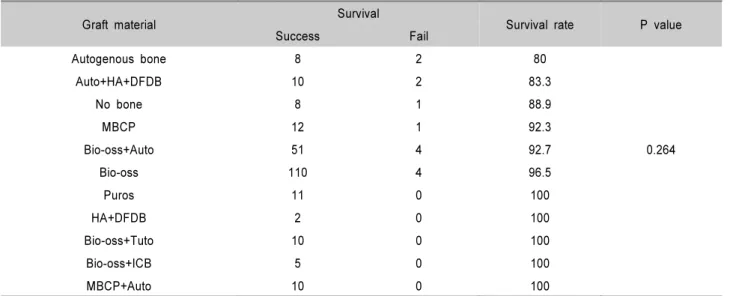
3. 이식재별 생존율
이식재 종류에 따른 분석 결과를 보면 단독 이식한 경우 에 자가골, MBCP™, Bio-ossⓇ, Puros™의 생존율은 80%, 92.3%, 96.5%, 100% 자가골 단독 이식한 경우 가장 낮은 생존율을 보였고 실패한 2개의 임플란트의 원인은 골소실이었 다. 혼합 이식의 경우 Auto+HA+DFDB, Bio-ossⓇ+Auto, HA+DFDB, Bio-ossⓇ+Puros™, Bio-ossⓇ+MBCP™, Bio- ossⓇ+ICBⓇ의 생존율은 83.3%, 92.7%, 100%, 100%, 100%, 100%, 100%로 나타났다. 골 이식 하지 않은 경우에서는
8개 중 1개가 실패하여 88.9%의 생존율을 볼 수 있었다. 이 식재별 생존율은 통계학적으로 유의하진 않았다(Table 9).
4. 임플란트 표면 처리별 생존율
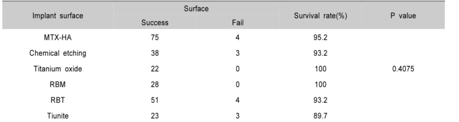
임플란트 표면 처리에 따라 Tiunite™, chemical etch- ing, RBT, MTX-HA, RBM, Tioblast™로 분류했을 때 임 플란트별 생존율은 각각 89.7%, 93.2%, 93.2%, 95.2%, 100%, 100%로 이 중 Tiunite™가 다소 낮은 결과를 보였다 (Table 10).
Table 7. Survival Rate According to Implant Width
Implant width Placed Failed Survival rate(%)
3.5 mm 2 0 100
3.7 mm, 3.8 mm, 4.0 mm 52 2 96.4
4.3 mm, 4.5 mm 45 3 93.9
4.7 mm, 5.7 mm 152 9 94.4
Total 251 14 94
Table 8. Survival Rate According to Implant Height
Implant Height Placed Failed Survival rate(%)
9 mm 2 0 100
10 mm, 10.5 mm, 11 mm, 11.5 mm 117 8 93.2
12 mm, 13 mm 132 6 95.4
Total 251 14 94
Table 9. Survival Rate According to Graft Bone
Graft material Survival
Survival rate P value
Success Fail
Autogenous bone 8 2 80
Auto+HA+DFDB 10 2 83.3
No bone 8 1 88.9
MBCP 12 1 92.3
Bio-oss+Auto 51 4 92.7 0.264
Bio-oss 110 4 96.5
Puros 11 0 100
HA+DFDB 2 0 100
Bio-oss+Tuto 10 0 100
Bio-oss+ICB 5 0 100
MBCP+Auto 10 0 100
5. 수술 방법에 따른 생존율
치조정 접근법, 측방 접근법(동시 식립), 측방 접근법(지 연 식립)에서 각각 93.6%, 97.1%, 77.8%의 생존율을 보였 다(Table 11).
6. 잔존골 고경에 다른 생존율
잔존골 고경에 따른 생존율은 1~3mm, 4~5 mm, 6~7 mm, 8mm 이상의 분류별로 각각 88.4%, 97%, 96.2%, 93.8%로 나타났고 1~3mm에서 낮은 생존율을 볼 수 있었 다(Table 12).
고찰
상악 구치부 골소실이 심하거나 상악동의 함기화가 심한 경우 골량이 불충분하여 임플란트 식립에 장애로 작용하게 되는데 이런 경우 상악동 거상술이 요구된다. 수술전 잔존 골 고경, 수술 방법, 이식재, 임플란트 선택 등 세부적인 준 비와 연구가 선행되었을 때 성공적인 임상적 결과를 기대할 수 있다. 성공적인 임플란트의 조건 중의 하나는 안정된 초 기 고정을 들 수 있다. 임플란트 식립 부위의 잔존골의 높이 는 임플란트 초기 고정을 얻고 또한 수술 방법 중 동시 식 립 또는 지연 식립을 결정하는 주요 요소가 될 수 있다.
Jensen과 Greek16)의 연구에서 상악동 거상술시 3mm 이하 Table 10. Survival Rate According to Implant Surface
Implant surface Surface
Survival rate(%) P value
Success Fail
MTX-HA 75 4 95.2
Chemical etching 38 3 93.2
Titanium oxide 22 0 100 0.4075
RBM 28 0 100
RBT 51 4 93.2
Tiunite 23 3 89.7
*HA:Hydroxyapatite, RBM:Resorbable blast media, RBT:Resorbable blast texturing.
Table 11. Survival Rate According to Operation Method
Op method Survival
Survival rate(%)
Success Fail
Crestal(93) 87 6 93.6
Lateral-simultaneous(140) 135 4 97.1
Letaral-staged(18) 15 4 79
Table 12. Survival Rate According to Bone Height
Bone height Survival
Survival rate(%) P value
success Fail
1~3 mm 38 5 88.4
4~5 mm 64 2 97 0.1223
6~7 mm 75 3 96.2
>8 mm 60 4 93.8
에서는 29%의 성공률로 저조하며 5mm 이상에서는 결과가 좋은 편이고 7 mm 이상에서는 매우 안정적이라 하였다.
Peleg 등17)은 잔존골 1~5mm에서 측방 접근법으로 동시 식 립한 2091개의 임플란트의 9년간의 cumulative survival rate를 97.9%로 발표하였고 이 중 20.4%가 잔존골이 1~2 mm였다고 하였다. Osteotome을 이용한 치조정 접근법에서 의 중요한 인자는 상악동저와 치조골정 사이의 잔존골 높이
이다18-19). Van den Bergh 등20)은 잔존골이 4mm 이하일
경우 이식재가 임플란트의 지지 역할을 못하여 초기 임플란 트의 안정성이 감소된다고 하였다. Ferrigno 등21)은 6~9 mm의 잔존골 높이에서 osteotome을 이용한 치조정 접근법 의 12년 누적 성공률을 94.8%로 발표하였고 Cavicchia 등22) 은 Summers method로 97개의 임플란트 식립 후 35개월의 평균 관찰 기간동안의 생존율은 88.6%로 발표하였다.
Toffler23)는 최근의 발표에서 평균 27.9개월 부하를 가한 276개의 임플란트의 생존율을 93.5%였고 4mm 이하의 잔 존골에서 73.3%로 떨어진다고 하였다. 이번 연구에서 치조 정 접근법에서 잔존골의 고경에 따른 생존율은 1~3mm의 고경에서 식립된 임플란트는 없었고 4~5mm에서 식립된 5 개의 임플란트가 모두 성공하였고 6~7 mm에서는 91.7%를 8mm 이상에서는 94.2%의 높은 성공률을 보였다. 측방 접 근법에서 잔존골 1~3mm 고경에서 동시 식립한 경우 96.3%의 높은 성공률로 Peleg의 연구와 비슷한 결과를 볼 수 있었다. 또한 측방 식립법 중 지연 식립의 경우 1~3mm 잔존골의 고경에서 16개의 임플란트 중 4개가 실패하여 가 장 낮은 75%를 나타냈다. 여기서 실패한 4개의 임플란트 중 2개의 임플란트의 원인이 골소실이었다. Herzberg 등24)은 4 mm 이하의 잔존골의 고경에서의 상악동 거상술 후 1년동 안의 marginal bone loss를 0.2mm를 기준으로 측정한 결 과 동시 식립에서는 94%, 지연 식립에서는 74%의 성공률을 발표하여 MBL(marginal bone loss)은 잔존골의 높이와 이 식재보다는 시간이 더 중요하게 작용한다고 하였다. 따라서 초기 고정만 얻을 수 있다면 지연 식립보다는 동시 식립이 추천된다고 발표하였다. 본 연구에서도 18개의 지연 식립된 임플란트 중 4개가 실패하였고 이 중 2개의 원인이 골소실 로 나타났다. 이는 위의 Herzberg 연구의 결과와 유사하게 나타남을 알 수 있고 계속적인 연구가 요구된다고 할 수 있 다. 초기 상악동 거상술의 연구에서는 자가골이 최상의 선 택 이식재이지만 공요부의 제한성과 비조절성 흡수율에 의 해 그 사용이 제한적임을 알 수 있다25). 또한 동물 실험에서
자가골을 이식재로 사용한 경우 임플란트 주위의 이식재가 점점 흡수되어 결국 상악동 내에 임플란트가 노출되었다26). 1999년 Academy of Osseointegration Sinus Consensus Conference에서는 자가골이 상악동 이식재로 유용하고 더 나아가 동종골, 이종골, 합성골 등의 다른 이식재들도 유용 하다 하였고 더 많은 연구가 필요하다고 발표되어 자가골 이외의 골이식재로 선택의 폭을 넓히는 계기가 되었다.
Schelege27)는 bio-ossⓇ가 6년동안 천천히 흡수되거나 거의 흡수가 되지 않는다고 하였고 Mcalliter 등28)은 bio-ossⓇ를 사용하여 상악동 거상술을 한 경우 안정된 결과를 보이고 생활력 있는 숙주골로 대체되어 함입된 소견을 관찰하였다 고 보고하였다. Hallman29)은 자가골, 20:80 자가골:이종 골 혼합 이식, 이종골 이식의 경우 각각 82.4%, 94.4%, 96%의 성공률을 발표한 바 있다. 본 연구에서의 동종골, 이 종골, 합성골 등의 단독 혹은 혼합 이식의 경우 높은 생존율 을 볼 수 있었다. Hallman의 연구 결과와 유사하게 이번 연구에서 이식재에 따른 성공률로 자가골, 자가골과 이종골 혼합이식, 이종골 이식의 경우 80%, 92.7%, 96.5%의 성공 률을 볼 수 있었고 Puros™ 등 동종골 이식의 경우도 100%
의 생존율로 긍정적인 평가를 내릴 수 있었다. 이 등30)과 김 등31)의 발표에 의하면 MBCP를 단독으로 사용하거나 그 외 의 이식재와 혼합해서 사용할 경우 예견성 있는 결과를 나타 내었다. Bone implant surface의 범위는 임플란트 표면의 거칠기가 증가할수록 증가한다. Titanium plasma sprayed implant는 30~40%의 골접촉률을, hydroxyapatite coated implant는 60~70% 골접촉률을 가지나 hydroxyapatite는 흡수의 양상을 보인다32). Wallace와 froum33)은 상악동 거상 술시 rough surface 임플란트와 Machined surface 임플란 트의 성공률을 비교할 때 95.2%, 82.4%로 rough surface 임플란트의 높은 성공률을 발표하였고 다른 rough surface 의 형태에 따른 통계적 유의성은 없었다고 하였다. 2005년 홍 등34)에 의해 상악동 거상술 및 BAOSFE를 동반하여 식 립된 Brnemark Tiunite와 ITI SLA 임플란트를 비교 관찰 하여 생존율에 대한 두 시스템간의 유의차가 없는 결과를 보여주었다. 이번 연구에서는 MTX-HA, chemical etch- ing, RBM, RBT, Tiunite,의 표면 처리가 된 6가지 임플란 트 시스템이 사용되었고 생존율의 통계적 유의성은 없었다.
이번 연구의 생존율은 251개의 임플란트 중 14개의 임플란 트가 실패하여 94%의 생존율을 나타내었고 실패한 임플란 트를 분석하자면 실패 시기는 stage 1이 50%로 가장 많았
고 실패한 원인으로는 골소실, 골유착 실패, 감염, 인접치의 치근단 병소 등을 들 수 있었다. 결과적으로 이식재나 임플 란트 표면 처리 등의 특징이 단독으로 상악동 거상술의 생 존율에는 영향을 미치지 않지만 잔존골의 고경과 임플란트 초기 고정의 상태에 따른 신중한 수술 방법의 선택 등이 중 요하다고 볼 수 있다. 이외에도 잔존골의 골질, 보철물의 loading 시기, 술 전 CT(computed tomography)에서 발견 할 수 있는 상악동 내의 병소 등이 생존율에 미치는 영향에 대한 추가적인 연구 등이 필요할 것이다.
참고문헌
1. Albrektsson T, Zarb G, Worthington P, Eriksson AR. The long-term efficacy of currently used dental implants:a re- view and proposed criteria of success. Int J Oral Maxillofac Implants 1986;1:11-25.
2. Chanavaz M. Maxillary sinus anatomy, physiology, surgery and bone grafting related to implantology-eleven years sur- gical experience(1979-1990). J Oral Implantol 1990;16:
199-209.
3. Boyne PJ, James RA. Grafting of the maxillary sinus floor with autogenous marrow and bone. J Oral Surg 1980;
38:613-616.
4. Tatum HJ. Maxillary and sinus implant reconstructions.
Dent Clin North Am 1986;30:207-229.
5. Summers RB. A new concept in maxillary implant sur- gery:the osteotome technique. Compend contin Educ Dent 1994;15:152-162.
6. Misch CE. Maxillary sinus augmentation for endosteal im- plants:organized alternative treatment plans. Int J Oral Implantol 1987;4:49-58.
7. Smiler DG, Holmes RE. Sinus lift procedure using porous hydroxyapatite:a preliminary clinical report. J Oral Implantol 1987;13:239-253.
8. Wood RM, Moore DL. Grafting of the maxillary sinus with intraorally harvested autogenous bone prior to implant placement. Int J Oral Maxillofac Implants 1988;3:209-214.
9. Jensen OT, Shulman LB, Block MS, Iacono VJ. Report of the sinus consensus conference of 1996. Int J Oral Maxillofac Implants 1998;13(suppl):11-45.
10. Hising P, Bolin A, Branting C. Reconstruction of severely
resorbed alveolar crests with dental implants using a bovine bone mineral for augmentation. Int J Oral Maxillofac Implants 2001;16:90-97.
11. Froum SJ, Tarnow DP, Wallace SS, Rohrer MD, Cho SC.
Sinus floor elevation using anorganic bovine bone materials with and without autogenous bone:a clinical, histologic, ra- diographic and histomorphometric analysis-Part 2 of an on- going prospective study. Int J Periodontics Restorative Dent 1998;18:528-543.
12. Lee JH, Jung UW, Kim CS, Choi CH, Cho KS. Maxillary sinus augmentation using calcium phosphate(MBCP):three case report with histologic evaluation. J Korean Acad Periodontol 2007;37:277-286.
13. Kim JS, Lee SK, Chae GJ et al. A radiographic evaluation of graft height changes after maxillary sinus augmentation and placement of dental implants. J Korean Acad Periodontol 2007;37:277-286.
14. Rosen PS, Summers R, mellado JR et al. The bone-added osteotome sinus floor elevation technique:multicenter retro- spective report of consecutively treated patients. Int J Oral Maxillofac Implants 1999;14:853-858.
15. Rosenberg ES, Cho SC, Elian N et al. A comparison of characteristics of implant failure and survival in perio- dontally compromised and periodontally healthy patients:a clinical report. Int J Oral maxillofac Implants 2004;19:
873-879.
16. Jensen OT, Greer R. Immediate placement of osseointegrat- ing implants into the maxillary sinus augmented with miner- alized cancellous allograft and Gore-Tex:second-stage surgi- cal and histologic findings. In: Laney WR, Tolman DE(eds).
Tissue Integration in Oral, Orthopedic, and Maxillofacial Reconstruction. Chicago:Quintessence Publishing Co Inc;
1992:321-333.
17. Peleg M, Garg AK, Mazor Z. Predictability of simultaneous implants placement in the severely atrophic posterior max- illa:a 9-year longitudinal experience study of 2,132 im- plants placed into 731 human sinus grafts. Int J Oral Maxillofac Implants 2006;21:94-102.
18. Nkenke E, Schelgel A, Schultze-Mosgau S, Neukam FW, Wiltfang J. The endoscopically controlled osteotome sinus floor elevation:a preliminary prospective study. Int J Oral Maxillofac Implants 2002;17:557-566.
19. Winter AA, Pollack AS, Odrich RB. Placement of implants in the severely atrophic posterior maxilla using localized
management of the sinus floor:a preliminary study. Int J Oral Maxillofac Implants 2002;17:687-695.
20. Van den Bergh JP, Ten Bruggenkate CM, Krekeler G, Tuinzing DB. Sinus floor elevation and grafting with au- togenous iliac crest bone. Clin Oral Implants Res 1998;9:
429-435.
21. Ferrigno N, Laureti M, Fanali S. Dental implants in con- juction with osteotome sinus floor elevation:a 12-year life-table analysis from a prospective study on 588 ITI implants. Clin Oral Implants Res 2006;17:194-205.
22. Cavicchia F, Bravi F, Petrelli G. Localized augmentation of the maxillary sinus floor through a coronal approach for the placement or implants. Int J Periodontics Restorative Dent 2001;21:475-485.
23. Toffler M. Osteotome-mediated sinus floor elevation:a clin- ical report. Int J Oral Maxillofac Implants 2004;19:
266-273.
24. Herzberg R, Dolev E, Schwarz-Arad D. Implant marginal bone loss in maxillary sinus grafts. Int J Oral maxillofac Implants 2006;21:103-110.
25. Aaboe M, Pinholt EM, Hjorting-Hansen E. Healing of ex- perimentally created defects:a review. Br J Oral Maxillofac Surg 1995;33:312-318.
26. Coombs CJ, Mutimer KL, Holmes AD et al. Osseointegration in sinus-forming bone. Plast and Reconstr Surg 1995;95:
866-875.
27. Schelgel AK. Long term results with Bio-oss bone replace- ment material. Schweiz Monatsschr Zahnmed 1996;106:
141-149.
28. McAllister BS, Marqolin MD, Cogan AG et al. Eighteen- month radiographic and histologic evaluation of sinus graft- ing with anorganic bovine in the chimpanzee. Int J Oral Maxillofac Implants 1999;14:361-368.
29. Hallman M, Sennerby L, Lundgren S. A clinical and histo- logic evaluation of implant integration in the posterior maxilla after sinus floor augmentation with autogenous bone, bovine hydroxyapatite, or a 20:80 mixture. Int J Oral maxillofac Implants 2002;17:635-643.
30. Lee JH, Jung UW, Kim CS et al. Maxillary sinus augmen- tation using macroporous biphasic calcium phosphate (MBCP):three case report with histologic evaluation. J Korean Acad Periodontol 2006;36:567-577.
31. Kim MS, Choi SH, Cho KS et al. A cumulative survival of implants installed on posterior maxilla augmented using MBCP after 2 years of loading:a retrospective clinical study.
J Korean Acad Periodontol 2008;38:669-678.
32. Buser D, Schenk RK, Steinemann S et al. Influence of sur- face characteristics on bone integration of titanium implants.
A histomorphometric study in miniature pigs. J Biomed Mater Res 1992;26:831-833.
33. Wallace SS, Froum SJ. Effect of maxillary sinus augmenta- tion on the survival of endosseous dental implants. A sys- temic review. Ann Periodontol 2003;8:328-343.
34. Hong SB, Chai GJ, Jung UW et al. Clinical evaluation of Brnemark Ti-unite implants and ITI SLA implant in the post maxillary area with sinus evaluation technique. J Korean Acad Periodontol 2005;35:813-822.