• 검색 결과가 없습니다.

Characterization of CCND1 and TWIST1 as Prognostic Markers with the Mortality Rate of Breast Cancer

N/A
N/A
Protected

Academic year: 2021

Share "Characterization of CCND1 and TWIST1 as Prognostic Markers with the Mortality Rate of Breast Cancer"

Copied!
11
0
0

로드 중.... (전체 텍스트 보기)

전체 글

(1)

Biomedical Science Letters 2018, 24(2): 76~86 https://doi.org/10.15616/BSL.2018.24.2.76 eISSN : 2288-7415

Characterization of CCND1 and TWIST1 as Prognostic Markers with the Mortality Rate of Breast Cancer

Sungwoo Ahn

1,§

, Sangjung Park

2,§

, Hye-Young Wang

3

, Sunyoung Park

1

, Jungho Kim

1

and Hyeyoung Lee

1,†

1

Department of Biomedical Laboratory Science, College of Health Sciences, Yonsei University, Wonju 26493, Korea

2

Department of Biomedical Laboratory Science, College of Life and Health Sciences, Hoseo University, Asan 31499, Korea

3

Optipharm M&D, Inc., Wonju Eco Environmental Technology Center, Wonju 26493, Korea

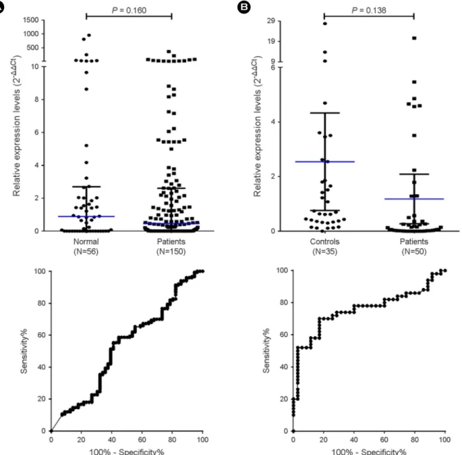
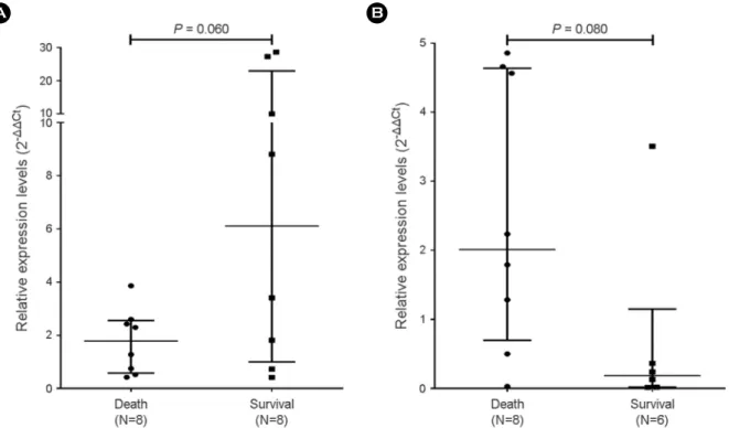
Breast cancer is one of the most common cancers affecting women worldwide. Although the survival rate of breast cancer has increased, breast cancer still results in a high mortality rate. Breast cancer deaths are caused by metastasis that occurs in organ dysfunction. Recently, there have been many studies on circulating tumor cells (CTCs), which are related to breast cancer metastasis in the blood. Recent studies have demonstrated that some CTCs do not express epithelial markers. Therefore, in this study, total RNA was extracted from blood without separating out the CTCs, and the characteristics of the CTCs were analyzed by RT-qPCR. Cyclin D1 and twist-related protein 1 (TWIST1) are well-known markers for predicting the prognosis of patients with breast cancer. However, few studies have demonstrated the use of CCND1 and TWIST1 in blood as diagnostic and prognostic markers of breast cancer. In this study, patients with late-stage breast cancer had overexpressed CCND1 and TWIST1 than patients with different stages of breast cancer (P < 0.001 and P < 0.01, respectively). The relative expression level of CCND1 in survivors was higher than in patients who died (P = 0.06). The relative expression level of TWIST1 in survivors was lower than in patients who died (P = 0.08). Overall CCND1 and TWIST1 were not useful as markers for the diagnosis of breast cancer through blood. However, we showed the possibility of using CCND1 and TWIST1 as prognostic markers, and a large-scale study is needed to confirm the usefulness of these prognostic markers.

Key Words: Breast cancer, CCND1, TWIST1, Circulating tumor cells, Blood, RT-qPCR, Prognosis

INTRODUCTION

Breast cancer is the most common cancer affecting women worldwide and is responsible for high mortality rate among all cancer (Ferlay et al., 2015). According to a World Health

Organization (WHO) report of 2015, there have been 1.6 million new cases per year and 520,000 deaths due to breast cancer (Torre et al., 2015).

Improvements in diagnostic and treatment methods have reduced the mortality rate of breast cancer. However, it re- mains the leading cause of mortality in female patients suf-

Original Article

*Received: May 3, 2018 / Revised: June 11, 2018 / Accepted: June 14, 2018

§Contributed equally to this work.

Corresponding author: Hyeyoung Lee. Department of Biomedical Laboratory Science, College of Health Sciences, Yonsei University, 1 Yonseidae-gil, Wonju, Gangwon 26493, Korea.

Tel: +82-33-760-2740, Fax: +82-33-760-2561, e-mail: [email protected]

CThe Korean Society for Biomedical Laboratory Sciences. All rights reserved.

CCThis is an Open Access article distributed under the terms of the Creative Commons Attribution Non-Commercial License (http://creativecommons.org/licenses/by-nc/3.0/) which permits unrestricted non-commercial use, distribution, and reproduction in any medium, provided the original work is properly cited.

(2)

fering from cancer (Miller et al., 2016). Mortality in patients with breast cancer is caused by metastases that cause dys- function of organs. Breast cancer is a systemic disease that causes micro-metastases, and also occurs in the very early stages of cancer (Hansen et al., 2009; Redig and McAllister, 2013). Tumor cells isolated from primary tumors have been known to metastasize to another organ through the blood- stream or lymph (Bonnomet et al., 2010).

Tumor cells entering the blood vessels are called cir- culating tumor cells (CTCs). CTCs can metastasize in two ways. The first is when the premalignant cells enter the blood vessels for metastasis to other tissues. The second is when there is the formation of primary cancer and the tumor cells simultaneously enter the blood vessels and transfer cancer to other tissues (Pantel and Speicher, 2016). Therefore, CTCs have been found not only in patients with metastatic breast cancer but also in those with early breast cancer (Barrière et al., 2012; Politaki et al., 2017).

To detect CTCs, the most widely used method for mea- suring the number of cells has been by capturing cells using epithelial cell adhesion molecule (EpCAM), which is a sur- face antigen. In particular, the CellSearch system (Veridex LLC, Raritan, NJ, USA) is the only CTC detection method approved by the US Food and Drug Administration (FDA).

It has been used to predict the outcome of breast, prostate, and colon cancers. In this system, epithelial cells are immune- magnetically separated and labeled with fluorescence, and then the number of nucleated (DAPI-positive) cells that are positive for EpCAM, as well as cytokeratin (CK) 8, 18, and 19, are estimated for the number of CTCs (Raimondi et al., 2014). Another method of studying CTCs involve immune- magnetic separation followed by multiplex RT-PCR of tumor markers such as human epithelial growth factor receptor 2 (HER2), mucin-1 (MUC-1), and GA773-2 (Müller et al., 2012).

However, CTCs in the blood vessels lose their characteristic of an epithelial cell and undergo epithelial-to-mesenchymal transition (EMT) during the process of entering the blood vessels, thereby adopting the characteristics of mesenchymal cells (Krebs et al., 2014). Because of this, there are many CTCs without surface antigen or EpCAM and various other characteristics. The CCND1 and TWIST1 are biomarkers

that have been researched to characterize CTC and breast cancer (Kim et al., 2014; Markiewicz et al., 2014).

The CCND1 is a gene that synthesizes cyclin D1, which regulates the G1 to S phase transition of the cell cycle (Motokura et al., 1991). Many studies have reported the oncogenic function of cyclin D1 (Ma et al., 2003; Oesterreich et al., 2003). In particular, over-expression of CCND1 is known to increase the synthesis of cyclin D1 and regulate the G1 to S phase, thereby contributing to cancer growth and affecting endocrine therapy resistance (Tian et al., 2007). The twist-related protein 1 (TWIST1) is a basic helix-loop-helix transcription factor that acts as an important regulator of cell migration and tissue reorganization during early embryo- genesis. However, TWIST1 is known to be reactivated in cancer (Ansieau et al., 2010). In cancer, TWIST1 is known to be an EMT-inducing factor that inhibits E-cadherin (Bonnomet et al., 2010). Therefore, TWIST1 causes cancer metastasis by inhibiting E-cadherin (Yang et al., 2004).

RT-qPCR is widely used in many diagnostic and prog- nostic marker research. RT-qPCR is a sensitive method, which can be used to detect small quantities of transcripts in the blood. Furthermore, each clinical sample can be easily nor- malized based on the expression levels of the reference genes.

In this study, an RT-qPCR assay targeting CCND1 and TWIST1 was developed and investigated to assess the re- lative expression levels of TWIST1 and CCND1 in the blood of breast cancer patients. The relationship between each marker and clinical information of breast cancer patients was analyzed. Furthermore, the association between the prognosis of patients and the two markers by comparing the relative expression levels of these genes in patients who survived versus in those who died.

MATERIALS AND METHODS

Breast cancer cell lines and cell culture

SKBR-3, MCF-7, BT-474, and MDA-MB-231 were used

for assay development and the generation of gene-specific

quantification calibrators. Human breast cancer cell lines,

SK-BR3 (KCLB No. 30030), MCF-7 (KCLB No. 30022),

BT-474 (KCLB No. 60062), and MDA-MB-231 (KCLB

No. 30026) were obtained from the Korean Cell Line Bank

(3)

(Seoul, Republic of Korea). The SKBR-3, MCF-7, BT-474, and MDA-MB-231 cell lines were grown at 37℃ in a humidified atmosphere with 5% CO

2

in the air.

SKBR-3, MCF-7, BT-474, and MDA-MB-231 cells were cultured in RPMI 1640 medium (Gibco-BRL, Carlsbad, CA, USA) supplemented with 10% fetal bovine serum (FBS), 100 U/mL of penicillin, and 100 μg/mL of streptomycin (Gibco-BRL, Carlsbad, CA, USA).

Study samples

A total of 152 patients with breast cancer and 56 healthy subjects were included in the study for evaluating CCND1, and a total of 51 patients with breast cancer and 38 healthy subjects were included in the study for evaluating TWIST1.

Patients with breast cancer were recruited from the Yonsei Severance Hospital (Seoul, Republic of Korea) from 2013 to 2014. Healthy subjects were recruited from Yonsei Severance Hospital and Yonsei University at Wonju. This study was approved by the Institutional Ethics Committee at Yonsei Severance Hospital (approval number 4-2011-0011 for patients with breast cancer and healthy donors) and Yonsei University at Wonju (approval number 1041849-201311-BM-020-02), and all subjects provided written informed consent.

The information on estrogen receptor (ER), progesterone receptor (PR), and human epidermal growth factor receptor 2 (HER2) was collected from the pathology reports of the Yonsei University Severance Hospital. ER and PR were posi- tive when over 10% of the cells were stained. HER2 status was determined based on the criteria from the American Society of Clinical Oncology/College of American Patholo- gists, as follows IHC 0 or 1+ considered as negative HER2 IHC, and 3+ was considered as positive HER2 IHC. When the HER2 IHC score was 2+, positivity and negativity were distinguished by gene amplification with fluorescence in situ hybridization (FISH).

Blood collection and total RNA extraction

All blood samples were obtained from a middle vein puncture. The blood cell samples were lysed with 1× ACK solution (0.15 M NH

4

Cl, 1 mM KHCO

3

, and 0.1 mM Na

2

EDTA). Total RNA was isolated using the Isol-RNA Lysis Reagent (5 Prime, Austin, TX, USA) according to the

manufacturer's instructions. The total RNA purity and con- centration were determined by measuring the absorbance at 260 and 280 nm using an Infinite 200

®

(Tecan, Salzburg, Austria) spectrophotometer. The preparation and handling of total RNA were conducted in a laminar flow hood under RNase-free conditions. The isolated total RNA was stored at -70℃.

Synthesis of cDNA and RT-qPCR assay

The cDNA was synthesized using an M-MLV Reverse Transcriptase Kit (Invitrogen, Carlsbad, CA, USA) with ran- dom hexamers (Invitrogen) according to the manufacturer's instructions. cDNA was synthesized at 25℃ for 10 min, then at 37℃ for 50 min, followed by at 70℃ for 15 min.

The relative expression of CCND1 and TWIST1 to gly- ceraldehyde-3-phosphate dehydrogenase (GAPDH) mRNA levels was measured with TaqMan probes in the CFX-96 real-time PCR system (Bio-Rad, Hercules, CA, USA). RT- qPCR amplification was performed with 10 μL of 2×

Thunderbird probe qPCR mix (Toyobo, Osaka, Japan), 3 μL of primer and TaqMan probe mixture, 2 μL of template cDNA, and distilled water to bring the final volume to 20 μL.

Each sample was tested in duplicate, and all PCR runs were performed twice. The reaction conditions were as followed:

95℃ for 3 min, followed by 40 cycles at 95℃ for 15 sec, and then at 55℃ for 30 sec. The relative gene expression was assessed using the comparative Ct method (ΔΔCt method).

The amount of target gene expression, normalized to an internal housekeeping gene and relative to a calibrator, was calculated by the 2

-ΔΔCt

method, which was normalized according to the following equation: ΔΔCt = [ΔCt

(test)

= Ct

(target test)

- Ct

(reference test)

] ― [ΔCt

(calibrator)

= Ct

(target calibrator)

― Ct

(reference calibrator)

]

The relative expression levels of CCND1 and TWIST1 were referred to a calibrator, and the healthy donor blood samples were used to represent the 1× relative expression.

The expression of CCND1 and TWIST1 were expressed as n-fold changes.

Statistical analysis

GraphPad PRISM software version 6.01 (GraphPad, La

Jolla, CA, USA) and the Statistical Package for the Social

(4)

Sciences software version 24 (SPSS Inc., Chicago, IL, USA) were used for statistical analysis. A receiver operating char- acteristic (ROC) curve analysis was performed to determine the optimal cut-off values. To determine whether the relative expression levels of CCND1 and TWIST1 were different according to the stage of breast cancer the Kruskal-Wallis test was performed because the number of patients per stage was too small for an analysis of variance (ANOVA) test.

The relative expression levels of CCND1 was analyzed by Student's t-test, and TWIST1 was analyzed by the Mann- Whitney test according to the results of IHC.

RESULTS

Clinical characteristics of breast cancer

The characteristics of the patients involved in this study are shown in Table 1. A total of 152 and 51 patients were in- volved in the marker development for CCND1 and TWIST1, respectively.

In the CCND1 set, the age of patients ranged from 25 to

79 years, and the median age ± standard deviation (SD) was 51 ± 10.16 years. Among the 152 patients, 46 had stage 0 cancer, 60 had early-stage cancer (stage I & II), 20 had late-stage cancer (stage III & IV), 23 were neoadjuvant (no stage), and 3 were unknown. Furthermore, 57 patients (37.5%) were HER2-positive, 87 (57.2%) were ER-positive, and 59 (38.8%) were PR-positive by IHC examination (Table 1). In the TWIST1 set, the age of patients ranged from 26 to 80 years, and the median age ± SD was 48 ± 12.64 years.

There were no stage 0 cancer in the TWIST1 set, and 29 (56.9%) had early stage cancer, 14 (27.5%) had late-stage cancer, and 8 (15.6%) were neoadjuvant. Among the 51 patients, 18 (35.3%) were HER2-positive, 29 (56.9%) were ER-positive, and 17 (33.3%) were PR-positive by IHC exam- ination (Table 1).

Comparative analyses of the relative expression levels of the CCND1 and TWIST1 markers between healthy sub- jects and patients with breast cancer

To compare the relative expression levels of CCND1 and Table 1. Clinicopathologic characteristics of patients with breast cancer in the CCND1 and TWIST1 sets

Characteristics No. of patients CCND1 set (N=152)

(percentage) No. of patients TWIST1 set (N=51) (percentage)

Age

< 50's 61 (40.1) 30 (58.8)

≥ 50's 69 (45.4) 19 (37.3)

Unknown 22 (14.5) 2 (3.9)

Cancer stage

0 46 (30.3) 0 (0)

I & II (early) 60 (39.5) 29 (56.9)

III & IV (late) 20 (13.1) 14 (27.5)

Neoadjuvant 23 (15.1) 8 (15.6)

Unknown 3 (2.0) 0 (0)

HER2 IHC

Positive 57 (37.5) 18 (35.3)

Negative 67 (44.1) 31 (60.8)

Unknown 28 (18.4) 2 (3.9)

ER IHC

Positive (≥ 10%) 87 (57.2) 29 (56.9)

Negative (< 10%) 39 (25.7) 20 (39.2)

Unknown 26 (17.1) 2 (3.9)

PR IHC

Positive (≥ 10%) 59 (38.8) 17 (33.3)

Negative (< 10%) 66 (43.4) 32 (62.8)

Unknown 27 (17.8) 2 (3.9)

Abbreviations; ER: estrogen receptor, PR: progesterone receptor, HER2: human epidermal growth factor receptor type 2

(5)

TWIST1 markers in blood samples from the patients with breast cancer and those from healthy subjects, a ROC curve analysis was used. Two groups were analyzed with a standard cut-off for the reference gene GAPDH. The GAPDH cut-off standard was set at 25, and the samples with over 25 thresh- old cycles (Ct) of GAPDH were excluded because the RNA was considered to be of poor quality. Of the 152 patients in

the CCND1 set, 2 samples had a Ct value of more than 25 for GAPDH, and this sample was excluded from further analysis. One out of the 51 patients in the TWIST1 set was excluded for the same reason.

As a result, CCND1 expression was not significantly dif- ferent between patients with breast cancer (mean ± SD, 7.55 ± 35.55) and the healthy subjects (mean ± SD, 39.71

Fig. 1. Comparison of the relative expression of the two markers in the blood samples from patients with breast cancer with those in the

blood samples from healthy subjects. (A) The relative expression levels of CCND1 showed no significant differences between patients with

breast cancer and the healthy subjects (AUC = 0.529, P = 0.160). (B) The relative expression levels of TWIST1 in healthy subjects was higher

than that in patients with breast cancer however, there was no statistically significant difference between healthy subjects and patients with

breast cancer using the Student's t-test (AUC = 0.758, P = 0.138).

(6)

± 167.7), where the AUC was 0.529 and P value was 0.160 (Fig. 1A). The relative expression levels of TWIST1 also showed no significant difference between patients with breast cancer (mean ± SD, 1.18 ± 3.20) and the healthy subjects (mean ± SD, 2.54 ± 5.20), where the AUC was 0.758 and P value was 0.138 (Fig. 1B).

Relative expression levels of CCND1 and TWIST1 mar- kers according to the stages of breast cancer

In the CCND1 set, patients with late-stage breast cancer had a higher CCND1 expression (mean ± SD, 5.96 ± 9.04) than patients with stage 0 breast cancer (mean ± SD, 0.73 ± 1.69). Moreover, patients with stage II breast cancer had over-expressed CCND1 (mean ± SD, 2.81 ± 3.05) than patients with stage 0 breast cancer. TWIST1 was over- expressed in patients with late-stage breast cancer (mean ± SD, 1.73 ± 1.90) than those with stage I (mean ± SD, 0.07 ± 0.09) and stage II (mean ± SD, 0.19 ± 0.42) breast cancer. There were no significant differences in the expression of TWIST1 between patients with stage I and stage II breast cancer, and in the expression of CCND1 in

the patients with stage 0 and stage, I breast cancer (Fig. 2).

The relationship between the relative expression levels of CCND1 and TWIST1 and the results of IHC

The association between CCND1 and TWIST1 markers and the results of IHC that used for hormone therapy in the patients with breast cancer were investigated.

Among the 150 patients in the CCND1 set, 123 patients had IHC data. There were no significant differences of be- tween the patients who were ER-negative (mean ± SD, 14.7 ± 59.9) and those who were ER-positive (mean ± SD, 6.1 ± 25.5, P = 0.26). The patients who were PR- negative had higher expression of CCND1 (mean ± SD, 9.5 ± 45.8) than those who were PR-positive (mean ± SD, 7.7 ± 30.5). However, this was not statistically significant (P = 0.80). Furthermore, there were no differences in the relative expression levels of CCND1 between patients who were HER2-negative (mean ± SD, 4.2 ± 12.0) and those who were HER2-positive (mean ± SD, 14.2 ± 56.7, P = 0.16) (Fig. 3).

Of the 50 patients in the TWIST1 set, 48 patients had

Fig. 2. Relative expression levels of (A) CCND1 and (B) TWIST1 according to breast cancer stages. In the CCND1 set, patients with late-

stage breast cancer showed higher expression of CCND1 than in those with stage 0 breast cancer (P = 0.002). Patients with stage II breast

cancer showed statistically higher expression of CCND1 compared with those with stage 0 breast cancer (P = 0.016). In the TWIST1 set

statistically significant higher expression of TWIST1 was seen in patients with late-stage breast cancer than in patients with stage II (P =

0.022) and stage I breast cancer (P = 0.005).

(7)

IHC data. There were no significant differences of between patients who were PR-negative (mean ± SD, 1.7 ± 3.9) and those who were PR-positive patients (mean ± SD, 0.3

± 0.5, P = 0.46). The relative expression levels of TWIST1

was higher in patients who were HER2-negative (mean ± SD, 1.5 ± 3.9) than in those who were HER2-positive (mean

± SD, 0.8 ± 1.6), but this was not statistically significant (P = 0.51). However, ER-negative patients with breast cancer Fig. 3. Relative expression levels of CCND1 markers in the blood of patients with breast cancer according to IHC results. The relative expression levels of CCND1 showed no statistically significant differences according to (A) ER IHC data, (B) PR IHC data, and (C) HER2 IHC data (Student's t-test, P = 0.26, P = 0.80, and P = 0.16, respectively).

Fig. 4. Relative expression levels of TWIST1 markers in the blood of patients with breast cancer according to IHC results. The relative expression levels of TWIST1 in (A) estrogen receptor (ER) negative patients with breast cancer were higher than that in ER-positive patients (P = 0.03). However, there was no significant difference in the immunohistochemistry data for (B) progesterone receptor (PR) (P

= 0.46), and (C) HER2 (P = 0.51).

(8)

showed higher relative expression of TWIST1 (mean ± SD, 2.6 ± 4.7) than those who were ER-positive (mean ± SD, 0.2 ± 0.5, P = 0.03) (Fig. 4).

Comparison of the relative expression levels of CCND1 and TWIST1 in patients with late-stage breast cancer grouped according to survival

To confirm the association of CCND1 and TWIST1 with prognosis in patients with breast cancer, the relative ex- pression levels of CCND1 and TWIST1 were analyzed by dividing the patient with late-stage breast cancer into two groups: patients who survived and those who died.

In the CCND1 set, 8 of the 16 patients with late-stage breast cancer died. The relative expression levels of CCND1 in the 8 patients who died (mean ± SD, 1.77 ± 1.22) was lower than that of the survivors (mean ± SD, 10.15 ± 11.56). However, there was no significant difference in the expression between the two groups (P = 0.06). In the TWIST1 set, 8 of the 14 patients with late-stage cancer died. In con- trast to CCND1, the relative expression levels of TWIST1

was higher in the patients who died (mean ± SD, 2.49 ± 1.95) than in the survivors (mean ± SD, 0.71 ± 1.38). It is also showed no significant difference in expression between the two groups (P = 0.08).

DISCUSSION

CCND1 and TWIST1 have been widely used as markers for the diagnosis and prognosis of breast cancer. However, the functions of these two markers, which have been asso- ciated with EMT or cell proliferation, have been studied using breast cancer cell lines. Also, studies that explored EMT-associated cancer metastasis at the animal model level predominate (Yang et al., 2004; Tian et al., 2007). However, gene expression levels of TWIST1 and CCND1 have rarely been applied to clinical samples of patients with breast cancer.

In particular, there have been few studies using blood as a marker for detecting CTCs that are highly related to EMT and metastasis.

CCND1 is overexpressed in 20% of the patients with

Fig. 5. Comparison of the relative expression levels of CCND1 and TWIST1 in breast cancer patients who survived and died. The relative

expression level of (A) CCND1 in the survivors was higher than that in breast cancer patients who died. The relative expression level of

(B) TWIST1 in the survivors was lower than that in breast cancer patients who died.

(9)

breast cancer, and Cyclin D1 has been reported to be elevated in 50% of patients with breast cancer (Reis-Filho et al., 2006).

Cyclin D1, which is involved in the cell cycle has been reported to promote tumor cell proliferation when overex- pressed (Bostner et al., 2007; Jares et al., 2007). A previous study reported that the increased expression of CCND1 in- creased the proliferation of cancer but decreased migration (Lehn et al., 2010). Furthermore, it has been reported that when CCND1 expression has decreased the proliferation of cancer cells decreased and their migration ability increased (Lehn et al., 2010). Another study reported that CCND1- negative patients with breast cancer had a lower survival rate than CCND1-positive patients with breast cancer (Barnes and Gillett, 1998). However, the results of this study showed that CCND1 expression was higher in patients with late- stage breast cancer than in those with stage 0 breast cancer.

To analyze the relationship between CCND1 expression and mortality, we analyzed the survival of patients with late- stage breast cancer and the expression of CCND1 in them.

The expression levels of CCND1 in patients who died was 5.7-fold lower than that of the surviving patients (Fig. 5, P

= 0.06). Also, higher expression of CCND1 in patients with stage II breast cancer and larger tumor sizes compared with that in patients with stage 0 breast cancer may be associated with tumor proliferation. Contrarily, the low expression levels of CCND1 may be associated with the poor prognosis of breast cancer.

TWIST1 has been known to be associated with tumor initiation, angiogenesis, invasion, metastasis, and drug re- sistance in a variety of cancers (Qin et al., 2012; Khan et al., 2013). In this study, the expression levels of TWIST1 did not differ between healthy individuals and patients with breast cancer. However, a stage analysis of patients with breast cancer showed higher expression levels of TWIST1 in patients with late-stage breast cancer than in those with early-stage breast cancer. Furthermore, the expression of TWIST1 has been reported to be highly expressed in invasive carcinoma (Yang et al., 2004). Tumor cells overexpressing TWIST1 in an animal model were more likely to have bone metastasis (Croset et al., 2014). Furthermore, patients with TWIST1 overexpression have been reported to have a poorer prog- nosis (Martin et al., 2005). To confirm this, we compared

the survival of 12 patients with late-stage breast cancer and the expression level of TWIST1 in them, which showed that the relative expression of TWIST1 was higher in the patients who died than in the survivors, but this was not statistically significant.

Furthermore, it has been reported that CCND1 is highly expressed in the ER-positive patients with breast cancer (Elsheikh et al., 2008). The expression of CCND1 has also been reported to be higher in the ER-positive patients than in the ER-negative patients. Additionally, compared with the ER-positive patients, both CCND1 and ER-positive patients with breast cancer (71%) had a better response to tamoxifen (67%) (Barnes and Gillett, 1998). In this study, however, there was no significant difference in the relative expression level of CCND1 between the ER-positive and ER-negative patients. Previous studies have analyzed the association of CCND1 and ER in cell lines and tumor tissue, but this study used blood samples. This may be owing to changes in the characteristics of the tumor cells by EMT when entering the blood vessel. The association of CCND1 with PR and HER2 has not been reported, and a statistical significance was not shown in this study.

Additionally, another previous study has demonstrated that TWIST1 acts as a negative regulator of ER expression and contributes to resistance to hormone therapy (Vesuna et al., 2012). The relative expression level of TWIST1 has been reported to be higher in PR-negative breast cancer patients than in those that were PR-positive (Zhang et al., 2015). Similar to the results of the previous study, the relative expression of TWIST1 was significantly higher in the ER- negative patients than in the ER-positive patients. TWIST1 expression was higher in the PR-negative group than in the PR-positive group, but not significantly. The total number of patients included in the TWIST1 set was small, but the proportion of patients with late-stage breast cancer was high.

In patients with late-stage breast cancer, including those with metastatic cancer, it has been reported that cancer cells can enter the blood vessels without EMT (Hou et al., 2012).

These results suggest that tumor cells with the same char-

acteristics of primary tumors are present in blood vessels,

and therefore, show a significant correlation with the IHC

results of primary tumors. The relative expression level of

(10)

TWIST1 was similar in both HER2-negative and HER2- positive patients with breast cancer.

The results of this study suggest that CCND1 and TWIST1 are not valuable diagnostic markers for the diagnosis of breast cancer. However, the possible prognostic value of the two markers was confirmed, and these markers should be further validated in a large-scale study on patients with breast cancer who died.

ACKNOWLEDGEMENTS None.

CONFLICT OF INTEREST

The authors have no conflicts of interest to disclose.

REFERENCES

Ansieau S, Morel AP, Hinkal G, Bastid J, Puisieux A. TWISTing an embryonic transcription factor into an oncoprotein. Onco- gene. 2010. 29: 3173-3184.

Barnes DM, Gillett CE. Cyclin D1 in breast cancer. Breast Cancer Research and Treatment. 1998. 52: 1-15.

Barrière G, Riouallon A, Renaudie J, Tartary M, Rigaud M. Mesen- chymal and stemness circulating tumor cells in early breast cancer diagnosis. BMC Cancer. 2012. 12: 114-120.

Bonnomet A, Brysse A, Tachsidis A, Waltham M, Thompson EW, Polette M, Gilles C. Epithelial-to-mesenchymal transitions and circulating tumor cells. Journal of Mammary Gland Biology and Neoplasia. 2010. 15: 261-273.

Bostner J, Ahnstrom Waltersson M, Fornander T, Skoog L, Nordenskjold B, Stal O. Amplification of CCND1 and PAK1 as predictors of recurrence and tamoxifen resistance in post- menopausal breast cancer. Oncogene. 2007. 26: 6997-7005.

Croset M, Goehrig D, Frackowiak A, Bonnelye E, Ansieau S, Puisieux A, Clezardin P. TWIST1 expression in breast cancer cells facilitates bone metastasis formation. J Bone Miner Res.

2014. 29: 1886-1899.

Elsheikh S, Green AR, Aleskandarany MA, Grainge M, Paish CE, Lambros MBK, Reis-Filho JS, Ellis IO. CCND1 amplification and cyclin D1 expression in breast cancer and their relation with proteomic subgroups and patient outcome. Breast Cancer Research and Treatment. 2008. 109: 325-335.

Ferlay J, Soerjomataram I, Dikshit R, Eser S, Mathers C, Rebelo

M, Parkin DM, Forman D, Bray F. Cancer incidence and mortality worldwide: sources, methods and major patterns in GLOBOCAN 2012. Int J Cancer. 2015. 136: 359-386.

Hansen NM, Grube B, Ye X, Turner RR, Brenner RJ, Sim MS, Giuliano AE. Impact of micrometastases in the sentinel node of patients with invasive breast cancer. J Clin Oncol. 2009.

27: 4679-4684.

Hou JM, Krebs MG, Lancashire L, Sloane R, Backen A, Swain RK, Priest LJ, Greystoke A, Zhou C, Morris K, Ward T, Blackhall FH, Dive C. Clinical significance and molecular characteristics of circulating tumor cells and circulating tumor microemboli in patients with small-cell lung cancer. J Clin Oncol. 2012.

30: 525-532.

Jares P, Colomer D, Campo E. Genetic and molecular pathogenesis of mantle cell lymphoma: perspectives for new targeted thera- peutics. Nat Rev Cancer. 2007. 7: 750-762.

Khan MA, Chen HC, Zhang D, Fu J. Twist: a molecular target in cancer therapeutics. Tumor Biology. 2013. 34: 2497-2506.

Kim MJ, Choi NY, Lee EK, Kang MS. Identification of novel markers that outperform EpCAM in quantifying circulating tumor cells. Cellular Oncology. 2014. 37: 235-243.

Krebs MG, Metcalf RL, Carter L, Brady G, Blackhall FH, Dive C.

Molecular analysis of circulating tumour cells-biology and bio- markers. Nat Rev Clin Oncol. 2014. 11: 129-144.

Lehn S, Tobin NP, Berglund P, Nilsson K, Sims AH, Jirstrom K, Harkonen P, Lamb R, Landberg G. Down-regulation of the oncogene cyclin D1 increases migratory capacity in breast cancer and is linked to unfavorable prognostic features. Am J Pathol. 2010. 177: 2886-2897.

Müller V, Riethdorf S, Rack B, Janni W, Fasching PA, Solomayer E, Aktas B, Kasimir-Bauer S, Pantel K, Fehm T. Prognostic impact of circulating tumor cells assessed with the CellSearch System

TM

and AdnaTest Breast

TM

in metastatic breast cancer patients: the DETECT study. Breast Cancer Research. 2012.

14: R118.

Ma XJ, Salunga R, Tuggle JT, Gaudet J, Enright E, McQuary P, Payette T, Pistone M, Stecker K, Zhang BM, Zhou YX, Varnholt H, Smith B, Gadd M, Chatfield E, Kessler J, Baer TM, Erlander MG, Sgroi DC. Gene expression profiles of human breast cancer progression. Proc Natl Acad Sci U S A.

2003. 100: 5974-5979.

Markiewicz A, Ksiazkiewicz M, Welnicka-Jaskiewicz M,

Seroczynska B, Skokowski J, Szade J, Zaczek AJ. Mesen-

chymal phenotype of CTC-enriched blood fraction and lymph

node metastasis formation potential. PLoS One. 2014. 9:

(11)

e93901.

Martin TA, Goyal A, Watkins G, Jiang WG. Expression of the tran- scription factors snail, slug, and twist and their clinical signifi- cance in human breast cancer. Ann Surg Oncol. 2005. 12:

488-496.

Miller KD, Siegel RL, Lin CC, Mariotto AB, Kramer JL, Rowland JH, Stein KD, Alteri R, Jemal A. Cancer treatment and sur- vivorship statistics, 2016. CA Cancer J Clin. 2016. 66: 271 -289.

Motokura T, Bloom T, Kim HG, Juppner H, Ruderman JV, Kronenberg HM, Arnold A. A novel cyclin encoded by a bcl1-linked candidate oncogene. Nature. 1991. 350: 512-515.

Oesterreich S, Deng W, Jiang S, Cui X, Ivanova M, Schiff R, Kang K, Hadsell DL, Behrens J, Lee AV. Estrogen-mediated Down- Regulation of E-cadherin in Breast Cancer Cells. J Cancer Research. 2003. 63: 5203-5208.

Pantel K, Speicher MR. The biology of circulating tumor cells.

Oncogene. 2016. 35: 1216-1224.

Politaki E, Agelaki S, Apostolaki S, Hatzidaki D, Strati A, Koinis F, Perraki M, Saloustrou G, Stoupis G, Kallergi G, Spiliotaki M, Skaltsi T, Lianidou E, Georgoulias V, Mavroudis D. A comparison of three methods for the detection of circulating tumor cells in patients with early and metastatic breast cancer.

Cell Physiol Biochem. 2017. 44: 594-606.

Qin Q, Xu Y, He T, Qin C, Xu J. Normal and disease-related bio- logical functions of Twist1 and underlying molecular mech- anisms. Cell Res. 2012. 22: 90-106.

Raimondi C, Gradilone A, Naso G, Cortesi E, Gazzaniga P. Clinical utility of circulating tumor cell counting through CellSearch ((R)): the dilemma of a concept suspended in Limbo. Onco Targets Ther. 2014. 7: 619-625.

Redig AJ, McAllister SS. Breast cancer as a systemic disease: a view of metastasis. Journal of Internal Medicine. 2013. 274:

113-126.

Reis-Filho JS, Savage K, Lambros MB, James M, Steele D, Jones

RL, Dowsett M. Cyclin D1 protein overexpression and CCND1 amplification in breast carcinomas: an immunohistochemical and chromogenic in situ hybridisation analysis. Mod Pathol.

2006. 19: 999-1009.

Tian X, Aruva MR, Zhang K, Shanthly N, Cardi CA, Thakur ML, Wickstrom E. PET imaging of CCND1 mRNA in human MCF7 estrogen receptor positive breast cancer xenografts with oncogene-specific [64Cu]chelator-peptide nucleic acid-IGF1 analog radiohybridization probes. J Nucl Med. 2007. 48: 1699 -1707.

Torre LA, Bray F, Siegel RL, Ferlay J, Lortet-Tieulent J, Jemal A.

Global cancer statistics, 2012. CA Cancer J Clin. 2015. 65: 87 -108.

Vesuna F, Lisok A, Kimble B, Domek J, Kato Y, van der Groep P, Artemov D, Kowalski J, Carraway H, van Diest P, Raman V.

Twist contributes to hormone resistance in breast cancer by downregulating estrogen receptor-alpha. Oncogene. 2012. 31:

3223-3234.

Yang J, Mani SA, Donaher JL, Ramaswamy S, Itzykson RA, Come C, Savagner P, Gitelman I, Richardson A, Weinberg RA. Twist, a master regulator of morphogenesis, plays an essential role in tumor metastasis. Cell. 2004. 117: 927-939.

Zhang YQ, Wei XL, Liang YK, Chen WL, Zhang F, Bai JW, Qiu SQ, Du CW, Huang WH, Zhang GJ. Over-expressed twist associates with markers of epithelial mesenchymal transition and predicts poor prognosis in breast cancers via ERK and Akt activation. PLoS One. 2015. 10: e0135851.

https://doi.org/10.15616/BSL.2018.24.2.76

Cite this article as: Ahn S, Park S, Wang HY, Park S,

Kim J, Lee H. Characterization of CCND1 and TWIST1

as Prognostic Markers with the Mortality Rate of Breast

Cancer. Biomedical Science Letters. 2018. 24: 76-86.

수치

Fig. 1. Comparison of  the relative expression of  the two markers in the blood samples from patients with breast cancer with those in the  blood samples from healthy subjects
Fig. 4. Relative expression levels of  TWIST1 markers in the blood of  patients with breast cancer according to IHC results
Fig. 5. Comparison of the relative expression levels of CCND1 and TWIST1 in breast cancer patients who survived and died

참조

관련 문서

cancer tissues. Moreover, lowered expression of FSTL1 was as- sociated with decreased survival in breast cancer patients. Ex- periments on the Fstl1 +/− mouse model established

Although patients with estrogen receptor-positive (ER+) and progesterone receptor-positive (PR+) breast cancer can be treated with endocrine therapy, patients with different

EZH2 ex- pression progressively increases from normal breast tissue to atypical ductal hyperplasia, ductal carcinoma in situ (DCIS), and invasive ductal carcinoma (IDC),

[28] reported that tissue adiponectin levels in patients with breast cancer were signifi- cantly higher than healthy individuals, and that high tissue adi- ponectin levels

Because, unlike lung cancer or esophageal cancer treatment where multiple beams are used, opposing tangential field arrange- ment was used in analyzed breast cancer patients..

Although some studies demonstrated that high sHER2 levels at diagnosis are associated with poor prognosis in patients with operable breast cancer [14–16], there were few

We performed a study on the Bfl-1 ex- pression in a series breast cancer samples to analyze and cor- relate the expression with the clinicopathological features of the patients

(21) have reported that the amplification of the HER2 gene in breast cancer assessed by Southern blot had been associated with poor prognosis and these results had been found only