INTRODUCTION
The prognosis of breast cancer is usually determined by the disease stage (TNM stage) after surgery that assesses the size of tumor (T), the status of metastasis to adjacent lymph nodes (N), and the presence or absence of distant metastasis to other organs (M). The prognosis of patients classified according to TNM stage is different even in the same stage.
In other words, in the same stage of breast cancer, the prog- nosis depends on the expression of estrogen or progesterone receptor (ER or PR) and the over-expression of HER2 pro- tein or the amplification of the gene. Therefore, some inves- tigators have proposed a new staging system for breast can- cer including such prognostic factors mentioned above (1).
So far, the over-expression of HER2 protein or the ampli- fication of the gene has been recognized as a prognostic fac- tor of breast cancer and to be associated with poor prognosis (2-7). However, it has been also claimed that the prognostic significance of HER2 is found not in all breast cancer, but only in advanced cases with lymph node metastasis (8). Thus, it is still a controversial issue. Recently, the therapeutic effect of the targeted therapy using trastuzumab for breast cancer
has been approved in adjuvant setting (9, 10). So, the clini- cal significance of HER2 is increased in the determination of indication for targeted therapy as well as of prognosis of breast cancer.
Test methods to assess HER2 status are diverse from gene level to protein level. Immunohistochemistry (IHC) for the detection of HER2 proteins is relatively easy, simple, cheap, and thus performed widely, but has a great shortcoming of the variable interpretation of the same results, i.e., lack of objectivity of results. Therefore, for a vague result obtained by immunohistochemical staining method, it is recommend- ed to assess the accurate status of HER2 gene amplification by performing fluorescence in-situ hybridization (FISH) because its accuracy and reliability are high, even though expensive (11-13). Therefore, the results by FISH are more significant than that of IHC in determination of effects of HER2 on the prognosis of breast cancer.
In this study, we performed FISH for the detection of HER2 gene amplification and investigated the prognostic signifi- cance of HER2 according to the stages of breast cancer.
414
Yong-Seok Kim, Yong Sung Won, Kyung Shin Park*, Byung Joo Song, Jeong Soo Kim, Se Jeong Oh, Hae Myung Jeon, Sang Seol Jung, and Woo-Chan Park
Departments of Surgery and Hospital Pathology*, College of Medicine, The Catholic University of Korea, Seoul, Korea
Address for correspondence Woo-Chan Park, M.D.
Department of Surgery, College of Medicine, The Catholic University of Korea, 62 Yeouido-dong, Youngdeungpo-gu, Seoul 150-713, Korea Tel : +82.2-3779-1035, Fax : +82.2-786-0802 E-mail : [email protected]
*This study was supported by Research Grant from Korean Breast Cancer Foundation.
DOI: 10.3346/jkms.2008.23.3.414
Prognostic Significance of HER2 Gene Amplification According to Stage of Breast Cancer
It is well known that the amplification of the HER2 gene is closely associated with poor prognosis of breast cancer. However, there is controversy about the clinical significance of HER2 according to lymph node status in breast cancer. The aim of this study was to identify the differences in the prognostic significance of HER2 gene amplification according to the stages of breast cancer. We prepared a tissue array for fluorescence in situ hybridization (FISH) with breast cancer specimens from the surgery in 1994 to 1999. Total 338 cases of breast cancer were enrolled and the median follow-up period was 6.3 yr. The detection rates of HER2 gene amplifica- tion were as follows: 10.3% in stage I, 22.3% in stage II, and 43.8% in stage III. On survival analyses HER2-positive groups showed worse prognosis in stage III of breast cancer, but not in stage I or II. Multivariate analyses with a Cox-regression model also revealed that HER2 amplification was an independent prognostic fac- tor only in stage III breast cancer. Regarding HER2 gene amplification as a prog- nostic factor of breast cancer, the clinical significance of the gene was found to be confined to advanced breast cancer.
Key Words : Breast Neoplasms; In Situ Hybridization, Fluorescence; Genes, erbB-2; Prognosis
Received : 14 June 2007 Accepted : 17 November 2007
MATERIALS AND METHODS Subjects
Among the cases of patients underwent mastectomy for breast cancer with invasive ductal carcinoma (NOS) at our institution from January 1994 to December 1999, 338 cases were enrolled in this study who were available for the retro- spective examination of medical records and the follow-ups as well as feasible for the amplification of HER2 gene test with good condition of the stored paraffin-embedded tissues.
The subjects had never been treated with trastuzumab before this study and had a sufficient follow-up period.
Methods
Preparation of tissue array
The area representing the histological finding of breast can- cer in the paraffin-embedded tissues of each case was assessed by microscope, and from the area, tissues were obtained using a punch 3 mm in diameter, and 30 cases of tissues per array block were aligned.
Test for amplification of HER2 gene by fluorescence in situ hybridization
The paraffin-embedded tissue array blocks were sectioned at 4 μm thickness, attached to slides, and after performing the deparaffinization and hydration process, using a commer- cialized HER2 FISH kit (Vysis Inc, Downers Grove, Ill), experiments were performed according to the guideline of the manufacturer. First, the samples were treated with dilut- ed wash buffer for 2 min, treated with pre-treatment solu- tion at 95-99℃for 10 min, and washed with wash buffer for 3 min twice. The samples were treated with pepsin reagent for 10 min, washed with wash buffer for 3 min twice, and dehydrated. Subsequently, the samples were treated with HER2/CEN-17 Probe Mix at 82℃for 5 min, incubated in a 45℃humidified hybridization chamber for 14-20 hr, and treated with stringent wash buffer at 65℃for 10 min. Wash- ed again with wash buffer for 3 min twice, the samples were dehydrated, and a fluorescent mounting solution containing 4′-6-diamidine-2-phenylindole (DAPI) was applied to slides and read. In normal cells, CEN-17 DNA probe bound to the centromere area of chromosome 17 and thus 1-2 green fluorescent signal was shown, and HER2 DNA probe bound to the HER2 gene and thus 1-2 red fluorescent signal was shown, and the cases whose HER2/CEN-17 ratio was high- er than 2 were considered to be positive for the amplifica- tion of HER2 gene.
Survival survey and survival analysis
Through the examination of the medical records of the sub- ject patients and the records of the visit to our outpatient clin- ic, the survival status of patients was assessed, and for patients
who did not have the record of the visit to hospital for longer than 6 months, the survival survey was performed by tele- phone. The analysis of the survival of patients was performed by obtaining the Kaplan-Meier survival curve. Comparing and analyzing survival data by Log-rank test. The determi- nation of independent prognostic factors on the survival was performed by multivariate analysis by applying Cox regres- sion model. A p value less than 0.05 was considered to be statistically significant.
RESULTS Characteristics of the subjects
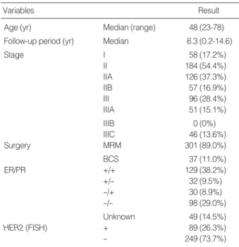
The median age of the entire subjects was 48 yr, and the median duration of follow-ups was 6.3 yr (Table 1). With regard to the distribution of the stages of breast cancer, the stage I was 58 cases (17.2%), the stage II was 184 cases (54.4
%), and the stage III was 96 cases (28.4%), and concerning surgical treatment, mastectomy was performed on 301 cases (89.0%), and postoperative adjuvant chemotherapy and hor- mone therapy were performed by considering the status of lymph node metastasis and hormone receptor test. In the status of hormone receptors of breast cancer, of 288 tested cases, 190 cases (66.0%) were found to be positive, and in the HER2 test by FISH of breast cancer, positive results meaning the amplification of the HER2 gene was shown in 89 cases of the total 388 cases (26.3%).
Variables Result
Age (yr) Median (range) 48 (23-78)
Follow-up period (yr) Median 6.3 (0.2-14.6)
Stage I 58 (17.2%)
II 184 (54.4%)
IIA 126 (37.3%)
IIB 57 (16.9%)
III 96 (28.4%)
IIIA 51 (15.1%)
IIIB 0 (0%)
IIIC 46 (13.6%)
Surgery MRM 301 (89.0%)
BCS 37 (11.0%)
ER/PR +/+ 129 (38.2%)
+/- 32 (9.5%)
-/+ 30 (8.9%)
-/- 98 (29.0%)
Unknown 49 (14.5%)
HER2 (FISH) + 89 (26.3%)
- 249 (73.7%)
Table 1. Clinicopathological characteristics of 338 cases of breast cancer
MRM, modified radical mastectomy; BCS, breast-conserving surgery;
ER, estrogen receptor; PR, progesterone receptor; FISH, fluorescence in situ hybridization.
Results of HER2 test according to the stage
In 89 cases showing the result of the amplification of HER2 gene in breast cancer, the positivity was examined according to the stage (Table 2). The positive result was detected in 6 cases of the stage I (10.3%), 41 cases in the stage II (22.3%), and 42 cases in the stage 3 (43.8%), hence, it was found that the positive rate became higher as the dis- ease stage progressed.
Survival analyses according to the stage
In the survival analysis based on the disease-free period, the 5-yr survival rate according to the amplification of HER2 gene showed different results among the stages of breast can- cer (Fig. 1). In the stage I breast cancer, the 5-yr disease-free survival (DFS) rate of the HER2 gene positive group was 100%, the survival rate of the negative group was 85%, and the survival rate of the positive group was higher, but it was not statistically significant (p=0.2053). In the stage II breast cancer, the 5-yr DFS rate of the positive group was 85%, that of the negative group was 87%, and a difference was not sig- nificant (p=0.3855). However, in the stage III breast cancer, the DFS rate of positive group was 41%, that of the nega- tive group was 70.5%, and the DFS rate of the HER2 gene- positive group was significantly lower (p=0.0090).
In the survival analysis based on the overall survival peri- od, similarly, the 5-yr survival rate according to the amplifi- cation of HER2 gene showed different results depending on the stages of breast cancer (Fig. 2). In the stage I breast can- cer, the 5-yr overall survival (OS) rate of the HER2-positive
FISH, fluorescence in situ hybridization.
Stage HER2 (FISH)
+ -
Stage I (n=58) 6 (10.3%) 52 (89.7%)
Stage II (n=184) 41 (22.3%) 143 (77.7%)
StageIII (n=96) 42 (43.8%) 54 (56.3%)
Table 2. HER2 gene amplification according to the stages of breast cancer
Cum survival
1.0
0.8
0.6
0.4
0.2
0.0
0.00 2.00 4.00 6.00 8.00 10.00 12.00 Duration (yr)
Stage I
Stage III
Stage II
Log-rank test p=0.2053
Fig. 1. Disease-free survival (DFS) curves according to the stages of breast cancer. (A) stage I (B) stage II (C) stage III. Statistical significance between HER2 gene amplification -positive group (solid line) and -negative group (dotted line) was noted only in stage III of breast cancer.
A
Cum survival
1.0
0.8
0.6
0.4
0.2
0.0
0.00 3.00 6.00 9.00 12.00 15.00
Duration (yr)
Log-rank test p=0.3855
B
Cum survival
1.0
0.8
0.6
0.4
0.2
0.0
0.00 2.00 4.00 6.00 8.00 10.00 12.00 14.00 Duration (yr)
Log-rank test p=0.0090
C
group was 100% and that of the negative group was 94%.
In the stage II breast cancer, the OS rate of the positive group was 88% and that of the negative group was 92.5%, and the difference was not significant (p=0.5927 in the stage I, p=0.8566 stage II). However, in the stage III breast cancer, the 5-yr OS rate of the HER2-positive group was 28% and that of the negative group was 60%, and the difference was significant (p=0.0034).
Prognostic factors influencing survival
To examine prognostic factors that independently influ- ence the disease-free survival and the overall survival, multi- variate analysis was performed by Cox regression model (Table 3). As an important prognostic factor influencing recurrence and death, the statistical significance of the posi- tive finding of HER2 was confirmed. The risk of recurrence of the HER2 gene amplification-positive group was 1.669 times higher than that of the negative group (p=0.036), and the risk of death was 2.394 times higher (p=0.003). On the other hand, in the same analysis performed according to the
disease stage, the amplification of HER2 gene did not show any significant effect on recurrence and survival in the dis- ease stage I and II breast cancer, but in the stage III breast cancer, increased the risk of recurrence by 2.678 times (p=
0.002) and the risk of death by 2.576 times (p=0.005). There- fore, the significance as a prognostic factor could be confirm- ed only in the stage III breast cancer (Table 4).
Stage I
Fig. 2. Overall survival (OS) curves according to the stages of breast cancer. (A) stage I (B) stage II (C) stage III. Statistical sig- nificance between HER2 gene amplification -positive group (solid line) and -negative group (dotted line) was noted only in stage III of breast cancer.
Cum survival
1.0
0.8
0.6
0.4
0.2
0.0
0.00 2.00 4.00 6.00 8.00 10.00 12.00 Duration (yr)
Log-rank test p=0.5927
A Stage III
Cum survival
1.0
0.8
0.6
0.4
0.2
0.0
0.00 2.00 4.00 6.00 8.00 10.00 12.00 14.00 Duration (yr)
Log-rank test p=0.0034
C
Stage II
Cum survival
1.0
0.8
0.6
0.4
0.2
0.0
0.00 3.00 6.00 9.00 12.00 15.00
Duration (yr)
Log-rank test p=0.8566
B
Variables DFS
RR 95% C.I. p
OS RR 95% C.I. p Tumor size (cm) 1.156 1.029-1.299 0.015 1.150 0.998-1.324 0.053 Node (number) 1.053 1.036-1.069 0.000 1.056 1.036-1.078 0.000 ER 0.997 0.634-1.570 0.991 0.777 0.448-1.349 0.370 HER2 (FISH) 1.669 1.035-2.690 0.036 2.394 1.384-4.140 0.002 Table 3. Multivariate analyses for disease-free survival and over- all survival in all cases (Cox regression model)
RR, relative risk; C.I., confident interval; DFS, disease-free survival; OS, overall survival; ER, estrogen receptor; FISH, fluorescence in situ hyb- ridization.
DISCUSSION
The significance of this study is that the amplification of the HER2 gene, which has been reported to be closely asso- ciated with the prognosis of breast cancer, was re-examined and the effect on the prognosis of the same stage breast can- cer was assessed by survival analysis based on the long-term follow-ups over than 5 yr. As mentioned already in the intro- duction, to raise the reliability of HER2 test, in our study, HER2 results were assessed by gene amplification test using FISH, which could be also considered to be greatly mean- ingful for the understanding of the significance HER2 as a prognostic factor. However, this study was performed retro- spectively and the number of patients in stage I was rela- tively small to that of other stages due to the retrospective selection of appropriate tissues of breast cancer for HER2 FISH test and survival analyses. These would be a limita- tion of this study.
Generally, the positive rate of HER2 in breast cancer has been reported to be 20-30% (14-17), and in our study, sim- ilarly, the positive rate of the amplification of HER2 was found to be 26.3%, and thus it could be confirmed. Never-
theless, HER2-positive rate showed a difference depending on the stage of breast cancer, and the positive result was shown in 10.3% in the stage I, 22.3% in stage II, and 43.8% in the stage III. This result suggests that as the disease stage of breast cancer is advanced, HER2-positive rate increases, and HER2 positivity is closely associated with high stage of breast can- cer, that is, poor prognosis (18-20).
According to the summary of HER2 of breast cancer report- ed until 1998 by Revillion et al. (18), HER2-positive result was a marker representing the aggressiveness of tumors and showed poor prognosis, nonetheless, because of the associa- tion with other strong prognostic factors, it did not have a clinically important significance. Slamon et al. (16) and Tan- don et al. (21) have reported that the amplification of the HER2 gene in breast cancer assessed by Southern blot had been associated with poor prognosis and these results had been found only in the lymph node positive group, but not in the lymph node negative group. These results are similar to our results in this study. We stratified the patients not by nodal status but by stages of breast cancer to exclude the influence of other prognostic factors such as tumor size, hor- monal status as well as nodal status. In this study the pure prognostic effect of HER2 gene amplification was noted only in stage III patients of breast cancer, not in stage I or II. We could suggest an explanation for these results that the ad- vance of breast cancer would be associated with the increas- ed amplification of the HER2 gene and the longer duration of HER2 gene action than in early breast cancer (22), there- fore, stage III of breast cancers in this study might have taken sufficient chances to express harmful effects on their survival not only by HER2 signal transduction pathway but also by crosstalks with other signal transduction pathways such as ER pathway.
On the other hand, Borg et al. (23) have reported that, with the result obtained by the same method, the amplification of HER2 gene did not influence prognosis in both lymph node -positive and -negative group. Afterwards, in numerous results applied immunohistochemical staining method, sim- ilar results were shown (24, 25), and until now, a clear con- clusion could not be reached and it is a controversial issue.
HER2 as a prognostic factor assessed by multivariate analysis, the amplification of the HER2 gene was confirmed to increase the risk of recurrence by 1.669 times (p=0.0036), and the risk of death by 2.394 times (p=0.002) in overall cases of breast cancer. However, our result showed that the effect of the amplification of HER2 gene differs depending on the disease stage. In other words, the prognosis of the HER2 gene amplification group was poor only in the stage III breast cancer, and in the stage I and II breast cancer, a statistically significant difference from the negative group was not detected. In multivariate analysis performed accord- ing to the stage of breast cancer, as the disease stage is ad- vanced, the amplification of the HER2 gene was associated with a high risk level of death, however, the statistical sig-
Variables DFS
RR 95% C.I. p
OS
RR 95% C.I. p
Stage I
Tumor size 0.286 0.059-1.387 0.120 1.466 0.003-805.141 0.905 (cm)
ER 1.001 0.992-1.010 0.814 0.690 0.312-1.528 0.361 HER2 (FISH) 0.000 0.000 0.991 0.000 0.000 0.996 Table 4. Multivariate analyses of disease-free survival and over- all survival according to the stages of breast cancer
Variables DFS
RR 95% C.I. p
OS
RR 95% C.I. p
Stage II
Tumor size 0.910 0.666-1.247 0.552 0.815 0.536-1.242 0.341 (cm)
Node (number) 1.236 1.090-1.402 0.001 1.307 1.133-1.508 0.000 ER 0.724 0.330-1.590 0.422 0.455 0.155-1.331 0.150 HER2 (FISH) 0.715 0.238-2.146 0.549 1.370 0.415-4.520 0.606
Variables DFS
RR 95% C.I. p
OS
RR 95% C.I. p
Stage III
Tumor size 1.209 1.063-1.374 0.004 1.131 0.975-1.313 0.103 (cm)
Node (number) 1.027 1.004-1.050 0.020 1.015 0.986-1.045 0.312 ER 1.302 0.708-2.394 0.396 0.987 0.512-1.902 0.969 HER2 (FISH) 2.678 1.437-4.991 0.002 2.576 1.339-4.959 0.005 DFS, disease-free survival; OS, overall survival; RR, relative risk; C.I., confidence interval; ER, estrogen receptor; FISH, fluorescence in situ hybridization.
nificance was noted only in the stage III breast cancer. There- fore, based on the result of our study, it could be confirmed that the amplification of the HER2 gene exerts a poor effect only on the prognosis of advanced breast cancer.
In conclusion, the amplification of the HER2 gene in breast cancer confirmed by FISH was associated with the progres- sion of the disease stage of breast cancer, and the clinical sig- nificance as a prognostic factor might be confined only in advanced breast cancer.
REFERENCES
1. Veronesi U, Viale G, Rotmensz N, Goldhirsch A. Rethinking TNM:
Breast cancer TNM classification for treatment decision-making and research. Breast 2006; 15: 3-8.
2. Dykins R, Corbett IP, Henry JA, Wright C, Yuan J, Hennessy C, Lennard TJ, Angus B, Horne CH. Long-term survival in breast can- cer related to overexpression of the c-erbB-2 oncoprotein: an immuno- histochemical study using monoclonal antibody NCL-CB11. J Pathol 1991; 163: 105-10.
3. Andrulis IL, Bull SB, Blackstein ME, Sutherland D, Mak C, Sidlof- sky S, Pritzker KP, Hartwick RW, Hanna W, Lickley L, Wilkinson R, Qizilbash A, Ambus U, Lipa M, Weizel H, Katz A, Baida M, Mariz S, Stoik G, Dacamara P, Strongitharm D, Geddie W, McCready D. neu/erbB-2 amplification identifies a poor-prognosis group of women with node-negative breast cancer. Toronto Breast Cancer Study Group. J Clin Oncol 1998; 16: 1340-9.
4. Hartmann LC, Ingle JN, Wold LE, Farr GH Jr, Grill JP, Su JQ, Maihle NJ, Krook JE, Witzig TE, Roche PC. Prognostic value of c-erbB2 overexpression in axillary lymph node positive breast cancer. Results from a randomized adjuvant treatment protocol. Cancer 1994; 74:
2956-63.
5. Press MF, Pike MC, Chazin VR, Hung G, Udove JA, Markowicz M, Danyluk J, Godolphin W, Sliwkowski M, Akita R, Paterson MC, Slammon DJ. Her-2/neu expression in node-negative breast cancer:
direct tissue quantitation by computerized image analysis and asso- ciation of overexpression with increased risk of recurrent disease.
Cancer Res 1993; 53: 4960-70.
6. Seshadri R, Firgaira FA, Horsfall DJ, McCaul K, Setlur V, Kitchen P. Clinical significance of HER-2/neu oncogene amplification in primary breast cancer. The South Australian Breast Cancer Study Group. J Clin Oncol 1993; 11: 1936-42.
7. Tetu B, Brisson J. Prognostic significance of HER-2/neu oncopro- tein expression in node-positive breast cancer. The influence of the pattern of immunostaining and adjuvant therapy. Cancer 1994; 73:
2359-65.
8. Bianchi S, Paglierani M, Zampi G, Cardona G, Cataliotti L, Bonardi R, Ciatto S. Prognostic significance of c-erbB-2 expression in node negative breast cancer. Br J Cancer 1993; 67: 625-9.
9. Romond EH, Perez EA, Bryant J, Suman VJ, Geyer CE Jr, David- son NE, Tan-Chiu E, Martino S, Paik S, Kaufman PA, Swain SM, Pisansky TM, Fehrenbacher L, Kutteh LA, Vogel VG, Visscher DW, Yothers G, Jenkins RB, Brown AM, Dakhil SR, Mamounas
EP, Lingle WL, Klein PM, Ingle JN, Wolmark N. Trastzumab plus adjuvant chemotherapy for operable HER2-positive breast cancer.
N Engl J Med 2005; 353: 1673-84.
10. Piccart-Gebhart MJ, Procter M, Leyland-Jones B, Goldhirsch A, Untch M, Smith I, Gianni L, Baselga J, Bell R, Jackisch C, Cameron D, Dowsett M, Barrios CH, Steger G, Huang CS, Andersson M, Inbar M, Lichinitser M, Lang I, Nitz U, Iwata H, Thomssen C, Lohrisch C, Suter TM, Ruschoff J, Suto T, Greatorex V, Ward C, Straehle C, McFadden E, Dolci MS, Gelber RD; Herceptin Adjuvant (HERA) Trial Study Team. Trastzumab after adjuvant chemotherapy in HER2- positive breast cancer. N Engl J Med 2005; 353: 1659-72.
11. Ellis CM, Dyson MJ, Stephenson TJ, Maltby EL. HER2 amplifica- tion status in breast cancer: a comparison between immunohisto- chemical staining and fuorescence in situ hybridization using man- ual and automated quantitative image analysis scoring techniques.
J Clin Pathol 2005; 58: 710-14.
12. Press MF, Bernstein L, Thomas PA, Meisner LF, Zhou JY, Ma Y, Hung G, Robinson RA, Harris C, El-Naggar A, Slamon DJ, Phillips RN, Ross JS, Wolman SR, Flom KJ. HER-2/neu gene amplification characterized by fluorescence in situ hybridization: poor prognosis in node-negative breast carcinomas. J Clin Oncol 1997; 15: 2894- 904.
13. Ratcliffe N, Wells W, Wheeler K, Memoli V. The combination of in situ hybridization and immunohistochemical analysis: an evaluation of Her2/neu expression in paraffin-embedded breast carcinomas and adjacent normal-appearing breast epithelium. Mod Pathol 1997;
10: 1247-52.
14. Kallioniemi OP, Kallioniemi A, Kurisu W, Thor A, Chen LC, Smith HS, Waldman FM, Pinkel D, Gray JW. ERBB2 amplification in breast cancer analyzed by fluorescence in situ hybridization. Proc Natl Acad Sci USA 1992; 89: 5321-5.
15. Slamon DJ, Clark GM, Wong SG, Levin WJ, Ullrich A, McGuire WL. Human breast cancer: correlation of relapse and survival with amplification of the HER-2/neu oncogene. Science 1987; 235: 177- 82.
16. Slamon DJ, Godolphin W, Jones LA, Holt JA, Wong SG, Keith DE, Levin WJ, Stuart SG, Udove J, Ullrich A, Press MF. Studies of the HER-2/neu proto-oncogene in human breast and ovarian cancer.
Science 1989; 244: 707-12.
17. Thor AD, Schwartz LH, Koerner FC, Edgerton SM, Skates SJ, Yin S, McKenzie SJ, Panicali DL, Marks PJ, Fingert HJ, Wood WC.
Analysis of c-erbB-2 expression in breast carcinomas with clinical follow-up. Cancer Res 1989; 49: 7147-52.
18. Revillion F, Bonneterre J, Peyrat JP. ERBB2 oncogene in human breast cancer and its clinical significance. Eur J Cancer 1998; 34:
791-808.
19. Ross JS, Fletcher JA. The HER-2/neu oncogene in breast cancer:
prognostic factor, predictive factor, and target for therapy. Stem Cells 1998; 16: 413-28.
20. Tagliabue E, Menard S, Robertson JF, and Harris L. c-erbB-2 expres- sion in primary breast cancer. Int J Biol Markers 1999; 14: 16-26.
21. Tandon AK, Clark GM, Chamness GC, Ullrich A, McGuire WL.
HER-2/neu oncogene protein and prognosis in breast cancer. J Clin Oncol 1989; 7: 1120-8.
. .
22. Mylonas I, Makovitzky J, Jeschke U, Briese V, Friese K, Gerber B.
Expression of Her2/neu, steroid receptors (ER and PR), Ki67 and p53 in invasive mammary ductal carcinoma associated with ductal carcinoma In Situ (DCIS) Versus invasive breast cancer alone. Anti- cancer Res 2005; 25: 1719-23.
23. Borg A, Tandon AK, Sigurdsson H, Clark GM, Ferno M, Fuqua SA, Killander D, McGuire WL. HER-2/neu amplification predicts poor survival in node-positive breast cancer. Cancer Res 1990; 50:
4332-7.
24. Rosen PP, Lesser ML, Arroyo CD, Cranor M, Borgan P, Norton L.
Immunohistochemical detection of HER-2/neu in patients with axil- lary lymph node negative breast carcinoma. A study of epidemio- logic risk factors, histologic features, and prognosis. Cancer 1995;
75: 1320-6.
25. Reed W, Hannisdal E, Boehler PJ, Gundersen S, Host H, Marthin J.
The prognostic value of p53 and C-erb-B2 immunostaining is over- rated for patients with lymph node-negative breast carcinoma?: a multivariate analysis of prognostic factors in 613 patients with a follow up of 14-30 years. Cancer 2000; 88: 804-13.