INTRODUCTION
In Korea, breast cancer ranks the second most common cancer after stomach cancer in women, and its rate is gradu- ally increasing (1). Despite having similar histological find- ings, breast cancer cases have distinctly different biological behaviors, so that it is difficult to evaluate the prognosis of breast cancer and to predict its response to treatment and the likelihood of recurrence. Traditionally, prognostic factors of breast cancer include axillary nodal status, tumor size, his- tological grade, pathologic subtype, cancer cell proliferation, and hormonal receptor status (2). The biological markers, for example c-erbB2, p53, Bcl-2, Ki-67 labeling index etc., have been intensively investigated as new prognostic or pre- dictive markers, and are currently applied in the management of breast cancer patients in many hospitals.
Apoptosis is the process of active cell death that is observed in the generation of multinuclear creatures and the preserva- tion of individuals (3). Recently, a great deal of interest has been devoted to the role of apoptosis in tumorigenesis. Bcl- 2 was first identified as repressor of apoptosis and Bcl-2-relat- ed genes are involved in the regulation of apoptosis that is implicated in a variety of cellular activities, including the transformation of a normal cell into a cancer cell (4). At pre-
sent, the reported Bcl-2-related genes are Bax, Bcl-x, mcl-1, A1, Bak, ced-9, Bfl-1, and BRAG-1. Among these, the Bfl- 1 gene, which is isolated from human fetal liver, is a mem- ber of the Bcl-2 gene family and was only recently described, inhibits p53-induced apoptosis and exhibits a potent coop- erative transforming activity (5, 6). Bfl-1 was also shown to be induced by inflammatory cytokines, TNF- or IL-1 , suggesting that it may play a protective role during inflam- mation (8). It has been reported that over-expression of the Bfl-1 homologous genes, Bcl-2, Bax, and Bcl-x has a relation- ship with prognosis of breast cancer, but there has been no reports on the prognostic relevance of the Bfl-1 gene in lit- erature.
The purpose of the present study was to compare the expression of the Bfl-1 gene by reverse transcriptase-poly- merase chain reaction (RT-PCR) analysis with other prog- nostic factors, such as the status of hormonal receptors, p53, c-erbB2, Bcl-2, and tumor infiltrating lymphocytes (TIL) and the most important clinicopathologic parameters.
PATIENTS AND METHODS
The study was performed on 30 breast cancer tissue sam- Ho-Sung Yoon, Sung-Hee Hong,
Hee-Jun Kang, Byung Kyun Ko, Sei-Hyun Ahn, Joo-Ryung Huh*
Department of Surgery and *Pathology, University of Ulsan, College of Medicine, Asan Medical Center and Ulsan University Hospital, Seoul, Korea
Address for correspondence Ho-Sung Yoon, M.D.
Department of Surgery, University of Ulsan, College of Medicine, Asan Medical Center, 388-1 Poongnap-dong, Songpa-gu, Seoul 138-736, Korea Tel : +82.2-3010-3480, Fax : +82.2-474-9027 E-mail : [email protected]
*This study was supported by a grant (2000-022) from the Asan Institute for Life Science, Seoul, Korea.
225
Bfl-1 Gene Expression in Breast Cancer
: Its Relationship with other Prognostic Factors
The Bfl-1 gene, which was isolated from human fetal liver and only recently de- scribed, is a member of the Bcl-2 gene family. Reverse transcriptase-polymerase chain reaction was performed on RNA drawn from 30 breast cancer tissues to com- pare the expression of the Bfl-1 gene with other prognostic factors. The median relative ratio was 3.0 (range, 0.12-26.83) and the Bfl-1 gene expression rate was 36.7% (11/30). There was no statistically significant relationship between the clini- copathologic parameters of patients and the expression value of Bfl-1 gene. The level of Bfl-1 gene expression was higher in more advanced breast cancers than in early cancers. There was no significant relationship between the expression val- ues and currently acknowledged prognostic factors, but a higher expression pattern was noticed in the groups of positive hormone receptors and negative p53 and neg- ative c-erbB2, albeit statistically not significant. It seems that the increased expres- sion of the Bfl-1 gene serves as a contributory factor in breast cancer, in the same way that another group of genes, the Bcl-2 family, contributes to apoptosis.
Key Words : Breast Neoplasms; Bfl-1; Prognosis; Reverse Transcriptase Polymerase Chain Reaction
Received : 9 October 2002 Accepted : 2 December 2002
ples. All the patients were enrolled at the Department of Surgery of Asan Medical Center in Korea. None of the breast cancer patients received preoperative radiation treatment or chemotherapy. Patients were treated by mastectomy or breast- conserving surgery. The median age of the patients was 47.5 yr (range, 32-83 yr). Nineteen (63.3%) patients were pre- menopausal and 11 (36.7%) were postmenopausal. During surgery, after the tumor had been cut in half, about 1 g of tumor tissue was frozen in liquid nitrogen immediately after surgical resection and stored at -80℃ until being processed.
The rest of the tissue was processed for histopathological examination, and immunohistochemical analysis was per- formed for the expression of the estrogen and progesterone receptors, p53, and c-erbB2, Bcl-2 using the antibody for individual proteins by routine ABC method. All the patients were staged according to the AJCC/UICC classification sys- tem, and each tumor was graded as a histologically by the Bloom and Richardson grading system.
Semiquantitative RT-PCR analysis
Total RNA was isolated from tissue samples using Tri- reagent (Sigma, T9424) after removal of contaminating chro- mosomal DNA with DNAse I treatment, and then was reverse transcribed by the Moloney murine leukemia virus reverse transcriptase (Gibco BRL, Gainthersberg, MD, U.S.A.) and oligo dT primers (Promega, Medison, WI, U.S.A.) to synthesize the first-strand complementary DNA (cDNA).
Three microliters of cDNA were then amplified by poly- merase chain reaction (PCR). The -globin gene served as internal control. The primer used were: Bfl-1 upstream primer: 5、-AGCTCAAGACTTTGCTCTCCACC-3、; Bfl-1 downstream primer: 5、-TGGAGTGTCCTTTCTGGTGA- CATTAAGG-3、; -globin upstream primer: 5、-GACA- CAACGTTCATAG 3、; -globin downstream primer: 5、- AGGGTAGACACCAGCAGC-3、. PCR was performed in a 30 L reaction volume with the following parameters: 94
℃ for 5 min; 94℃ for 45 sec; 60℃ for 45 sec; 72℃ for 30 sec for 28 cycles; and 72℃ for 10 min. PCR products were electrophoresed on 2% agarose gels and visualized by stain- ing with ethidium bromide (9). The gel was scanned, and the band intensity was measured by a computerized densito- metric analysis device. We calculated the ratio of the inten- sity of the Bfl-1 bands to the intensity of the -globin bands.
We considered any ratio greater than 5 to indicate expres- sion of the Bfl-1 gene.
Measurement of Tumor Infiltrating Lymphocytes (TIL)
The 5- mthick sections of the formalin-fixed, paraffin- embedded blocks were used for hematoxylin-eosin stains.
Using a microscope, a total of three areas of tumor with infil- tration by TILs were selected for the counting of TILs in the following order: (a) TIL1, the area of the maximal number of
TILs; (b) TIL2, the area of tumor with a median number of TILs in each case; and (c) TIL3, the area with the lowest num- ber of TILs as judged by examination at low-power magni- fication (×100). For the counting of TILs, a high-powered magnification (×200) image was captured by Image Pro Plus (version 4.0, Media Cybernetics), and printed out on a devel- oping paper. The TILs in the entire image were counted using grids, and we calculated the mean number of the three areas.
The infiltration of TILs was divided into 3 groups: patients with a low number of TIL infiltration (<100 cells/highpow- er field), a moderate number (100< and <300), and a high number (>300).
Statistical Analysis
The Chi-squared test, Fisher’s exact test, the Mann-Whit- ney nonparametric test, or Pearson’s correlation test was used to analyze the expression of the Bfl-1 levels according to the clinicopathological characteristics, other prognostic factors and TIL, as appropriate. Those with p<0.05 were considered significant.
RESULTS
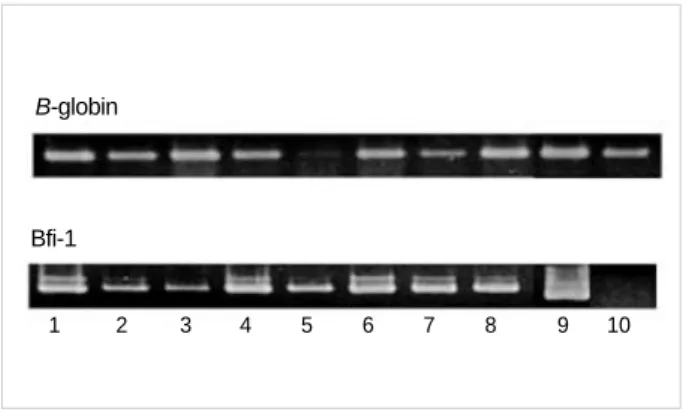
The median relative ratio was 3.0 (range, 0.12-26.83) and the Bfl-1 gene expression rate was 36.7% (11/30) (Fig. 1).
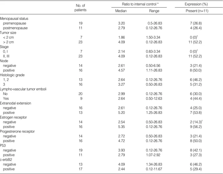
No statistically significant relationship was found between the clinicopathologic parameters of patients: menopausal sta- tus, tumor size, axillary nodal status, stage, histologic grade, lympho-vascular invasion of tumor, and the relative ratio numerical values of gene expression. However, the median value was 2.14 (range, 0.83-3.34) in the group with stage 0 and stage I, but the value was 4.09 (range; 0.12-26.83) in the group with stage II or over. Therefore, a lower level of expres- sion of the Bfl-1 gene was shown in cases of the early stage of breast cancer. Compared with other cases, higher intensi- ty of expression was displayed in cases with larger tumors and in those with metastasis to axillay lymph nodes showed high-
Fig. 1.RT-PCR analysis of Bfl-1 mRNA in breast cancer tissues.
lanes 1-8, patient number: lane 9, positive control (B-cell lympho- ma): lane 10, negative control (liver tissue).
1 2 3 4 5 6 7 8 9 10
B-globin
Bfi-1
er intensity of expression (1.86 versus 4.06, 2.61 versus 4.57).
These observations showed that the Bfl-1 gene was highly expressed in more advanced breast cancers than in early can- cers. There was no significant relationship between expression values and currently acknowledged prognostic factors, that is estrogen and progesterone receptors, p53, and c-erbB2, but a higher expression pattern was noticed in the groups of positive hormone receptors and negative p53 and negative c-erbB2, albeit without statistical significance. Regarding the gene expression rate, none of the group of 7 patients with tumors smaller than 2 cm showed expression, while 52.2%
(11/23) of those in the group with tumors larger than 2 cm (p=0.029) did. This trend could also be applied to patients with breast cancer of stage 0 or I and of stage II: the former group was markedly different from the latter (p=0.02). In the group with only positive estrogen receptor among other prog- nostic factors, 9 out of 16 cases showed expression (56.3%) compared to 14.3% (2/14) of the negative cases (p=0.017) (Table 1). In the group with positive expression of the Bcl-2
No. of Ratio to internal control * Expression (%)
patients Median Range Present (n=11)
Menopausal status
premenopause 19 3.20 0.5-26.83 7 (26.8)
postmenopause 11 2.79 0.12-26.76 4 (26.4)
Tumor size
< 2 cm 7 1.86 1.50-3.34 0 (0)�
> 2 cm 23 4.09 0.12-26.83 11 (52.2)
Stage
0, I 7 2.14 0.83-3.34 0 (0)�
II, III 23 4.09 0.12-26.83 11 (52.2)
Node
negative 14 2.61 0.50-6.56 3 (21.4)
positive 16 4.57 1.11-26.83 8 (50.0)
Histologic grade
1, 2 13 2.64 0.12-26.76 6 (46.2)
3 16 3.27 0.50-26.83 5 (31.2)
Lympho-vascular tumor emboli
No 20 2.99 0.12-26.76 6 (30.0)
Yes 9 2.64 0.50-12.63 4 (44.4)
Extranodal extension
negative 16 2.61 0.12-26.76 4 (25.0)
positive 13 5.20 1.25-26.83 7 (53.8)
Estrogen receptor
negative 14 2.54 0.50-26.83 2 (14.3)�
positive 16 5.35 0.12-26.76 9 (56.2)
Progestrerone receptor
negative 14 2.72 0.50-26.83 3 (21.4)
positive 16 4.72 0.12-26.76 8 (50.0)
P53
negative 19 3.93 0.12-26.76 8 (42.1)
positive 11 2.79 1.07-2.92 3 (27.3)
c-erbB2
negative 13 4.09 1.34-26.83 6 (46.2)
positive 17 2.44 0.12-11.67 5 (29.4)
*: The difference between subgroup, as determined by the Mann-Whitney U test, was not significant (p>0.05); �: Statistically significant (p<0.05).
Table 1.Clinical data of patients and result of Bfl-1 gene expression
Fig. 2.Box and whiskers plots showing the distribution of Bfl-1 values, expressed as relative ratio in the category of Bcl-2 pro- tein expression-the median values are indicated as a line across each box.
Bfl-1 value(relative ratio), Log
4 3 2 1 0 -1
-2 16 14
negative positive
Bcl-2 expressiorn p=0.04
protein, the expression value of the Bfl-1 gene ranged between 0.83 and 26.76 (median value, 5.93), and the gene expression rate was 64.3% (9/14). On the other hand, in the group with negative Bcl-2 expression, the value ranged from 0.12 to 26.83 (median value, 2.29) and 12.5% (2/16). This obser- vation suggests that there would be a significant relationship between the two genes (p<0.04) (Fig. 2). Infiltration of TILs was present in the tumor stroma in all examined cases. The distribution of TILs was uneven with the heaviest infiltration at the periphery of the tumor in most cases. The TIL infil- tration ranged from 0 cells per high-power field to 530 cells per high-power field in cancerous tissue, with a mean num- ber ranging between 1 and 502 (total mean number, 150.4).
For the grade of TIL density, the rates of the low, moderate, high density groups were 46.7%, 30.0%, and 23.3%, retro- spectively. However, a correlation between the density dif- ference of tumor infiltrating lymphocytes and the expression of the Bfl-1 gene was not found.
DISCUSSION
Despite the gradual increase in the incidence of breast can- cer worldwide, there has been only substantial improvement in the management of breast cancer that has contributed to the substantial fall in its mortality since 1990. A major con- tributor to this decrease is clearly the widespread application of hormonal and adjuvant chemotherapy after surgery. How- ever it is essential to understand the characteristics of breast cancer, including the carcinogenic process itself and its patho- genesis (2). In recent years, there have been important clini- cal researches on the genes, c-erbB2, p53, Bcl-2, etc., and their association with breast cancer, including their roles as prog- nostic or predictive factors (10-16). Bcl-2 has been studied as a cell survival-promoting protein in a variety of human tumors with contradictory results. For instance, in accordance with its anti-apoptotic nature, Bcl-2 has been shown to be associated with a poorer prognosis in prostate and colon can- cers, and neuroblastoma (17). In breast cancer, on the other hand, Bcl-2 expression has been reported to be associated with better outcomes and improved disease-free survival, although Bcl-2 was not an independent prognostic variable (19). Complex interactions among the various Bcl-2 family members determine the outcome in terms of cell progression toward either apoptotic cell death or survival (20, 21). The Bfl-1 gene is one of the Bcl-2 related genes recently identified by computer analysis of the expressed sequenced tag databas- es constructed by random cDNA clones from a human fetal liver (5, 23). The Bfl-1 gene, as a member of the Bcl-2 fami- ly, was reported to have 72% homology to the murine A1 gene, one of the Bcl-2-related genes. The Bfl-1 gene contains the BH1 and BH2 domains, which are known to be impor- tant regulators for apo-ptosis in Bcl-2-related proteins (22).
Recently, some investigators demonstrated that the Bfl-1 is
a direct transcriptional target of NF- B, and thus the activa- tion of Bfl-1 may be the means by which NF- B‚ promotes oncogenesis and cell resistance to anticancer therapy. The expression patterns of other Bcl-2 related genes are very dis- tinctive (6). The Bfl-1 gene was highly expressed in bone marrow and was also present in hematopoietic cell lines, such as Raji and HL60, and in some normal adult tissues includ- ing the lung, spleen, and esophagus. It was not, however, detected in the heart, testis, thyroid, or brain (7). Conflict- ing data have been reported on the Bfl-1 gene expression in different tumor tissues: a high rate of expression in gastric (86%) and colon cancer (93%) specimens, a low rate of expres- sion in breast cancer (33%), bone and soft tissue sarcoma, and ovarian cancer specimens, and a very low rate in some human cancer cell lines (7). We performed a study on the Bfl-1 ex- pression in a series breast cancer samples to analyze and cor- relate the expression with the clinicopathological features of the patients and other prognostic factors, including Bcl-2 and tumor infiltrating lymphocytes. The latter is considered to reflect the host’s effort to resist tumor growth and has predictive values also (26). In our study, Bfl-1 was expressed in 36.7% as a mRNA band on sensitive RT-PCR. These data were in agreement with the previous observation by Park et al. who demonstrated the Bfl-1 gene was expressed at a low rate (33.3%) in breast cancer (7).
There have been reports that the function of Bfl-1 is dis- tinct from that of Bcl-2 because Bfl-1 permits cell prolifera- tion by its structural variation (6), while the two molecules have a similar function, inhibition of apoptosis. Bcl-2 ex- pression has been reported to be associated with better out- comes and factors that have favorable prognosis, although Bcl-2 was not an independent prognostic variable (19). In the present study, expression of the Bcl-2 protein on immu- nohistochemistry has a significant association with the hor- monal receptors and c-erbB2 (data not shown). Indeed, these patterns were similar to the results for Bfl-1. In the patient groups with the higher level of Bf1-1 expression intensity, the tumors were positive for hormonal receptors and nega- tive for p53 or c-erbB2, which are known as good prognos- tic markers. A good correlation was found between the ex- pression of the Bfl-1 gene and the estrogen receptor only.
When the intensities of Bfl-1 gene expression were com- pared with the characteristics, including existing prognostic factors, there were no statistically significant parameters.
But considering the trend of higher expression in the cancer tissues of patients with larger tumors, axillary nodal metas- tasis, or extranodal tumor ex-tension, this observation sug- gests that Bfl-1 could possibly play a role in advanced cases of breast cancer. Interestingly, there were no significant asso- ciation of the expression of the Bcl-2 protein with the tumor size or staging as showing a significant difference on the expression of the Bfl-1 gene. Breast cancer cells are often surrounded by inflammatory cell infiltrates as a sign of tumor- host interaction and the presence of lymphocyte infiltrates
is considered to reflect the host’s effort to resist tumor growth (26). Some authors have related the presence of tumor infil- trating lymphocytes to favorable prognosis (27). A study in gastric carcinoma suggests that inflammatory cells may be the major source of Bfl-1 gene ex-pression. In a study that utilized the mRNA in situ hybridization technique, Ha et al. (8) showed that the signal for Bfl-1 was localized within the inflammatory cells, including neutrophils and eosinophils.
In the present study, we examined whether the expression of the Bfl-1 gene has a correlation with the infiltration of lymphocytes in breast cancer as preliminary research. We found no association between the expression of Bfl-1 and the densities of tumor infiltrating lymphocytes, although further studies are needed before drawing a definite conclu- sion.
In conclusion, it seems that the increased expression of the Bfl-1 gene serves as an contributory factor in breast can- cer, in the same way that another group of genes, the Bcl-2 family, contribute to apoptosis. The Bfl-1 gene was related to more advanced breast cancer and to factors that have favorable prognosis. However in order to know if the Bfl-1 gene may be influential in the clinical manifestation mecha- nism and outcomes of breast cancer, long-term follow-up of patients with further research on the role of the gene is needed.
REFERENCES
1. Ministry of Health and Welfare, Republic of Korea. Annual Report of the Central Cancer Registry in Korea 2002 (2000. 1.-2000. 12.).
2. Page DL, Jensen RA, Simpson JF. Routinely available indicators of prognosis in breast cancer. Breast Cancer Res Treat 1998; 51: 195- 208.
3. White E. Life, death, and the persuit of apoptosis. Genes Dev 1996; 10:
1-15.
4. Kapranos N, Karaiosifidi H, Valavanis C, Kouri E, Vasilaros S. Prog- nostic significance of apoptosis related proteins Bcl-2 and Bax in node-negative breast cancer patients. Anticancer Res 1997; 17:
2499-505.
5. Choi SS, Park IC, Yun JW, Sung YC, Hong SI, Shin HS. A novel Bcl-2 related gene, Bfl-1, is overexpressed in stomach cancer and preferentially expressed in bone marrow. Oncogene 1995; 11:
1693-8.
6. D’Sa-Eipper C, Subramanian T, Chinnadurai G. Bfl-1, a Bcl-2 homo- logue, suppresses p53-induced apoptosis and exhibits potent cooper- ative transforming activity. Cancer Res 1996; 56: 3879-82.
7. Park IC, Lee SH, Whang DY, Hong WS, Choi SS, Shin HS, Choe TB, Hong SI. Expression of a novel Bcl-2 related gene, Bfl-1, in various human cancers and cancer cell lines. Anticancer Res 1997;
17: 4619-22.
8. Ha HJ, Kim D, Lee SB, Hong SI, Park SY, Huh JR, Kim CW, Kim SS, Lee Y, Choi SS, Shin HS. Expression of Bfl-1 in normal and tumor tissues: Bfl-1 overexpression in cancer is attributable to its
preferential expression in infiltrating inflammatory cells. Hum Pathol 1998; 29: 723-8.
9. Shim YH, Byun EK, Lee MJ, Huh JR, Kim CW. Anti-apoptotic role of Bfl-1 in staurosporine-treated B-lymphoblastic cells. Int J Hema- tol 2000; 72: 484-90.
10. Korean Breast Cancer Society. Clinical characteristics of Korean breast cancer patients in 1998. J Korean Med Sci 2000; 15: 569-79.
11. Hamilton A, Piccart M. The contribution of molecular markers to the prediction of response in the treatment of breast cancer: a review of the literature on HER-2, p53 and Bcl-2. Ann Oncol 2000; 11:
647-63.
12. Bottini A, Berruti A, Bersiga A, Brizzi MP, Brunelli A, Gorzegno G.
p53 but not Bcl-2 immunostaining is predictive of poor clinical com- plete response to primary chemotherapy in breast cancer patients. Clin Cancer Res 2000; 6: 2751-8.
13. Daidone MG, Luisi A, Veneroni S, Benini E, Silvestrini R. Clinical studies of Bcl-2 and treatment benefit in breast cancer patients. En- docr Relat Cancer 1999; 6: 61-8.
14. Nakopoulou L, Michalopoulou A, Giannopoulou I, Tzonou A, Ker- amopoulos A, Lazaris AC. Bcl-2 protein expression is associated with a prognostically favorable phenotype in breast cancer irrespective of p53 immunostaining. Histopathology 1999; 34: 310-9.
15. Railo M, Nordling S, von Voguslawsky K, Leivonen M, Kyllonen L, von Smitten K. Prognostic value of Ki-67 immunolabelling in prima- ry operable breast cancer. Br J Cancer 1993; 68: 579-83.
16. Kobayashi S, Iwase H, Ito Y, Yamashita H, Iwata H, Yamashita T.
Clinical significance of Bcl-2 gene expression in human breast can- cer tissues. Breast Cancer Res Treat 1997; 42: 173-81.
17. Reed JC. Bcl-2 and the regulation of programmed cell death. J Cell Biol 1994; 124: 1-6.
18. Doglioni C, Dei Tos AP, Laurino L, Chiarelli C, Barbareschi M, Viale G. The prevalence of BCL-2 immunoreactivity in breast carcinomas and its clinicopathological correlates, with particular reference to oest- rogen receptor status. Virchows Archiv 1994; 424: 47-51.
19. Silvestrini R, Benini E, Veneroni S, Daidone MG, Tomasic G, Sal- vadori B. p53 and Bcl-2 expression correlates with clinical outcome in a series of node positive breast cancer patients. J Clin Oncol 1996;
14: 1604-10.
20. Krajewski S, Thor AD, Edgerton SM, Moore DH, Reed JC. Immuno- histological analysis of Bax and Bcl-2 in p53-immunopositive breast cancer. Clin Cancer Res 1997; 3: 199-208.
21. Reed JC, Miyashita T, Takayama S, Wang HG, Sato T, Krajewski S, Aime-Sempe C. Bcl-2 family proteins: regulator of cell death involved in the pathogenesis of cancer and resistance to therapy. J Cell Biochem 1996; 60: 23-32.
22. D’Sa-Eipper C, Chinnadurai G. Functional dissection of Bfl-1, a Bcl- 2 homology: anti-apoptosis, oncogene-cooperation and cell prolif- eration activities. Oncogene 1998; 16: 3105-14.
23. Choi SS, Park SH, Kim UJ, Shin HS. Bfl-1, a Bcl-2-related gene, is the human homolog of the murine A1, and maps to chromosome 15q 24.3. Mamm Genome 1997; 8: 781-2.
24. Cheng Q, Lee HH, Li Y, Parks TP, Cheng G. Upregulation of Bcl-x and Bfl-1 as a potential mechanism of chemoresistance, which can be overcome by NF-kappa B inhibition. Oncogene 2000; 19: 4936-40.
25. Zong WX, Edelstein LC, Chen C, Bash J, Gelinas C. The prosurvival Bcl-2 homolog Bfl-1/A1 is a direct transcriptional target of NF-kappa B that blocks TNFalpha-induced apoptosis. Genes Dev 1999; 13:
382-7.
26. Aaltomaa S, Lipponen P, Eskelinen M, Kosma V.-M, Marin S , Alha- va E. Lymphocyte infiltration as a prognostic variable in female breast
cancer. Eur J Cancer 1992; 28A: 859-64.
27. Yasunaga M, Tabira Y, Nakano K, Iida S, Ichimaru N, Nakamato N.
Accelerated growth signal and tumor infiltrating lymphocyte level predict poor outcome in T4 esophageal squamous cell carcinoma. Ann Thorac Surg 2000; 70: 1634-40 .