© 2017 Korean Breast Cancer Society. All rights reserved. http://ejbc.kr | pISSN 1738-6756
INTRODUCTION
Recently, the incidence of breast cancer has increased dras- tically, and breast cancer has become the most malignant tu- mor among women worldwide. With the development of ba- sic and clinical research, the treatment of breast cancer is cur- rently combined therapy that includes surgery, radiotherapy, chemotherapy, endocrine therapy, and targeted therapy, and so forth. The therapeutic concept is also gradually evolving as
new findings and therapeutic approaches become available.
Endocrine therapy of breast cancer, especially of steroid-de- pendent breast cancer, has an irreplaceable advantage. The ap- plication of endocrine therapy ranges from salvage treatment of advanced breast cancer, to adjuvant therapy of early breast cancer, to prophylactic treatment of high-risk populations.
Given its many benefits, it is hopeful that endocrine therapy could cure patients with breast cancer [1].
Although patients with estrogen receptor-positive (ER+) and progesterone receptor-positive (PR+) breast cancer can be treated with endocrine therapy, patients with different ex- pression levels of hormone receptors have different sensitivity to endocrine therapy [2,3]. Some previous studies showed that the efficacy of tamoxifen for ER+ breast cancer with high PR expression was better than that of patients with low PR ex- pression, in both the adjuvant setting and the treatment of metastatic breast cancer [4,5]. Data from the large ATAC (Arimidex, Tamoxifen, Alone or in Combination) adjuvant
Prognostic Impact of Progesterone Receptor Status in Chinese Estrogen Receptor Positive Invasive Breast Cancer Patients
Nan Yao, Zhenchuan Song1, Xinle Wang1, Shan Yang1, Heng Song1
Department of General Surgery, Aerospace Central Hospital, Beijing; 1Breast Diseases Treatment Center, Fourth Hospital of Hebei Medical University, Shijiazhuang, China
ORIGINAL ARTICLE
Purpose: Estrogen receptor (ER) and progesterone receptor (PR) have been used as indicators of endocrine system status since the mid-1970s in the clinical management of breast cancer. The predictive role of ER in endocrine therapy is undisputed, but the prognostic value of PR is still debated. The aim of this study was to investigate the clinical characteristics and prognosis of ER positive breast cancer with different PR expression levels.
Methods: A population cohort of 3,030 primary invasive ER posi- tive breast cancer patients from a single cancer center under- went surgery and received adjuvant endocrine therapy from 2004 to 2010. The clinical and biological features of these pa- tients with high PR-expressing tumors were compared with those of patients with low PR-expressing tumors. The follow-up data for disease-free survival (DFS), overall survival (OS), and breast cancer specific survival (BCSS) was obtained from 2,778 patients. Cox regression analysis was used to correlate biomark- ers and tumor characteristics with DFS, OS, and BCSS. Results:
Tumors with low PR expression had more invasive pathological features and biological indexes than those with high PR expres- sion. Low PR expression was an independent poor prognostic factor for DFS (p=0.014; hazard ratio [HR], 0.781; 95% confi- dence interval [CI], 0.641–0.950), OS (p=0.002; HR, 0.699; 95%
CI, 0.560–0.873), and BCSS (p=0.005; HR, 0.714; 95% CI, 0.566–0.902). Furthermore, in low PR expressing tumors, pa- tients who received chemotherapy had better DFS (p=0.002;
HR, 0.449; 95% CI, 0.268–0.751), OS (p<0.001; HR, 0.341;
95% CI, 0.192–0.606), and BCSS (p<0.001; HR, 0.292; 95% CI, 0.156–0.549) than patients who did not received chemotherapy.
Conclusion: Patients with ER positive invasive breast cancer with low PR expressing tumors have a worse prognosis than those with high PR expressing tumors, and these patients can benefit from chemotherapy.
Key Words: Breast neoplasms, Progesterone receptor, Prognosis, Tamoxifen
Correspondence to: Zhenchuan Song
Breast Diseases Treatment Center, Fourth Hospital of Hebei Medical University, No.169 Tianshan Street, Shijiazhuang 050035, China Tel: +86-311-66696310, Fax: +86-311-66696310
E-mail: [email protected]
This study was supported by Hebei Province Natural Science Foundation (H2012206169) and Wu Jieping Medical Foundation for Clinical Scientific Research (320.6750.13295).
Received: October 18, 2016 Accepted: January 24, 2017
Cancer
trial, a worldwide clinical trial comparing the efficacy of tamoxifen with that of the aromatase inhibitor (AI) anastro- zole, showed that patients with ER+/PR+ tumors had a lower recurrence rate than those with ER+/PR– tumors (7.6% vs.
14.8%, respectively) [6]. In this study, we analyzed cases of pa- tients who were diagnosed with primary breast cancer and treated with surgery in Breast Diseases Treatment Center of Hebei Province from 2004 to 2010. By summarizing their dis- tribution and clinical characteristics, and performing a prog- nostic analysis of ER+ breast cancer with different PR expres- sion profiles, we were able to identify a novel prognostic indi- cator, which could provide a future reference for the individu- alized treatment of breast cancer.
METHODS
Patient selection and data collection
The patient population comprised a consecutive series of all patients with invasive and operable breast cancer who pre- sented to a single regional cancer center between January 2004 and June 2010. The median age was 50 years (range, 22–
88 years). In our trial, all patients who underwent curative surgery were discussed postoperatively at a multidisciplinary meeting, and appropriate adjuvant endocrine therapy was prescribed according to national guidelines. Adjuvant tamoxi- fen was administered to pre-menopausal patients, and adju- vant AI was administered to postmenopausal patients. A total of 1,263 premenopausal patients received tamoxifen therapy, 1,022 postmenopausal patients received AI, and 493 patients switched from tamoxifen to an AI after 2 or 3 years, because they became postmenopausal during the years of tamoxifen treatment. The patients’ clinical information had been collect- ed previously and stored in a database. Specific patient con- sent was not required because retrospective data from the medical records of patients who had previously signed infor- mation release documents were used in this study. This re- search was approved by the Ethics Committee of Fourth Hos- pital of Hebei Medical University (number: SCXK2014-0031).
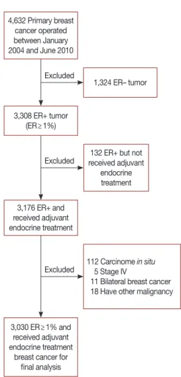
A total of 4,632 women with invasive carcinoma were iden- tified. The inclusion criteria were women who (1) had under- gone mastectomy or breast conserving surgery; (2) were ER+
and had been administered standard endocrine therapy after surgery; (3) had no severe concomitant diseases; (4) had com- plete immunohistochemistry data including ER, PR, and hu- man epidermal growth factor receptor 2 (HER2); and (5) had invasive breast carcinoma. Patients who were diagnosed with bilateral tumors or distant metastases at the preoperative work-up were excluded. At last, a total of 3,030 patients en- tered the final analysis (Figure 1).
All the immunohistochemistry slides for ER/PR/HER2 were reviewed again by two independent pathologists. Immu- nohistochemistry staining of 4-mm sections of formalin-fixed paraffin-embedded tissue was performed with anti-ER (clone SP1; Roche, Basel, Switzerland), anti-PR (clone 1E2; Roche), anti-HER2 (clone 4B5; Roche) primary monoclonal antibodies.
The ER and PR were visualized and classified based on the percentage of positive cells according to a semi-quantitative system. The slides were scored by counting the number of positive cells regardless of the staining intensity versus the total number of cells and calculating the percentage of positive cells (positive cells/total cells in one field). The positivity of several fields was averaged and presented as the ratio of positive cells per field to total cells per field: <1%, negative (–); 1% to 25%, weakly positive (+); 26% to 50%, positive (++); and >50%, strongly positive (+++) [7]. For PR expression profiling, a cutoff point of 25% was used to distinguish between low- expression (≤25%) and high-expression tumors (>25%).
4,632 Primary breast cancer operated between January 2004 and June 2010
3,308 ER+ tumor (ER≥1%)
3,176 ER+ and received adjuvant endocrine treatment
112 Carcinome in situ 5 Stage IV
11 Bilateral breast cancer 18 Have other malignancy
3,030 ER≥1% and received adjuvant endocrine treatment
breast cancer for final analysis
132 ER+ but not received adjuvant
endocrine treatment 1,324 ER– tumor
Figure 1. Flow chart of patients selection for final analysis.
ER=estrogen receptor.
Excluded
Excluded
Excluded
This cutoff point value is similar to a consensus 20% suggested at the St. Gallen International Breast Cancer Conference in 2013 [8]. HER2 scoring was categorized as 0, 1+, 2+, or 3+. A score of 3+ was considered positive, a score of 2+ was considered equivocal, and a score of 0/1+ was defined as negative [9].
Tumors with a HER2 score of 2+ were usually further ana- lyzed by fluorescence in situ hybridization (FISH). If not, these tumors were considered equivocal. The FISH results were considered positive, equivocal, or negative according to HER2 copy number or the HER2/chromosome 17 centromere (CEP17) ratio [10]. Any ambiguity in the reports was resolved by discussion with senior pathologists.
Follow-up
The follow-up, which involved a hospital visit, telephone, or mail interview, began from the first day after surgery. The starting point of the follow-up was the date of operation. The finishing point of the follow-up was June 30, 2014. For pa- Table 1. The relationship between PR expression and clinicopathologi- cal characteristics
Characteristic
Low expression of PR
(n=1,083) No. (%)
High expression of PR
(n=1,947) No. (%)
p-value
Age (yr) <0.001
≤35 61 (5.6) 134 (6.9)
>35, <50 341 (31.5) 900 (46.2)
≥50 681 (62.9) 913 (46.9)
Menopause status <0.001
Premenopausal 485 (44.8) 1,207 (62.0) Postmenopausal 598 (55.2) 740 (38.0)
Tumor size (cm) 0.014
≤2 393 (42.2) 794 (48.2)
>2, ≤5 492 (52.8) 780 (47.3)
>5 47 (5.0) 75 (4.5)
Pathological type 0.030
IDC 731 (67.5) 1,243 (63.8)
ILC 196 (18.1) 431 (22.1)
Others 156 (14.4) 273 (14.0)
Grade 0.011
I 423 (39.0) 680 (34.9)
II 533 (49.2) 1,069 (54.9)
III 127 (11.7) 198 (10.2)
Lymph node status 0.004
0 542 (50.1) 1,033 (53.1)
1–3 272 (25.1) 531 (27.3)
≥4 269 (24.8) 383 (19.7)
HER2 <0.001
Negative 597 (55.1) 1,318 (67.7) Positive 274 (25.3) 273 (14.0)
Unknown 212 (19.6) 356 (18.3)
PR=progesterone receptor; IDC=invasive ductal carcinoma; ILC=invasive lobular carcinoma; HER2=human epidermal growth factor receptor 2.
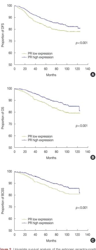
Figure 2. Univariate survival analysis of the estrogen receptor-positive (ER+)/progesterone receptor (PR) low expression patients and the ER+/
PR high expression patients with disease-free survival (DFS), overall survival (OS) and breast cancer specific survival (BCSS). (A) DFS, (B) OS, and (C) BCSS. The figure show that low expression of PR expres- sion was associated with significantly poorer DFS, OS, and BCSS in ER+ patients treated with any endocrine therapy.
100
90
80
70
60
50
0 20 40 60 80 100 120 140 Months
Proportion of DFS
A PR low expression
p<0.001
PR high expression
100
90
80
70
60
50
0 20 40 60 80 100 120 140 Months
Proportion of OS
B p<0.001 PR low expression
PR high expression
100
90
80
70
60
50
0 20 40 60 80 100 120 140 Months
Proportion of BCSS
C PR low expression
p<0.001
PR high expression
tients who died, the date and cause of death was recorded; all deaths not attributable to breast cancer were censored at the date of death. The primary outcome in this analysis was time to breast cancer death; time to death by any cause and time to recurrence (first episode, local and/or distant) were also ana- lyzed. Accordingly, the primary endpoints were disease-free survival (DFS), overall survival (OS), and breast cancer spe- cific survival (BCSS).
Statistical analysis
The distribution of steroid receptor status and other cate- gorical variables were compared using the standard chi-square test. Survival curves were constructed with the Kaplan-Meier method. A multivariate Cox regression model was used to de- termine the association of clinical pathological characteristics with DFS, OS, and BCSS in patients treated with endocrine therapy. Hazard ratios (HRs) for DFS, OS, and BCSS were es- timated using a Cox proportional hazards regression through
Table 2. Multivariate analyses of disease-free survival, overall survival, and breast cancer specific survival
DFS OS BCSS
HR (95% CI) p-value HR (95% CI) p-value HR (95% CI) p-value
Age (yr) 0.001 0.031 0.030
≤35 1 1 1
>35, <50 0.514 (0.359–0.735) 0.589 (0.382–0.908) 0.572 (0.370–0.883)
≥50 0.664 (0.441–0.998) 0.787 (0.485–1.274) 0.737 (0.451–1.203)
Menopause status 0.269 0.238 0.433
Premenopausal 1 1 1
Postmenopausal 1.176 (0.882–1.568) 1.216 (0.879–1.682) 1.145 (0.816–1.608)
Tumor size (cm) 0.016 0.072 0.097
≤2 1 1 1
>2, ≤5 1.315 (1.049–1.648) 1.383 (1.065–1.796) 1.376 (1.045–1.812)
>5 1.765 (1.203–2.591) 1.560 (1.002–2.431) 1.584 (1.004–2.499)
Unknown 1.106 (0.746–1.638) 1.192 (0.749–1.897) 1.301 (0.805–2.103)
Pathological type 0.677 0.691 0.379
IDC 1 1 1
ILC 1.090 (0.867–1.370) 1.118 (0.867–1.441) 1.090 (0.867–1.370)
Others 0.940 (0.668–1.322) 1.019 (0.686–1.513) 0.940 (0.668–1.322)
Surgery 0.415 0.263 0.117
BCS 1 1 1
Others 1.344 (0.661–2.732) 1.701 (0.672–4.304) 2.557 (0.790–8.282)
Lymph node status <0.001 <0.001 <0.001
0 1 1 1
1–3 1.746 (1.245–2.450) 1.947 (1.308–2.898) 2.152 (1.407–3.290)
≥4 4.117 (2.819–6.012) 5.075 (3.274–7.868) 5.757 (3.614–9.171)
Grade 0.110 0.123 0.101
I 1 1 1
II 0.941 (0.732–1.209) 1.098 (0.821–1.469) 1.071 (0.790–1.452)
III 1.249 (0.915–1.705) 1.419 (0.995–2.024) 1.437 (0.995–2.074)
PR 0.014 0.002 0.005
High expression 1 1 1
Low expression 1.280 (1.051–1.558) 1.431 (1.146–1.787) 1.400 (1.109–1.767)
HER2 0.588 0.762 0.791
Negative 1 1 1
Positive 1.138 (0.889–1.457) 1.104 (0.835–1.459) 1.103 (0.824–1.476)
Unknown 1.028 (0.795–1.329) 0.992 (0.739–1.332) 1.000 (0.735–1.361)
Chemotherapy 0.006 <0.001 <0.001
No 1 1 1
Yes 0.607 (0.425–0.867) 0.435 (0.293–0.646) 0.418 (0.273–0.640)
Radiotherapy 0.034 0.050 0.060
No 1 1 1
Yes 1.406 (1.027–1.926) 1.433 (1.000–2.053) 1.435 (0.985–2.090)
DFS=disease-free survival; OS=overall survival; BCSS=breast cancer specific survival; HR=hazard ratio; CI=confidence interval; IDC=invasive ductal carcino- ma; ILC=invasive lobular carcinoma; BCS=breast-conservation surgery; PR=progesterone receptor; HER2=human epidermal growth factor receptor 2.
a multivariate analysis. All the statistical tests were performed using SPSS Statistics version 19.0 (IBM Corp., Armonk, USA) with a two-sided significance level of 5%. Survival rates and HRs were presented with their 95% confidence intervals (CIs).
RESULTS
Clinicopathologic characteristics
The clinicopathological features of 3,030 ER+ patients with invasive breast carcinoma were analyzed. The clinical and bio- logic tumor characteristics are summarized in Table 1. Over-
all, low PR expression occurred more often in the postmeno- pausal group (55.2% vs. 38.0%, p<0.001) and older age group (aged 50 and above, 62.9% vs. 46.9%, p<0.001). However, the tumor size was larger (>2 cm) in the low PR expression group than in the high PR expression group (57.8% vs. 51.8%, re- spectively; p=0.014). In addition, lymph node metastasis was more prevalent (≥4) in the low PR expression group than in the high PR expression group (24.8% vs. 19.7%, respectively;
p=0.004). Finally, there was a higher percentage of invasive ductal carcinoma (67.5% vs. 63.8%, respectively; p=0.030) in the low PR expression group than in the high PR expression
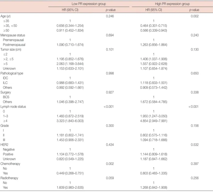
Table 3. Multivariate analyses of disease-free survival in low and high PR expression group
Low PR expression group High PR expression group
HR (95% CI) p-value HR (95% CI) p-value
Age (yr) 0.246 0.002
≤35 1 1
>35, <50 0.656 (0.344–1.254) 0.464 (0.301-0.715)
≥50 0.911 (0.452–1.834) 0.566 (0.339-0.943)
Menopause status 0.694 0.240
Premenopausal 1 1
Postmenopausal 1.090 (0.710–1.674) 1.263 (0.856–1.864)
Tumor size (cm) 0.101 0.130
≤2 1 1
>2, ≤5 1.195 (0.852–1.676) 1.406 (1.037–1.906)
>5 2.063 (1.168–3.644) 1.557 (0.922–2.628)
Unknown 1.153 (0.633–2.101) 1.107 (0.654–1.874)
Pathological type 0.998 0.650
IDC 1 1
ILC 0.988 (0.683–1.431) 1.118 (0.833–1.501)
Others 0.992 (0.592–1.661) 0.909 (0.573–1.442)
Surgery 0.927 0.338
BCS 1 1
Others 1.046 (0.398–2.747) 1.672 (0.584–4.785)
Lymph node status <0.001 <0.001
0 1 1
1–3 1.483 (0.872–2.519) 1.950 (1.247–3.050)
≥4 3.323 (1.840–6.003) 4.854 (2.949–7.991)
Grade 0.300 0.156
I 1 1
II 1.181 (0.802–1.741) 0.802 (0.575–1.116)
III 1.453 (0.906–2.331) 1.094 (0.718–1.666)
HER2 0.434 0.532
Negative 1 1
Positive 1.104 (0.772–1.578) 1.144 (0.809–1.618)
Unknown 0.820 (0.549–1.225) 1.187 (0.847–1.662)
Chemotherapy 0.002 0.397
No 1 1
Yes 0.449 (0.268–0.751) 0.803 (0.483–1.335)
Radiotherapy 0.059 0.256
No 1 1
Yes 1.609 (0.983–2.635) 1.268 (0.842–1.908)
PR=progesterone receptor; HR=hazard ratio; CI=confidence interval; IDC=invasive ductal carcinoma; ILC=invasive lobular carcinoma; BCS=breast-conserva- tion surgery; HER2=human epidermal growth factor receptor 2.
group. In terms of the association between PR expression level and molecular markers, compared with the high PR expres- sion group, the low PR expression group exhibited higher HER2 expression (25.3% vs. 14.0%, respectively; p<0.001) (Table 1).
Survival analysis of ER+ breast cancer patients between the high PR expression group and low PR expression group
The follow-up data for DFS, BCSS, and OS were obtained for 2,778 patients (91.7%). During the follow-up period, 329 patients died; 28 patients died from causes not attributable to
breast cancer, and 425 patients had recurrence and metastasis.
The median follow-up period of all patients was 70 months (range, 42–125 months). Among all the patients who were in- volved in this study, 2,656 (96.1%) were treated with mastec- tomy and 122 (4.4%) were treated with breast-conserving sur- gery. 85.9% of them (2,387/2,778) received chemotherapy, and approximately one-third of the patients (1,047/2,778) received radiotherapy. In our study, few of the participants with HER2 positive tumors were treated with trastuzumab because of the expensive charge, so targeted therapy was not included in the analysis of prognosis.
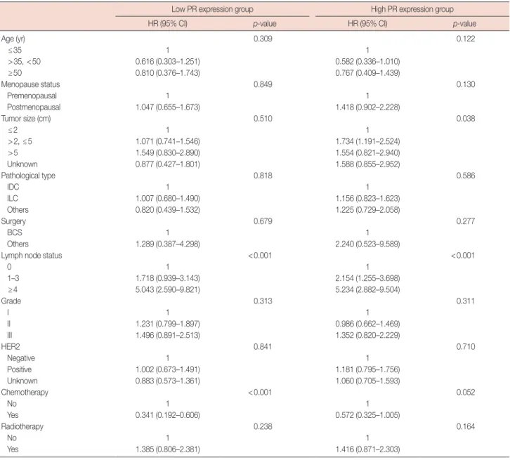
Table 4. Multivariate analyses of overall survival in low and high PR expression group
Low PR expression group High PR expression group
HR (95% CI) p-value HR (95% CI) p-value
Age (yr) 0.309 0.122
≤35 1 1
>35, <50 0.616 (0.303–1.251) 0.582 (0.336–1.010)
≥50 0.810 (0.376–1.743) 0.767 (0.409–1.439)
Menopause status 0.849 0.130
Premenopausal 1 1
Postmenopausal 1.047 (0.655–1.673) 1.418 (0.902–2.228)
Tumor size (cm) 0.510 0.038
≤2 1 1
>2, ≤5 1.071 (0.741–1.546) 1.734 (1.191–2.524)
>5 1.549 (0.830–2.890) 1.554 (0.821–2.940)
Unknown 0.877 (0.427–1.801) 1.588 (0.855–2.952)
Pathological type 0.818 0.586
IDC 1 1
ILC 1.007 (0.680–1.490) 1.156 (0.823–1.623)
Others 0.820 (0.439–1.532) 1.225 (0.729–2.058)
Surgery 0.679 0.277
BCS 1 1
Others 1.289 (0.387–4.298) 2.240 (0.523–9.589)
Lymph node status <0.001 <0.001
0 1 1
1–3 1.718 (0.939–3.143) 2.154 (1.255–3.698)
≥4 5.043 (2.590–9.821) 5.234 (2.882–9.504)
Grade 0.313 0.311
I 1 1
II 1.231 (0.799–1.897) 0.986 (0.662–1.469)
III 1.496 (0.891–2.513) 1.352 (0.820–2.229)
HER2 0.841 0.710
Negative 1 1
Positive 1.002 (0.673–1.491) 1.181 (0.795–1.756)
Unknown 0.883 (0.573–1.361) 1.060 (0.705–1.593)
Chemotherapy <0.001 0.052
No 1 1
Yes 0.341 (0.192–0.606) 0.572 (0.325–1.005)
Radiotherapy 0.238 0.164
No 1 1
Yes 1.385 (0.806–2.381) 1.416 (0.871–2.303)
PR=progesterone receptor; HR=hazard ratio; CI=confidence interval; IDC=invasive ductal carcinoma; ILC=invasive lobular carcinoma; BCS=breast-conserva- tion surgery; HER2=human epidermal growth factor receptor 2.
Univariate analysis using the Kaplan-Meier method showed that the patients with low PR-expressing tumors had worse DFS, BCSS, and OS than those with high PR-expressing tu- mors. The log-rank test showed a significant difference in the time to relapse and death between the two groups, with a shorter DFS (p<0.001), OS (p<0.001), and BCSS (p<0.001) in the low PR expression group (Figure 2). To readdress these imbalances and investigate whether PR status was an inde- pendent and significant predictor of DFS, OS, and BCSS among patients who received adjuvant endocrine therapy, a multivariate analysis was performed comparing the high and low PR expression groups with adjustment of all significant
variables. In general, the multivariate analyses demonstrated that high PR expression was still significantly associated with a better prognosis in terms of DFS (p=0.014; HR, 0.781; 95%
CI, 0.641–0.950), OS (p=0.002; HR, 0.699; 95% CI, 0.560–
0.873), and BCSS (p=0.005; HR, 0.714; 95% CI, 0.566–0.902) (Table 2). These results indicate that patients with low PR ex- pression have a higher recurrence risk and a higher chance of death than patients with high PR expression. In this study, we determined that chemotherapy was also a protective factor of DFS (p=0.006; HR, 0.607; 95% CI, 0.425–0.867), OS (p<
0.001; HR, 0.435; 95% CI, 0.293–0.646), and BCSS (p<0.001;
HR, 0.418; 95% CI, 0.273–0.640) (Table 2). To determine
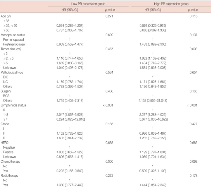
Table 5. Multivariate analyses of breast cancer specific survival in low and high PR expression group
Low PR expression group High PR expression group
HR (95% CI) p-value HR (95% CI) p-value
Age (yr) 0.271 0.116
≤35 1 1
>35, <50 0.591 (0.289–1.207) 0.561 (0.323-0.975)
≥50 0.787 (0.363–1.707) 0.688 (0.362-1.308)
Menopause status 0.699 0.137
Premenopausal 1 1
Postmenopausal 0.909 (0.559–1.477) 1.433 (0.892–2.300)
Tumor size (cm) 0.467 0.093
≤2 1 1
>2, ≤5 1.110 (0.747–1.650) 1.632 (1.109–2.402)
>5 1.669 (0.880–3.165) 1.434 (0.742–2.772)
Unknown 1.040 (0.497–2.176) 1.584 (0.835–3.006)
Pathological type 0.534 0.654
IDC 1 1
ILC 1.169 (0.783–1.744) 1.171 (0.826–1.661)
Others 0.783 (0.399–1.537) 1.126 (0.648–1.956)
Surgery 0.466 0.165
BCS 1 1
Others 1.715 (0.402–7.317) 4.152 (0.555–31.048)
Lymph node status <0.001 <0.001
0 1 1
1–3 2.047 (1.067–3.928) 2.277 (1.288–4.026)
≥4 6.224 (3.023–12.816) 5.677 (3.035–10.622)
Grade 0.185 0.477
I 1 1
II 1.152 (0.728–1.825) 0.986 (0.653–1.487)
III 1.605 (0.941–2.737) 1.282 (0.762–2.156)
HER2 0.885 0.683
Negative 1 1
Positive 1.003 (0.659–1.527) 1.199 (0.797–1.804)
Unknown 0.896 (0.567–1.416) 1.069 (0.701–1.631)
Chemotherapy 0.000 0.098
No 1 1
Yes 0.292 (0.156–0.549) 0.599 (0.326–1.100)
Radiotherapy 0.272 0.178
No 1 1
Yes 1.380 (0.777–2.449) 1.414 (0.854–2.342)
PR=progesterone receptor; HR=hazard ratio; CI=confidence interval; IDC=invasive ductal carcinoma; ILC=invasive lobular carcinoma; BCS=breast-conserva- tion surgery; HER2=human epidermal growth factor receptor 2.
whether the expression of PR was predictive of chemotherapy efficacy in breast carcinomas, a multivariate analysis was per- formed in both the high PR expression group and the low PR expression group. The results showed that patients with low PR expression who received chemotherapy had a better DFS (p=0.002; HR, 0.449; 95% CI, 0.268–0.751), OS (p<0.001;
HR, 0.341; 95% CI, 0.192–0.606), and BCSS (p<0.001; HR, 0.292; 95% CI, 0.156–0.549) (Tables 3-5) than those who did not receive chemotherapy. However, in the high PR expres- sion group, we found that chemotherapy was not associated with DFS (p=0.397; HR, 0.803; 95% CI, 0.483–1.335), OS (p =0.052; HR, 0.572; 95% CI, 0.325–1.005), or BCSS (p=0.098; HR, 0.599; 95% CI, 0.326–1.100) (Tables 3-5).
DISCUSSION
Breast cancer is one of the most common cancers among women worldwide [11]. The incidence of breast cancer con- tinues to rise, and more than 15% of patients develop incur- able disease [12]. It is important to identify those nonrespon- sive breast cancers and develop individualized therapies.
Breast cancer gene expression profiling has gained significant advances in recent years. The combinatorial origin, the het- erogeneity of malignant cells, and the variability of the host background create distinct molecular subgroups of tumors.
However, patients with different molecular subgroups do not respond the same to endocrine therapy [13]. In addition, hor- mone receptor status can provide prognostic assessment for the effect of specific endocrine therapy for patients with breast cancer [14].
PR is an important molecular marker that can predict the prognosis of breast cancer and its response to endocrine ther- apy, especially in ER+ breast cancers. Progestogens have been shown to oppose estrogen-stimulated growth of an ER+/PR+
patient-derived xenograft in a previous study [15]. In addi- tion, the expression of PR can hinder estrogen-mediated pro- liferation and ER transcriptional activity in ER+ breast cancer cells [16]. Furthermore, tumor metastasis could partly be in- hibited by high levels of PR in early-stage disease, and admin- istration of a progesterone injection prior to surgery can pro- vide improved clinical benefit [17]. The above-mentioned re- sults indicate that PR activation can have an anti-tumorigenic effect in the context of ER+ breast cancer. At the 2013 St. Gal- len International Breast Cancer Conference, the expression level of PR was defined in the molecular classification criteria.
The luminal A type was defined as ER+/PR+ with a PR cell number greater than 20% [8]. In our study, the cutoff value was 25%, which is similar to the consensus 20%. We investi- gated PR expression and its relationship to other clinical and
pathological parameters and studied the expression of other molecular markers in patients with invasive breast cancer.
Our results showed that patients with low PR expression were mostly aged 50 or above compared to patients with high PR expression. The association between age and PR expression level that we found in this study is consistent with Rakha et al.’s study [18]. The number of patients with low PR expression who were postmenopausal was significantly higher than that of patients with high PR expression; one previous report also showed this result [19].
In our study, we found that patients with low PR-expressing tumors had worse clinical and biologic characteristics than those with high PR-expressing tumors. The differences in tumor characteristics between the two groups might be related to hormone levels, which is currently widely recognized [20].
As the results indicate, compared to tumors with high PR ex- pression, low PR-expressing tumors had a larger tumor size and more lymph node metastasis. Our findings also showed that low PR expression breast cancer is more likely than high PR expression breast cancer to be HER2-positive. Similar re- sults were found in others research studies [2,20].
Adjuvant systemic therapy can significantly decrease breast cancer recurrence and mortality rates [21]. Endocrine therapy is recommended for most ER+ or PR+ patients due to its effi- cacy and favorable safety profile [21]. Some patients might re- ceive endocrine therapy as their only adjuvant therapy. How- ever, many unsolved questions prevent oncologists from se- lecting the appropriate endocrine therapeutic regimen for the distinct breast cancer subtypes. The role of ER expression level as a predictor of patients’ response to endocrine treatment has been consistently recognized, but the role of PR status in the management of breast cancer remains controversial. To date, relatively few studies have been performed to find an associa- tion between PR status and prognosis of breast cancer. Even fewer studies exist of Chinese female patients with breast can- cer. This study is a large and comprehensive evaluation of the prognosis of breast cancer in Chinese women with ER+ and different PR-expressing tumors. Furthermore, the relationship between PR status and chemotherapy effect was analyzed herein.
In our study, we found significant differences in DFS, BCSS, and OS through a multivariate analysis and each of these vari- ables was an independent predictor of PR status, chemothera- py, lymph node metastasis, and factors, which is similar to re- sults of previous studies [22-25]. Kakugawa et al. [26] suggest- ed that decreased PR expression might lead to excessive pro- liferation of glandular cells, which can cause cancer and meta- static lesions. In our study, all of the subjects were ER+ and treated with any endocrine therapy. Several studies have also
indicated that PR might be a marker for predicting the sensi- tivity of endocrine therapy [2]. Another study showed that PR is synthesized by tumor cells that are stimulated by estrogens through an interaction with ER [27]. Based on the above evi- dence, we can conclude that absence of PR expression might result in the loss of normal ER pathway function, which would account for the relative unresponsiveness to endocrine therapy.
Chemotherapy is also an important part of the treatment for breast cancer, and patients with different molecular sub- types have different chemosensitivity [28]. Few studies have been done to find an association between PR status and che- mosensitivity. In our study, we found that patients with low PR-expressing tumors could benefit from chemotherapy.
However, in patients belonging to the high PR expression group, the prognostic significance of chemotherapy was mod- est. In one study, HER2 was proven a useful marker to identify the chemosensitivity of breast cancer. Tumors with a higher expression of HER2 might be more sensitive to chemotherapy [29]. Furthermore, a previous study showed that PR loss cor- relates with HER2 overexpression in ER+ breast cancer [30].
The same results were obtained from our study. Taken togeth- er, we can infer that patients with low PR expression tumors might derive a greater benefit from adjuvant chemotherapy.
Further studies are warranted to find possible methods to prove the results.
In clinical practice, it is very complex to use PR as a biological marker. Despite progress in understanding the structure and function of PR, it is still not widely used as either a predictive or prognostic marker in the treatment of cancer. When adjuvant treatment decisions are made with individual patients, especial- ly when endocrine therapy alone or endocrine therapy com- bined with chemotherapy is considered, PR status might be an important additional consideration. Assessment of the PR sta- tus should be a mandatory part of assessing the prognosis of breast cancer patients.
CONFLICT OF INTEREST
The authors declare that they have no competing interests.
REFERENCES
1. Palmieri C, Patten DK, Januszewski A, Zucchini G, Howell SJ. Breast cancer: current and future endocrine therapies. Mol Cell Endocrinol 2014;382:695-723.
2. Bae SY, Kim S, Lee JH, Lee HC, Lee SK, Kil WH, et al. Poor prognosis of single hormone receptor-positive breast cancer: similar outcome as triple-negative breast cancer. BMC Cancer 2015;15:138.
3. Viale G, Regan MM, Maiorano E, Mastropasqua MG, Dell’Orto P,
Rasmussen BB, et al. Prognostic and predictive value of centrally re- viewed expression of estrogen and progesterone receptors in a random- ized trial comparing letrozole and tamoxifen adjuvant therapy for post- menopausal early breast cancer: BIG 1-98. J Clin Oncol 2007;25:3846- 52.
4. Arpino G, Weiss H, Lee AV, Schiff R, De Placido S, Osborne CK, et al.
Estrogen receptor-positive, progesterone receptor-negative breast can- cer: association with growth factor receptor expression and tamoxifen resistance. J Natl Cancer Inst 2005;97:1254-61.
5. Elledge RM, Green S, Pugh R, Allred DC, Clark GM, Hill J, et al. Estrogen receptor (ER) and progesterone receptor (PgR), by ligand-binding assay compared with ER, PgR and pS2, by immuno-histochemistry in pre- dicting response to tamoxifen in metastatic breast cancer: a Southwest Oncology Group Study. Int J Cancer 2000;89:111-7.
6. Dowsett M, Cuzick J, Wale C, Howell T, Houghton J, Baum M. Retro- spective analysis of time to recurrence in the ATAC trial according to hormone receptor status: an hypothesis-generating study. J Clin Oncol 2005;23:7512-7.
7. Hammond ME, Hayes DF, Wolff AC, Mangu PB, Temin S. American Society of Clinical Oncology/College of American Pathologists guide- line recommendations for immunohistochemical testing of estrogen and progesterone receptors in breast cancer. J Oncol Pract 2010;6:195- 7.
8. Goldhirsch A, Winer EP, Coates AS, Gelber RD, Piccart-Gebhart M, Thürlimann B, et al. Personalizing the treatment of women with early breast cancer: highlights of the St Gallen international expert consensus on the primary therapy of early breast cancer 2013. Ann Oncol 2013;
24:2206-23.
9. Wolff AC, Hammond ME, Hicks DG, Dowsett M, McShane LM, Allison KH, et al. Recommendations for human epidermal growth factor receptor 2 testing in breast cancer: American Society of Clinical Oncology/College of American Pathologists clinical practice guideline update. J Clin Oncol 2013;31:3997-4013.
10. Chao WR, Lee MY, Lin WL, Chen CK, Lin JC, Koo CL, et al. HER2 amplification and overexpression are significantly correlated in muci- nous epithelial ovarian cancer. Hum Pathol 2014;45:810-6.
11. Parkin DM, Bray F, Ferlay J, Pisani P. Global cancer statistics, 2002. CA Cancer J Clin 2005;55:74-108.
12. de Bono JS, Tolcher AW, Rowinsky EK. The future of cytotoxic therapy:
selective cytotoxicity based on biology is the key. Breast Cancer Res 2003;5:154-9.
13. Carey LA, Perou CM, Livasy CA, Dressler LG, Cowan D, Conway K, et al. Race, breast cancer subtypes, and survival in the Carolina Breast Cancer Study. JAMA 2006;295:2492-502.
14. Li FY, Wu SG, Zhou J, Sun JY, Lin Q, Lin HX, et al. Prognostic value of Ki-67 in breast cancer patients with positive axillary lymph nodes: a retrospective cohort study. PLoS One 2014;9:e87264.
15. Kabos P, Finlay-Schultz J, Li C, Kline E, Finlayson C, Wisell J, et al. Patient- derived luminal breast cancer xenografts retain hormone receptor het- erogeneity and help define unique estrogen-dependent gene signatures.
Breast Cancer Res Treat 2012;135:415-32.
16. Zheng ZY, Bay BH, Aw SE, Lin VC. A novel antiestrogenic mechanism in progesterone receptor-transfected breast cancer cells. J Biol Chem 2005;280:17480-7.
17. Mohammed H, Russell IA, Stark R, Rueda OM, Hickey TE, Tarulli GA,
et al. Progesterone receptor modulates ERalpha action in breast cancer.
Nature 2015;523:313-7.
18. Rakha EA, El-Sayed ME, Green AR, Paish EC, Powe DG, Gee J, et al.
Biologic and clinical characteristics of breast cancer with single hor- mone receptor positive phenotype. J Clin Oncol 2007;25:4772-8.
19. Liu S, Chia SK, Mehl E, Leung S, Rajput A, Cheang MC, et al. Progester- one receptor is a significant factor associated with clinical outcomes and effect of adjuvant tamoxifen therapy in breast cancer patients. Breast Cancer Res Treat 2010;119:53-61.
20. Huang HJ, Neven P, Drijkoningen M, Paridaens R, Wildiers H, Van Limbergen E, et al. Association between tumour characteristics and HER-2/neu by immunohistochemistry in 1362 women with primary operable breast cancer. J Clin Pathol 2005;58:611-6.
21. Early Breast Cancer Trialists’ Collaborative Group (EBCTCG), Davies C, Godwin J, Gray R, Clarke M, Cutter D, et al. Relevance of breast cancer hormone receptors and other factors to the efficacy of adjuvant tamoxifen: patient-level meta-analysis of randomised trials. Lancet 2011;378:771-84.
22. Xue C, Wang X, Peng R, Shi Y, Qin T, Liu D, et al. Distribution, clinico- pathologic features and survival of breast cancer subtypes in Southern China. Cancer Sci 2012;103:1679-87.
23. Rosa Mendoza ES, Moreno E, Caguioa PB. Predictors of early distant metastasis in women with breast cancer. J Cancer Res Clin Oncol 2013;
139:645-52.
24. Xing P, Li JG, Jin F, Zhao TT, Liu Q, Dong HT, et al. Prognostic signifi-
cance of body mass index in breast cancer patients with hormone re- ceptor-positive tumours after curative surgery. Clin Invest Med 2013;
36:E297-305.
25. Dirier A, Burhanedtin-Zincircioglu S, Karadayi B, Isikdogan A, Aksu R.
Characteristics and prognosis of breast cancer in younger women. J BUON 2009;14:619-23.
26. Kakugawa Y, Minami Y, Tateno H, Inoue H, Fujiya T. Relation of serum levels of estrogen and dehydroepiandrosterone sulfate to hormone receptor status among postmenopausal women with breast cancer.
Breast Cancer 2007;14:269-76.
27. Arafah BM, Finegan HM, Roe J, Manni A, Pearson OH. Hormone dependency in N-nitrosomethylurea-induced rat mammary tumors.
Endocrinology 1982;111:584-8.
28. Chen XS, Wu JY, Huang O, Chen CM, Wu J, Lu JS, et al. Molecular sub- type can predict the response and outcome of Chinese locally advanced breast cancer patients treated with preoperative therapy. Oncol Rep 2010;23:1213-20.
29. Yao L, Zhang J, Liu Y, Ouyang T, Li J, Wang T, et al. Association between HER2 status and response to neoadjuvant anthracycline followed by paclitaxel plus carboplatin chemotherapy without trastuzumab in breast cancer. Chin J Cancer Res 2015;27:553-61.
30. Kim HJ, Cui X, Hilsenbeck SG, Lee AV. Progesterone receptor loss correlates with human epidermal growth factor receptor 2 overexpres- sion in estrogen receptor-positive breast cancer. Clin Cancer Res 2006;12(3 Pt 2):1013s-8s.