Perinatology Vol. 30, No. 4, December, 2019 https://doi.org/10.14734/PN.2019.30.4.240
Case report
Perinatology
pISSN 2508-4887•eISSN 2508-4895
Min Jae Kang, MD1,
Seong Yeon Hong, MD, PhD1, Bo Young Kwon, MD1, Ji Eun Jeong, MD2, Jin Young Bae, MD, PhD1 Departments of 1Obstetrics and Gynecology, 2Pediatrics, School of Medicine, Catholic University of Daegu, Daegu, Korea
Cardiac rhabdomyoma is common cardiac mass found during the fetal period. Cardiac rhabdomyoma and tuberous sclerosis have significant associations. Tuberous sclerosis in newborns can cause disability in nearly all organs. Prenatal diagnosis of fetal tuberous sclerosis enables early evaluation and mana
gement of the affected infant. In Daegu Catholic University Hospital, a total of three cases of fetal intracranial tuberous sclerosis were diagnosed among five cases of fetal cardiac rhabdomyoma. The diagnosis in all three cases was confirmed by postnatal brain magnetic resonance imaging. Intra
cranial lesions appeared as multiple small, round and relatively hyperechoic masses on prenatal ultrasonography. Prenatal ultrasonography using transabdominal and transvaginal probes in various angles is helpful. As tuberous sclerosis is not observed in a single instance, regular followup examina
tions are necessary. Fetuses with cardiac rhabdomyoma require detailed prenatal evaluations for tuberous sclerosis, especially in the brain. For this, prenatal ultrasonography is a very useful technique.
Key Words: Heart neoplasms, Ultrasonography, Prenatal diagnosis, Tuberous sclerosis
Introduction
Cardiac rhabdomyoma (CR) is the most common cardiac mass found in heart, with an inci
dence of about 60%. Notably, 90% of CR cases involve multiple tumors that are found in the ventricular septum or cardiac valves. Most CRs are benign; however, fetal cardiac function can be altered according to the size, number, and location of the tumor mass, which in turn affects the prognosis of the newborn.1
CR and tuberous sclerosis complex (TSC) have significant associations. Fetal CRs detected with prenatal fetal echocardiography are usually the earliest signs of TSC. TSC is a multi
organ involvement disorder and generally affects the proliferation of normal cells in the skin, brain, kidney, heart and lungs, resulting in the dysfunction of each organ. TSC in newborns can cause disability in most organs of the body, including skin, eyes and kidney as well as neurological systems. Therefore, prenatal diagnosis of fetal TSC is important because it enables early evaluation and management of the affected infants. There were many reports described fetal brain lesions associated with TSC. However, most of these brain lesions were detected by prenatal or postnatal brain magnetic resonance imaging (MRI), not prenatal brain ultrasonography (USG).2 Therefore, we report the fetal brain lesions associated with TSC detected by fetal USG in fetuses who have CRs.
Case
From January 2015 to December 2018, there were five cases diagnosed with fetal CR at Received: 18 June 2019
Revised: 15 July 2019 Accepted: 24 July 2019 Correspondence to Jin Young Bae, MD, PhD Department of Obstetrics and Gynecology, School of Medicine, Catholic University of Daegu, 33 Duryugongwonro 17gil, Namgu, Daegu 42472, Korea
Tel: +82536504078 Fax: +82536504078 E-mail: [email protected] Copyright© 2019 by The Korean Society of Perinatology
This is an Open Access article distributed under the terms of the Creative Com
mons Attribution NonCommercial License (http://creativecommons.org/
license/bync/4.0/), which permits unrestricted noncommercial use, distribution, and reproduction in any medium, provided that the original work is properly cited.
Fetal Ultrasonography for Prenatal Detec
tion of Tuberous Sclerosis Associated with
Cardiac Rhabdomyoma
2019 December;30(4):240-243
www.e-kjp.org
https://doi.org/10.14734/PN.2019.30.4.240241
Perinatology
Daegu Catholic University Hospital. A retrospective review of medical records was conducted. Data on maternal characteristics, USG features of fetal CR and TSC, delivery, TSC gene mutations, and the clinical course; postnatal evaluation results, including neonatal transthoracic echocardiography (TTE) and brain MRI results were collected.
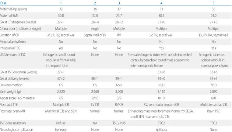
Table 1 shows the clinical features of the five fetuses. A total of four fetuses showed multiple CRs on prenatal USG and intra
cranial TSC findings were observed in three fetuses. Additionally, two patients were simultaneously diagnosed with CR and TSC, and one patient was diagnosed with TSC 6 weeks after the diag
nosis of CR. Case no. 1 showed intracranial tuberous nodules appear as echogenic round masses. In case no. 4, several echo
genic tubers with echogenic nodules and round tumor mass suspected with subependymal giant cell astrocytoma (SEGA) were revealed. In case no. 5, multiple echogenic nodules were present. The delivery method was determined regardless of the presence of CR or TSC. Four of the fetuses were delivered at
our clinic, and one was delivered at another hospital.
Postnatal TTE and brain MRI were performed in all patients, and abdominal USG was performed in four cases born at Daegu Catholic University Hospital. The prenatal diagnosis in all cases of intracranial TSC was confirmed by postnatal brain MRI. Case no. 4 was diagnosed as having a SEGA on postnatal brain MRI and subsequent surgery. Two fetuses who had not been diagnosed with intracranial TSC on prenatal examination showed normal postnatal brain MRI findings. On postnatal abdominal USG, case no.1 showed a small renal cyst, and the findings were normal in case no. 24. Four newborns underwent genetic evaluation (case no. 25), and TSC2 gene mutation was found in two cases.
Case no. 3 who had not revealed intracranial TSC lesion showed TSC2 gene mutation with variation of unknown significance (VUS). Parents of case no. 1 refused genetic evaluation.
Case no. 5 was diagnosed with multiple giant CRs in the right ventricle, obstructing the right ventricle outflow tract and com
pressing the left ventricle. The blood flow from the right ventricle
Table 1. Clinical Features of the Five Patients with Cardiac Rhabdomyomas
Case 1 2 3 4 5
Maternal age (years) 32 36 37 31 36
Maternal BMI 30.8 32.0 23.7 30.1 24.0
GA at CR diagnosis (weeks) 27+1 26+4 26+2 31+6 27+3
CR number (multiple or single) Multiple Single Multiple Multiple Multiple
Location of CR LV, LA, RV, septal wall Septal wall of LV RV LV, RV, septal wall LV, RV, RA, septal wall
Prenatal arrhythmia No No No No No
Intracranial TSC Yes No No Yes Yes
USG features of TSC Echogenic small round nodule in frontal lobe, intemporal lobe
None None Several echogenic tuber with nodule in cerebral cortex, hyperechoic round mass adjacent to interhemispheric fissure
Echogenic tuberous sclerosis nodule in cerebral parenchyme
GA at TSC diagnosis (weeks) 27+1 31+6 33+6
GA at delivery (weeks) 37+2 38+1 39+1 39+0 36+6
Delivery method CS CS NSD NSD NSD
Birth weight (g) 2,820 2,960 3,000 3,110 2,890
Apgar score (1/5 minutes) 7/9 9/10 8/9 8/10 6/8
Postnatal TTE Multiple CR LV CR RV CR RV, ventricular septum CR Multiple cardiac CR
Postnatal brain MRI Multifocal CTs and SEN Normal Normal Enhancing mass near foramen Monro r/o SEGA, small SEN near ventricle, CTs
Brain TSC
TSC gene mutation Refuse NA TSC2 VUS TSC2 TSC2
Neurologic complication Epilepsy None None Epilepsy None
Abbreviations: BMI, body mass index; GA, gestational age; CR, cardiac rhabdomyoma; LV, left ventricle; LA, Left atrium; RV, right ventricle; TSC, tuberous sclerosis; USG, ultrasonography; CS, cesarean section; NSD, normal spontaneous delivery; TTE, transthoracic echocardiography; MRI, magnetic resonance imaging; CTs, cortical tubers;
SEN, subependymal nodule; r/o, rule out; SEGA, subependymal giant cell astrocytoma; NA, not appear; VUS, variation of unknown significance.
Kang MJ, et al. Fetal ultrasonography for prenatal detection of tuberous sclerosis associated with cardiac rhabdomyoma
242
https://doi.org/10.14734/PN.2019.30.4.240www.e-kjp.org
Perinatology
no. 1 and 4 and anticonvulsants were administered. Case no. 1 exhibited seizure symptoms from 3 months of age and is cur
rently under medication, but the seizures are not well controlled.
Case no. 4 exhibited seizure symptoms for about 7 months after birth, and a SEGA was surgically removed at 11 months after birth.
Discussion
During the prenatal examination, TSC can be found in more than half of the fetuses with CR and can cause organ dysfunction.3 TSC is an autosomaldominant inheritance and is expressed in at least one mutation of TSC1 on chromosome 9q34 and TSC2 on chromosome 16p13.3 gene.4 Mutation of the TCR gene causes overactivation of mTORC1 and leads to dysregulation of the signaling pathway, leading to neurodegeneration, epilepsy, dia
betes mellitus and cancer.57 Currently, multiple CRs are thought to be related with TSC.3 Tworetzky et al.8 reported that three of nine patients (33.3%) with single CR had TSC and 30 of 33 pa
tients (91%) with multiple CRs had TSC.
At the second International Tuberous Sclerosis Complex Consensus Conference held in 2012, experts updated the recom
mendations for the evaluation and management for patients with TSC.3 Brain lesions associated with TSC are cortical tuber (CT), subependymal nodule (SEN), and SEGA. Intracranial CT and SEN lesions usually appear as small, multiple, round, and relatively hyperechoic masses in prenatal USG. In case no. 1, intracranial tuberous nodules appear as echogenic round masses in the left frontal lobe and right temporal lobe (Fig. 1E). In case no. 4, several echogenic tubers with echogenic nodules sus
pected to be TSC were revealed in the cerebral cortex. We also discovered another major diagnostic finding associated with TSC in the abovementioned case, namely SEGA. It appeared as a relatively hypoechoic, round mass compressing the cavum septum pellucidum, and the right frontal horn was located just adjacent to the interhemispheric fissure (Fig. 1BD, F).
Case no. 2 and 3 didn’t show intracranial TSC lesion in prenatal and postnatal evaluation. In genetic test, TSC2 gene mutation was not found in case no. 2 and case no. 3 just showed mutation with VUS. However, according to the Consensus Conference in 2012,3 possible diagnosis can be determined with either one toward the pulmonary artery was not seen and retrograde flow
via ductus arteriosus was observed. Therefore, functional pul
monary atresia was suspected due to obstruction of the right ventricular outflow by large CR in case no. 5 (Fig. 1A). The fetus was delivered at another hospital for postnatal cardiac surgery.
Postnatal brain MRI of the newborn revealed intracranial TSC findings, however, the baby died because of respiratory failure on the 19th day after birth.
Followup for 24 months revealed normal growth and develop
ment of case no. 2 and 3. Epileptic disorder was observed in case
F E
Fig. 1. (A) Fetal echocardiography showed multiple cardiac rhabdo- myomas in the right ventricle, the left ventricle and the interventricular septum. (B) The para-sagittal view of the fetal brain showed multiple cortical tubers at the cerebral cortex as hyperechoic dots and round masses (arrowheads). (C) The coronal view of the fetal brain showed a single subependymal giant cell astrocytoma (SEGA) as a hypoechoic round mass adjacent to the foramen of Monro (asterisk). (D) The sagittal view of the fetal brain showed a subependymal nodule as an echogenic spot along the ependymal lining of the lateral ventricle (arrow). (E) Postnatal brain magnetic resonance imaging (MRI) of case no. 1. Hy- perintensity of cortical and subcortical tubers in T1-weighted images (black arrows). (F) Postnatal brain MRI of case no. 4. SEGA within the lateral ventricle near the foramen of Monro (asterisk).
2019 December;30(4):240-243
www.e-kjp.org
https://doi.org/10.14734/PN.2019.30.4.240243
Perinatology
major feature (CR) or more than two minor features. Therefore, patients with possible diagnosis need regular followup also.
Nowadays, several physicians prefer prenatal MRI for the eva
luation of fetuses, especially in the case of fetal brain anomalies.
Prenatal MRI may be helpful under many circumstances. How
ever, it is expensive and causes more discomfort to the mothers than on USG. Furthermore, when the fetus moves during exa
mination, interpretation of the image is difficult, or almost impos
sible, in some cases.9 In our study, we proved the accuracy and usefulness of prenatal brain USG and the antenatal diagnosis was accord to postnatal brain MRI findings.
Prenatal USG is costeffective and reduces the discomfort experienced by the mother. Physicians can examine various angles using USG, the findings of which are free from fetal motion artifact. Occasionally, the examiner cannot get a pre
cise image of the fetal head by transabdominal USG because of deep engagement of the fetal head into the pelvic cavity. In this situa tion transvaginal USG is appropriate, with adjustment of angles. Mild fundal pressure and changing the maternal position could also be helpful. In our cases of prenatal TSC, all three fetuses were diagnosed with transvaginal USG upon vertex presentation; sagittal view and coronal view brain ima
ges were also especially helpful.
In conclusion, fetuses with CR require detailed prenatal eva
luation for TSC, especially in the brain. Newborn cardiac dys
function from various aspects of CR needs to be prepared, also.
In that respect, we emphasize importance and usefulness of prenatal USG. Furthermore, postnatal cardiac computer tomo
graphy, brain MRI, and TSC gene mutation tests may be helpful.
References
1) Yuan SM. Fetal primary cardiac tumors during perinatal period. Pediatr Neonatol 2017;58:205-10.
2) Dragoumi P, O'Callaghan F, Zafeiriou DI. Diagnosis of tuberous sclerosis complex in the fetus. Eur J Paediatr Neurol 2018;22:1027-34.
3) Northrup H, Krueger DA, International Tuberous Sclerosis Complex Con- sensus Group. Tuberous sclerosis complex diagnostic criteria update:
recommendations of the 2012 international tuberous sclerosis complex consensus conference. Pediatr Neurol 2013;49:243-54.
4) Saxena A, Sampson JR. Epilepsy in tuberous sclerosis: phenotypes, me- chanisms, and treatments. Semin Neurol 2015;35:269-76.
5) Kapoor A, Girard L, Lattouf JB, Pei Y, Rendon R, Card P, et al. Evolving strategies in the treatment of tuberous sclerosis complex-associated angiomyolipomas (TSC-AML). Urology 2016;89:19-26.
6) Wang CC, Wang CY, Lai YJ, Chang TY, Su HY. Prenatal diagnosis of tube- rous sclerosis complex using fetal ultrasonography and magnetic re- sonance imaging and genetic testing. Taiwan J Obstet Gynecol 2018;
57:163-5.
7) Ekmekci E, Ozkan BO, Yildiz MS, Kocakaya B. Prenatal diagnosis of fetal cardiac rhabdomyoma associated with tuberous sclerosis: a case report.
Case Rep Womens Health 2018;19:e00070.
8) Tworetzky W, McElhinney DB, Margossian R, Moon-Grady AJ, Sallee D, Goldmuntz E, et al. Association between cardiac tumors and tuberous sclerosis in the fetus and neonate. Am J Cardiol 2003;92:487-9.
9) Saada J, Hadj Rabia S, Fermont L, Le Bidois J, Bernardes LS, Martinovic J, et al. Prenatal diagnosis of cardiac rhabdomyomas: incidence of asso- ciated cerebral lesions of tuberous sclerosis complex. Ultrasound Obstet Gynecol 2009;34:155-9.