I. 서론
Titanium은 다른 금속재료와 비교하여 우수한 생 체친화성을 가지고 있어 생체내 이식재료로 널리 사 용되고 있는 금속 중 하나이다1. Titanium의 생체이 식에 있어서 장점으로는 corrosion에 대한 저항성이 크며, 표면의 물리화학적 특성을 바꾸는 것이 상대적 으로 용이하다는 점이다2. Titanium 표면의 물리화학 적 특성은 생체조직과의 반응에 있어서 결정적인 역 할을 하는 것으로 알려져 있다3. 이식체는 골조직과 긴밀한 접촉을 이루고 있기 때문에, 표면의 특성이 골조직과의 상호반응에 있어 중요한 역할을 할 것이 라는 것은 당연히 예상되어진다.
Titanium재료가 가지는 생체친화성은 표면에 형 성되는 oxide layer에 의해 얻어지는 것으로 알려져 있다. Titanium표면에 형성되는 oxide layer는 대략 5nm정도의 두께로 nonstoichiometric TiO2로 구성되 어 있으며, 대부분 amorphous한 형태로 존재한다2,4. 또한 titanium oxide는 막이 파괴되더라도 대기중이 나 물에 노출되었을 때 빠르게 재형성되는 특징을 가진다5-7. 이러한 특징외에도 thermal oxidation이나 electrochemical oxidation에 의해 산화막의 두께와 결정화의 정도에 변화를 주는 것이 가능하다8.
Titanium implant는 뛰어난 생체친화성에도 불구 하고 osseointegration이 일어나는 과정이 느리게 진 행되고, 기능중에 osseointegration이 파괴되기도 한 다. 이러한 이유들로 인해 implant표면을 물리적, 화 학적으로 변화시켜 osseointegration을 질적으로 향 상시키려는 연구들이 진행되고 있다9,10. 골조직과 implant간의 계면에서 양질의 접촉을 얻기 위하여, 초기에는 골조직과의 반응을 촉진하기 위해 HA coating을 이용하거나 표면적을 증가시키기 위해 titanium plasma spray 방법이 사용되었다11-13. 또한 최근에는 sandblasting이나 acid etching을 통해 표면 적의 증가뿐만 아니라 표면의 microtexture를 변화시 킴으로써 osseointegration을 개선하려는 연구가 진 행되어왔다. 최근의 많은 보고들에 의하면 표면의 roughness와 topography가 in vitro에서 생물학적 반 응-subcellular biomolecule의 표면흡착, cell attach- ment, cell proliferation, cell differentiation, protein synthesis-에 영향을 주며, 임상적으로 removal torque의 증가나 bone contact area의 증가를 보였다 고 하였다14-30. 특히 sandblasting에 의한 표면조도의 증가가 osseointegration이 일어나는 기간 중 조기에 미치는 영향이 큰 것으로 보고되었다31. 그러므로 implant 식립 후 loading까지 걸리는 시간을 줄일 수
Titanium 표면처리 방법이 Osteoblast-like Cells의 부착 및 증식에 미치는 영향
김도영1·설양조1·김우진2·류인철1·백홍구2·허성주3·한종현4 김명호5·최용창6·전흥재7·함병도1·권수경8·정종평1·최상묵1
1서울대학교 치과대학 치주과학교실, 2연세대학교 공과대학 금속공학과, 3서울대학교 치과대학 보철학교실,
4영동세브란스병원 보철학교실, 5(주)우리동명, 6카톨릭대 여의도 성모병원 치과
7연세대학교 공과대학 기계공학과 , 8을지의과대학 치과학교실
대한치주과학회지 : Vol. 30, No. 3, 2000
* This study was supported by a grant (HMP-98-G-2-035-B) of the HAN(highly advanced National) Project, Ministry of Health &
Welfare, R.O.K
있으며, 임상적 성공률에도 영향을 미칠 것으로 보인 다.
그러나 표면특성이 어떤 방식으로 세포에 전달되 는 지에 대해서는 거의 알려져 있지 않다. 실제 implant를 식립하였을 때 최초로 일어나게 되는 반 응은 implant표면과 serum, tissue fluide간에 반응이 다. 세포와 재료간 상호작용은 표면의 microstruc- ture에 의해 생체분자가 표면에 흡착되고 배열되는 방식에 의해 매개되며, 결과적으로 세포가 focal attachment를 일으켜서 세포의 phenotypic expres- sion을 야기하며, 세포형태의 변화는 integrin recep- tors를 통한 세포외 signal이 세포내로 transduction되 는 방식을 변화시킬 수 있다32,33.
본 실험에서는 titanium표면을 thermal oxidation 함으로써 oxide layer의 변화를 유도하여, 이런 변화 가 생체친화성에 미치는 영향을 기존에 이용되고 있 는 표면처리방법과 비교하고자 하였다.
II. 연구재료 및 방법
1. 세포배양이 실험에서는 백서의 경골과 대퇴골에서 채취한 osteoblast-like cells을 사용하였다. 과정을 간단하게 기술하면, 100g의 몸무게를 가진 백서로부터 경골과
대퇴골을 분리한 후 말단부를 잘라내었다. α-
MEM(minimal essential medium)을 syringe에 담아 한쪽 끝에서 주입하여 반대쪽으로 나오는 부유액을 10% FBS(fetal bovine serum)와 1% 항생제를 포함한 α-MEM을 이용하여 24시간 배양하였다. 24시간 후 남아있는 혈액세포를 제거하기 위하여 부유액을 흡 입하고 HBSS로 3회에 걸쳐 세척하였다. 바닥에 붙 어있는 osteoblast-like cells을 0.05% trypsin-0.02%
EDTA용액으로 30분간 처리하여 세포부유액을 만든 후, 원심분리하여 다시 배지에 2회 계대배양하였다.
2. Titanium disc 제작
표면처리를 위한 실험재료로 지름 15mm, 두께
3mm인 pure titanium disc를준비하여, 실험목적에 따라 다음과 같이 군을 나누어 표면처리를 시행하였 다. (1) machined(M) (2) sandblasted with Al2O3 (MB) (3) sandblasted and etched (MBE) (4) blasted and etched following thermal oxidation at 400℃ (O- 400) (5) blasted and etched following thermal oxida- tion at 600℃ (O-600) (6) blasted and etched follow- ing thermal oxidation at 800℃ (O-800).
Sandblast 표면처리의 경우 평균 입자 크기가 50μ m인 Al2O3를 5kgf/cm2압력을 유지하면서 분사하여 시편 전면에 걸쳐 표면처리를 시행하였다. Etching 은 NH4OH : H2O2 : H2O를 1:1:5의 비율로 혼합한 용 액을 이용하여 90℃에서 1분간 표면처리하였다.
Thermal oxidation은 진공관상로를 이용하여, 시편 이 장입된 후 약 3×10⁻⁶Torr까지 진공배기시킨 후 진 공관상로에 순수 산소를 불어넣어 대기압과 동일한 압력하에서 oxidation을 실시하였다. 400℃, 600℃, 800℃에서 2시간 행하였으며, 10℃/min의 승온과 냉 각속도를 유지하였다.
실험에 앞서, 각 군으로 나뉘어진 시편들을 ethanol하에서 5분간 초음파세척을 하고다시 DDW 에서 5분간 초음파세척을 한후 건조하였다. 건조된 시편들을 24 well plate에 위치시킨 후 EO gas멸균을 하였다.
3. Titanium disc의 표면조도 측정
Disc의 표면조도를 측정하기 위하여 profilometer 를 사용하였다. 측정은 각 군으로부터 2개의 시편을 사용하였으며, 시편당 2부위에서 측정을 시행하였다.
4. Osteoblast like cells의 부착에 미치는 영향
밀생상태의 osteoblast like cells를 각각의 disc당 1
×105cell, 100㎕를 접종하고 여기에α-MEM 1ml를 첨가하여 시편이 완전히 잠기게 하였다. 그리고 37
℃, 5% CO2 환경하에서 4시간, 8시간, 24시간 배양하 였다. 측정은 전체세포수에 대한 부착세포수의 백분 율로 나타내었다. 먼저 부유액을 흡입하여 미부착세
포의 수를 hemocytometer를 이용하여 측정하였다.
시 편 표 면 에 부 착 된 세 포 는 0.05% trypsin- 0.02%EDTA용액으로 30분간 처리하여 세포부유액 을 만든 다음 hemocytometer를 이용하여 세포수를 측정하였다.
5. Osteoblast like cells의 증식에 미치는 영향
Osteoblast like cells를 각각의 disc당 6.4×10⁴cell, 100㎕를 접종하고 4시간 배양한 후, α-MEM배지 1ml 를 첨가하여 1일, 3일, 7일에 걸쳐 배양하였다. 측정 은 세포부착에서와 같은 방법으로 시편에 부착된 세 포의 수를 측정하였다.
6. SEM 관찰
7일간 배양한 시편을 PBS(phosphate buffer solu- tion)로 세척한 후 0.01M HBSS하에서 2.5% glu- taraldehyde로 60분간 고정하였다. 그리고 1% osmi- um tetroxide로 2차고정한 후 critical point drying을 하고 gold sputter coating을 시행하였다. SEM 관찰 은 osteoblast like cells의 부착형태에 관해서 시행하 였다.
7. 통계분석
세포부착과 세포증식에 대한 군간 비교에서는
Mann-Whitney test(P<0.05)를 시행하였으며, 세포증 식에 대한 시간에 따른 군내 비교는 Wilcoxon signed rank test(P<0.05)를 시행하였다.
III. 연구결과
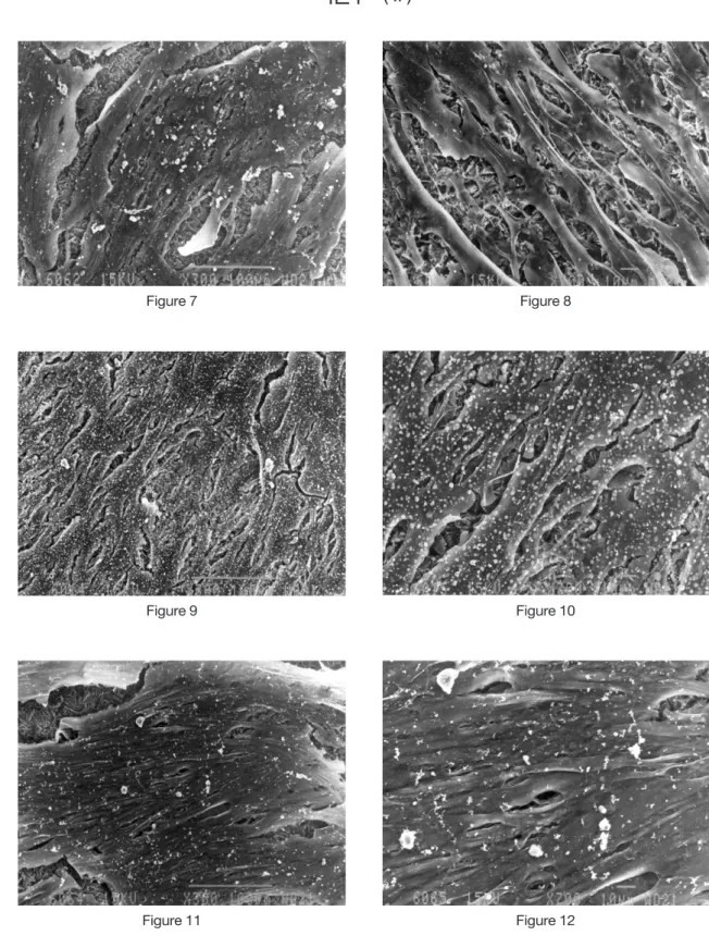
1. 표면조도 측정Machined surface의 경우 Ra값이 0.28㎛로 최소값 을 나타냈으며, 나머지 군에서는 Ra값이 0.7-0.9㎛로 유사한 표면조도를 보였다.
2. Osteoblast like cells의 부착에 미치는 영향
시간경과에 따라 모든 군에서 세포부착이 증가하 였으며, 배양시간에 따른 군간 부착수준은 유사하게 나타났다. 배양 8시간째 안정적인 세포부착을 보였 으며, 배양 24시간째와 비교하여 더 이상의 부착증가 를 보이지 않았다. MBE 군에서 다른 군에 비해 24시 간 후 약간 낮은 부착수준을 나타내었으나 통계학적 유의성은 없었다.
3. Osteoblast like cells의 증식에 미치는 영향
세포 증식은 시간의 경과에 따라 대부분 유의성있 게 증가하였다. 다만 O-800군에서 3일째와 7일째를 비교하였을 때 통계학적으로 유의한 차이가 없었다.
Table 1. Measurements of surface roughness(㎛) on two sample discs from each group.
Group Ra±SD Rq±SD Rt±SD
Machined 0.28±0.073+ 0.37±0.10 2.12±0.90
Sandblasted 0.75±0.075 0.98±0.078 6.32±0.64
Blasted and etched 0.84±0.094 1.06±0.12 6.29±0.76
Oxidation at 400℃ 0.80±0.074 1.00±0.091 4.84±2.16
Oxidation at 600℃ 0.88±0.065 1.11±0.084 6.85±0.70
Oxidation at 800℃ 0.84±0.026 1.05±0.060 6.07±1.28
+: means±SD
Ra : mean height deviation from peak to valley Rq : root mean square value of the surface departures
Rt : extreme value, the distance between the highest peak and the lowest valley
7일째 군간 비교에서 MB군과 O-800군에서 다른 군 과 비교하여 통계학적으로 낮은 세포증식을 보였다.
4. SEM관찰소견
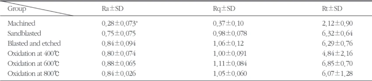
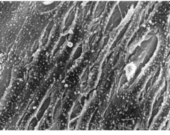
배양 7일째, 시편에 부착된 세포들은 dendritic extension에 의해 길게 늘어진 형태를 보였으며, 시 편의 중앙부위에서는 세포간 구분이 어려울 정도로 세포들이 밀생하여 있었다. 세포들은 여러 층에 걸 쳐 부착된 모습을 보였고, 가장자리로 증식해 나가는 양상을 나타내었다. 세포들의 표면에서는 작은 white spot들이 관찰되었으며, 부분적으로 융합되어 크기가 증가하는 것으로 보여졌다. White spot들은 시편 전체에 걸쳐 고르게 분포하였으나, 부분적으로 드문 양상을 보였다. 군간 부착된 세포들의 양상에 는 큰 차이가 없었다.
IV. 총괄 및 고안
식립된 implant가 성공적인 osseointegration을 형 성하기 위해서는 초기에 조골세포의 부착, 증식 및 분화가 필수적인 과정으로 여겨진다.17이 실험에서 는 titanium표면을 몇 가지 방법으로 처리함에 따른 표면특성의 변화가 osteoblast like cells의 반응에 미 치는 영향을 평가하고자 하였다.
실험에서 사용된 표면처리 방법은, 표면의 micro- topography와 roughness를 변화시키기 위해서 기존 에 많이 이용되고 있는 sandblast를 이용한 표면처리 방법과 blasting후 etching하는 방법을 사용하였고, oxide layer의 변형을 주는 방법으로 thermal oxida- tion을 사용하였다. 표면조도 측정결과, machined surface의 경우 0.28㎛로 가장 평활한 표면을 나타냈 으며, sandblasting에 의해 표면조도가 0.75㎛로 거칠 Table 2. Effect of Titanium Disc Surface on Cell Attachment(%)
Time(hours)
Group 4hr 8hr 24hr
Machined 26.98±7.67+ 83.45±2.96 73.58±7.21
Sandblasted 33.30±4.09 74.14±11.10 65.09±8.85
Blasted and etched 29.75±9.54 74.19±15.75 50.77±14.38
Oxidation at 400℃ 42.20±15.72 79.15±4.83 60.39±24.85
Oxidation at 600℃ 29.99±15.56 79.18±4.20 74.82±8.86
Oxidation at 800℃ 34.48±16.57 66.61±14.35 69.89±10.51
+: mean±SD
Table 3. Effect of Titanium Disc Surface on Cell Proliferation(×104cell) Time(days)
Group 1d 3d 7d
Machined 3.12±0.56+ 9.68±1.05 15.40±1.24
Sandblasted 1.52±0.55 7.40±1.28 10.85±1.65#
Blasted and etched 1.52±0.42 8.59±1.96 14.55±1.89
Oxidation at 400℃ 3.04±0.29 7.44±1.96 14.50±1.32
Oxidation at 600℃ 3.73±0.46 8.44±1.88 13.85±1.86
Oxidation at 800℃ 2.48±0.72 10.93±1.09 10.25±1.17*#
+: mean±SD
* no significantly difference at P<0.05, 3day versus 7day
# significantly difference at P<0.05, versus machined surface
어졌다. 그러나 etching이나 thermal oxidation에 의 해 표면조도가 sandblasted surface보다 조금 증가하 기는 하였으나 큰 차이를 보이지는 않았다. 이는 etching이나 thermal oxidation이 peak와 valley에 고 르게 작용하기 때문에 결과적으로 유사한 표면조도 를 보이는 것으로 사료된다.
세포부착을 위한 실험에서는 배양된 세포부유액 을 disc 표면에 접종한 후 1ml의 배양액을 첨가하여 disc 표면이 완전히 잠기게 하였다. 그렇게 함으로써 표면 전체에 걸쳐서 세포부착이 일어나도록 고안하 였다. 그러나 세포증식을 위한 실험에서는 100㎕의 세포부유액을 disc 가운데에 접종한 후 4시간 후에 세포배양액 1ml를 첨가하여 배양하는 방식으로 실 험을 진행하였다. 이렇게 함으로써 osteoblast like cells이 초기 4시간동안 disc 표면에 한정되어 부착이 일어나도록 하여 plastic well 표면에 부착되어 증식 하는 것을 최소로 줄일 수 있도록 하였다. 실험의 결 과, 세포배양 24시간에 걸친 세포부착은 모든 군간에 유사한 부착비율을 보였으며, 배양시간 8시간 후 안 정적인 세포부착을 보였다. 좀더 지연된 반응을 나 타내는 세포증식에 미치는 영향에 있어서는 시간의 경과에 따라 통계학적으로 유의하게 세포수가 증가 하였으며, 측정시기에 따른 군간 비교에서는 대부분 유의한 차이를 보이지는 않았다.
표면조도의 증가가 세포부착이나 증식에 미치는 영향에 대해서는 논쟁의 여지가 있는 것으로 보여진 다. Michaels 등과 Bowers 등은 표면이 거칠수록 부 착된 세포의 비율이 높았다고 보고한 반면14,15, Lincks 등은 표면조도가 증가할수록 낮은 세포증식 을 보인다고 하였다16. 또한 Martin 등과 Cooper 등 은 표면조도가 세포부착과 증식에 유의한 영향을 미 치지 않는다고 보고하였다17,18. 그러나 표면조도의 증가가 in animal study에서 histomorphometric analysis에 의한 implants-bone contact area의 증가 를 보이며, removal torque가 증가한다는 데에 대해 서는 별다른 이견이 없는 것 같다19-28. 이렇게 in vitro 실험과 in vivo 실험에서 얻어지는 결과가 상이한 이 유는 식립된 implants가 osseointegration을 형성하는 과정이 많은 요인들-Blood나 tissue fluid내에 존재하
는 biomolecules이나 ions과의 최초의 상호작용, 세 포의 부착 및 증식, 분화, 기질의 석회화, 조골세포의 활성도에 영향을 주는 local factors의 생산 등-이 포 함된 동적인 상태이기 때문이다6,34. 초기 세포부착이 나 증식이 감소된 양상을 보이더라도 ALPase나 matrix synthesis, mineralization이 증가된다면 결과 적으로 우수한 osseointegration을 이룰 수 있다17,35. 또 다른 이유로는 식립된 implants 표면에 골이 형성 되는 방식의 차이에 의해 설명되어질 수 있다. 평활 한 표면을 가진 implants에서는 새로이 형성되는 골 조직이 기존에 존재하는 골표면으로부터 형성되어 implants표면으로 성장해 들어가는implantopetal bone growth 또는 distance osteogenesis가 일어나는 반면에, 거친 표면을 가진 implants에서는 신생골이 implants 표면에서 직접 형성되는 implantofugal bone growth 또는 contact osteogenesis가 일어나게 된다27,36.
SEM 관찰결과, 세포표면에서 작은 white nodule 들이 산재해있는 것이 관찰되었다. 이것은 matrix vesicle이 석회화 과정에 있는 것으로 평가된다37. Matrix vesicle의 형성은 조골세포의 활성도를 평가 하는 하나의 기준이 되며, 골조직과의 bonding이 우 수한 implant 재료에서 높은 밀도로 관찰된다38,39.
Thermal oxidation에 의한 효과는 oxide layer의 두 께를 증가시키며, oxide crystallinity를 증가시킨다
4,8,40. Titanium표면에 형성되는 oxide의 morphology
와 microstructure는 하부에 존재하는 금속의 microstructure와 grain structure, chemical composi- tion 등과 관련이 있는 것으로 보고되었다2. 이러한 이유로 thermal oxidation에 의한 표면의 변화는 oxi- dation을 시행하기 전에 어떠한 방식으로 표면을 형 성하는 가에 의해 크게 달라지게 된다. 두꺼운 ther- mal oxides나 anodic oxides의 경우 heterogeneous 한 형태를 가지며, 부위에 따라 porosity와 morphol- ogy가 다양하게 나타난다8. Titanium oxide의 결정 화정도와 두께는 implant가 주위조직과의 생물학적 반응에 영향을 줄 수 있다41-43. Oxide layer의 두께가 증가할수록 corrosion resistance도 증가하며, titani- um ion의 생체내 유출도 줄어든다44-46. 거친 조도를
가진 표면이나 porous한 표면에서 metal ion의 유출 이 증가하며, 결과적으로 세포의 활동에 영향을 미칠
수 있다47,48. 또한 증가된 oxide layer는 Implant 식립
시 외상으로 인한 염증반응에서 발생하는 hydroxyl radicals의 유해한 효과를 중화시키는 작용도 있는 것 으로 보고되었다5,49. 그러나 Oxide layer의 두께가 과도하게 증가하는 것은 바람직하지 않을 수 있다.
두꺼운 oxide layer는 self tapping implants와 같은 높은 shear stress하에서 titanium으로부터 oxide layer가 분리될 수도 있기 때문이다40,50.
본 실험에서 사용된 thermal oxidation 방식은 순 수한 산소환경 하에서 시행하였다. 이렇게 함으로써 일반 대기 중에서 oxide layer가 형성될 때 hydrocar- bon을 포함한 불순물들이 포함되는 것을 최소로 방 지할 수 있다8. 이러한 불순물들은 모든 implants에 미량 포함되어 있으며, 완전히 제거하는 것은 불가능 하다. 이들이 생체내 반응에 미치는 영향에 대해서 는 정확히 알려진 바가 없으나, oxide layer의 특성에 영향을 주는 것으로 알려져 있다2.
이번 실험에서는 thermal oxidation에 의한 oxide layer의 변화가 세포의 부착과 증식에 큰 영향을 미 치지 않는 것으로 나타났다. 그리고 SEM관찰에서도 다른 표면처리를 한 군들과 비교하여 부착양상의 변 화를 보이지 않았다. 이 결과로부터 세포들의 생물 학적 반응에 영향을 주는 주된 요소로써 oxide layer 의 특성보다는 표면조도가 미치는 영향이 더 큰 것 으로 평가된다8,51. 그러나 위에서도 언급한 바와 같 이 in vitro 실험에서는 많은 요소들이 배제되어 있기 때문에 thermal oxidation이 osseointegration에 미치 는 영향을 평가하기 위해서는 동물실험을 포함한 더 많은 연구들이 필요할 것으로 사료된다.
V. 결론
1. 표면조도 측정결과, machined surface에서 Ra 값이 0.28㎛로 가장 평활한 표면을 나타냈으며, 나머지 표면처리를 시행한 군에서는 Ra값이 0.7-0.9㎛로 유사한 표면조도를 보였다.
2. 시간경과에 따라 모든 군에서 세포부착이 증가
하였으며, 배양시간에 따른 군간 부착수준은 유 사하게 나타났다.
3. 세포 증식은 시간의 경과에 따라 대부분 유의성 있게 증가하였다. 7일째 군간 비교에서 MB군과 O-800군에서 다른 군과 비교하여 통계학적으로 낮은 세포증식을 보였다.
4. 배양 7일째, 시편에 부착된 세포들은 dendritic extension에 의해 길게 늘어진 형태를 보였으며, 세포들의 표면에서는 작은 white spot들이 관찰 되었다. White spot들은 시편 전체에 걸쳐 고르 게 분포하였으며, 군간 부착된 세포들의 양상에 는 큰 차이가 없었다.
5. 세포들의 생물학적 반응에 영향을 주는 주된 요 소로써 oxide layer의 특성보다는 표면조도가 미 치는 영향이 더 큰 것으로 사료된다.
V. 참고문헌
1. Steflik DE, Parr GR, Sisk AL, Lake FT et al.
Osteoblast activity at the dental implant-bone interface: Transmission electron microscopic and high voltage electron microscopic observations.
J Periodontol 1994; 65: 404-413.
2. Kasemo B, Lausmaa J. Biomaterial and implant surfaces: A surface science approach. Int J Oral Maxillofacial Implants 1988; 3: 247-259.
3. Zreiqat H, Howlett CR. Titanium substrata com- position influences osteoblastic phenotype: In vitro study. J Biomed Mater Res 1999; 47: 360- 366.
4. McAlarney ME, Oshiro MA, McAlarney CV.
Effect of titanium dioxide passive film crystal structure, thickness, and crystallinity on C3 adsorption. Int J Oral Maxillofac Implants 1996;
11: 73-80.
5. Eliades T. Passive film growth on titanium alloys: Physicochemical and biologic considera- tions. J Oral Maxillofacial Implants 1997; 12: 621- 627.
6. Hanawa T, Asami K, Asaoka K. Repassivation of titanium and surface oxide film regenerated in simulated bioliqid. J Biomed Mater Res 1998; 40:
530-538.
7. Hanawa T, Asami K, Asaoka K. Repassivation of titanium and surface oxide film regenerated in simulated bioliquid. J Biomed Mater Res 1998;
40: 530-508.
8. Larsson C, Thomsen P, Lausmaa J, Podahl M, Kasemo B, Ericson LE. Bone response to surface modified titanium implants: studies on electrop- olished implants with different oxide thicknesses and morphology. Biomaterials 1994; 15: 1062- 1074.
9. Nanci A, Wuest JD, Peru L, Brunet P, Sharma V, Zalzal S, McKee MD. Chemical modification of titabium surfaces for covalent attachment of bio- logical molecules. J Biomed Mater Res 1998; 40:
324-335.
10. Taborelli M, Jobin M, Francois P, Vaudaux P, Tonerri M, Szmukler-Moncler S, Simpson P, Descouts P. influence of surface treatments developed for oral implants on the physical and biological properties of titanium. (I)Surface char- acterization. Clin Oral Impl Res 1997; 8: 208-216.
11. Wheeler SL. Eight-year clinical retrospective study of titanium plasma-sprayed and hydroxya- patite-coated cylinder implants. J Oral Maxillofacial Implants 1996; 11: 340-350.
12. Gross KA, Berndt CC, Goldschlag DD, Iacono VJ. In vitro changes of hydroxyapatite coatings.
J Oral Maxillofacial Implants 1997; 12: 589-597.
13. Chang YL, Stanford CM, Wefel JS, Keller JC.
Osteoblastic cell attachment to hydroxyapatite- coated implant surfaces in vitro. J Oral Maxillofacial Implants 1999; 14: 239-247.
14. Michaels C, Keller J, Stanford C, Solursh M. In vitro cell attachment of osteoblast-like cells to titanium. J Dent Res 1989; 68: 276-281.
15. Bowers KT, Keller JC, Randolph BA et al.
Optimization of surface micromorphology for enhanced osteoblast responses in vitro. Int J Oral Maxillofacial Implants 1992; 7: 302-310.
16. Lincks J, Boyan BD, Blanchard CR, Lohmann CH et al. Response of MG63 osteoblast-like cells to titanium and titanium alloy os dependent of sur- face roughness and composition. Biomaterials 1998; 19: 2219-2232.
17. Martin JY, Dean DD, Cochran DL, Simpson J, Boyan BD, Schwartz Z. Proliferation, differentia- tion, and protein synthesis of human osteoblast- like cells(MG63) cultured on previously used titanium surfaces. Clin Oral Impl Res 1996; 7: 27- 37.
18. Cooper LF, Masuda T, Whitson SW, Yliheikkila P, Felton DA. Formation of mineralizing osteoblast cultures on machined, titanium oxide grit-blasted, and plasma-sprayed titanium sur- faces. Int J Oral Maxillofacial Implants 1999; 14:
37-47.
19. Wennerberg A, Albrektsson T, Andersson B, Krol JJ. A histomorphometric and removal torque study of screw-shaped titanium implants with three different surface toporaphies. Clin Oral Impl Res 1995; 6: 24-30.
20. Wennerberg A, Albrektsson T, Andersson B.
Bone tissue response to commercially pure tita- nium implants blasted with fine and coarse parti- cles of aluminum oxide. Int J Oral Maxillofacial Implants 1996; 11: 38-45.
21. Wennerberg A, Ektessabi A, Albrektsson T, Johansson C, Andersson B. A 1-year follow-up of implants of differing surface roughness placed in rabbit bone.Int J Oral Maxillofacial Implants 1997; 12: 486-494.
22. Wennerberg A, Hallgren C, Johansson C, Danelli S. A histomorphometric evaluation of screw- shaped implants each prepared with two surface
roughnesses. Clin Oral Impl Res 1998; 9: 11-19.
23. Klokkevold PR, Nishimura RD, Adachi M, Caputo A. Osseointegration enhanced by chemi- cal etching of the titanium surface: A torque removal study in the rabbit. Clin Oral Impl Res 1997; 8: 442-447.
24. Ericsson I, Johansson CB, Bystedt H, Norton MR.
A histomorphometric evaluation of bone-to- implant contact on machine-prepared and roughened titanium dental implants: A pilot study in the dog. Clin Oral Impl Res 1994; 5:
202-206.
25. Hure G, Donath K, Lesourd M, Chappard D, Basle MF. Does titanium surface treatment influ- ence the bone-implant interface? SEM and histo- morphometry in a 6-month sheep study. Int J Oral Maxillofacial Implants 1996; 11: 506-511.
26. Han CH, Johansson CB, Wennerberg A, Albrektsson T. Quantitative and qualitative investigations of surface enlarged titanium and titanium alloy implants. Clin Oral Impl Res 1998;
9: 1-10.
27. Piattelli A, Manzon L, Scarano A, Paolantonio M, Piattelli M. Histologic and histomorphometric analysis of the bone response to machined and sandblasted titanium implants: An experimental study in rabbits. Int J Oral Maxillofacial Implants 1998; 13: 805-810.
28. Gotfredsen K, Nimb L, Hjorting-Hansen E, Jensen JS, Holmen A. Histomorphometric and removal torque analysis for TiO2-blasted titani- um implants: An experimental study on dogs.
Clin Oral Impl Res 1992; 3: 77-84.
29. Francois P, Vaudaux P, Taborelli M, Tonerri M, Lew DP, Descouts P. influence of surface treat- ments developed for oral implants on the physi- cal and biological properties of titanium.
(II)Adsorption isotherms and biological activity of immobilized fibronectin. Clin Oral Impl Res
1997; 8: 217-225.
30. Walivaara B, Aronsson BO, Rodahl M, Lausmaa J, Tengvall P. Titanium with different oxides: in vitro studies of protein adsorption and contact activation. Biomaterials 1994; 15: 827-834.
31. Gotfredsen K, Wennergberg A, Johansson C, Skovgaard LT, Hjorting-Hansen E. Anchorage of TiO2-blasted, HA-coated, and machined implants: an experimental study with rabbits. J Biomed Mater Res 1995; 29:1223-1231.
32. Boyan BD, Hummert TW, Dean DD, Schwartz Z. Role of material surfaces in regulating bone and cartilage cell response. Biomaterials 1996;
17: 137-146.
33. Brunette DM. The effects of implant surface topography on the behavior of cells. J Oral Maxillofacial Implants 1988; 3: 231-246.
34. Nygren H, Tengvall P, Lundstrom I. The initial reactions of Tio2 With blood. J Biomed Mater Res 1997; 34: 487-492.
35. Lohmann CH, Sagun R, Sylvia V, Cochran DL, Dean DD, Boyan BD, Schwartz Z. Surface roughness modulates the response of MG63 osteoblast-like cells to 1,25-(OH)2D3 through regulation of phospholipase A2activity and acti- vation of protein kinase A. J Biomed Mater Res 1999; 47: 139-151.
36. Piattelli A, Scarano A, Piattelli M, Calabrese L.
Direct bone formation on sand-blasted titanium implants. An experimental study. Biomaterials 1996; 17: 1015-1018.
37. Pan J, Liao H, Leygraf C, Thierry D, Li J.
Variation of oxide films on titanium induced by osteoblast-like cell culture and the influence of an H2O2pretreatment. J Biomed Mater Res 1998;
40: 244-256.
38. Marshall TS, Schwartz Z, Swain LD, Amir D, Sela J et al. Matrix vesicle enzyme activity in endosteal bone following implantation of bond-
ing and non-bonding implant materials. Clin Oral Impl Res 1991; 2: 112-120.
39. Braun G, Kohavi D, Amir D, Luna M, Caloss R et al. Markers of primary mineralization are corre- lated with bone-bonding ability of titanium or stainless steel in vivo. Clin Oral Impl Res 1995; 6:
1-13.
40. Kim H, Miyaji F, Kokubo T, Nishiguchi S, Nakamura T. Graded surface structure of bioac- tive titanium prepared by chemical treatment. J Biomed Mater Res 1999; 45:100-107.
41. Sunny MC, Sharma CP. Titanium-protein interac- tion: changes with oxide layer thickness. J Biomater Appl 1991; 6: 89-98.
42. Huang N, Chen YR, Luo JM, Yi J, Lu R et al. In vitro investigation of blood compatibility of Ti with oxide layers of rutile structure. J Biomater Appl 1994; 8: 404-412.
43. Pan J, Liao H, Leygraf C, Thierry D, Li J.
Variation of oxide films on titanium induced by osteoblast-like cell culture and the influence of an H2O2 pretreatment. J Biomed Mater Res 1998; 40: 244-256.
44. Pan J, Leygraf C, Thierry D, Ektessabi AM.
Corrosion resistance for biomaterial applications of TiO2 films deposited on titanium and stainless steel by ion- beam-assisted sputtering. J Biomed Mater Res 1997; 35: 309-318.
45. Chen G, Wen X, Zhang N. Corrosion resistance
and ion dissolution of titanium with different surface microroughness. Biomed Mater Eng 1998; 8: 61-74.
46. Wisbey A, Gregson PJ, Peter LM, Tuke M. Effect of surface treatment on the dissolution of titani- umj-based implant materials. Biomaterials 1991;
12: 470-473.
47. Smith DC, Lugowski S, McHugh A, Deporter D, Watson PA, Chipman M. Systemic metal ion lev- els in dental implant patients. J Oral Maxillofacial Implants 1997; 12: 828-834.
48. Maurer AM, Merritt K, Broun SA. Cellulat uptake of titanium and vanadium from addition of salts or fretting corrosion in vitro. J Biomed Mater Res 1994; 28: 241-246.
49. Taylor GC, Waddington RJ, Moseley R, Williams KR, Embery G. Influence of titanium oxide and titanium peroxy gel on the breakdown of hyaluronan by reactive oxygen species.
Biomaterials 1996; 17: 1313-1319.
50. Taira Y, matsumura H, Yoshida K, Tanaka T, Atsuta M. Influence of surface oxidation of titani- um on adhesion. J Dent 1998; 26: 69-73.
51. Larsson C, Thomsen P, Aronsson BO, Rodahl M, Lausmaa J et al. Bone response to surface-modi- fied titanium implants: studies on the early tissue response to machinedand electropolished implants with different oxide thicknesses.
Biomaterials 1996; 17: 605-616.
사진부도 설명
Figure 1. Machined surface군의 주사전자현미경 사진(×300) Figure 2. Machined surface군의 주사전자현미경 사진(×700) Figure 3. Sandblasted surface군의 주사전자현미경 사진(×300) Figure 4. Sandblasted surface군의 주사전자현미경 사진(×700)
Figure 5. Sandblasted and etched surface군의 주사전자현미경 사진(×300) Figure 6. Sandblasted and etched surface군의 주사전자현미경 사진(×700) Figure 7. Thermal oxidation at 400℃군의 주사전자현미경 사진(×300) Figure 8. Thermal oxidation at 400℃군의 주사전자현미경 사진(×700) Figure 9. Thermal oxidation at 600℃군의 주사전자현미경 사진(×300) Figure 10. Thermal oxidation at 600℃군의 주사전자현미경 사진(×700) Figure 11. Thermal oxidation at 800℃군의 주사전자현미경 사진(×300) Figure 12. Thermal oxidation at 800℃군의 주사전자현미경 사진(×700)
사진부도( I )
Figure 1 Figure 2
Figure 3 Figure 4
Figure 5 Figure 6
사진부도( II )
Figure 7 Figure 8
Figure 9 Figure 10
Figure 11 Figure 12
-Abstract-
The Effect of TitaniumI Surface Treatment on Osteoblast-Like Cell Attachment and Proliferation
Do-Yung Kim1, Yang-Jo Seol1, Woo-Jin Kim2, In-Cheul Rhyu1, Hong-Koo Baik2, Seong-Joo Heo3 Chong-Hyun Han4, Myung-Ho Kim5, Yong-Chang Choi6, Heoung-Jae Chun7, Byung-Do Hahm1,
Soo-Kyoung Kwon8, Chong-Pyoung Chung1, Sang-Mook Choi1
1Dept. of Periodontology, College of Dentistry, Seoul National University
2Dept. Material Science and Engineering,Yonsei University,
3Dept. of Prosthdontics, College of Dentistry, Seoul National University
4Dept. of Prosthdontics, College of Dentistry, Yonsei University, 5We dongMyung, Co., Ltd
6Dept. of Dentistry, St. Mary's hospital, 7School of Electrical & Mechanical Engineering, Yonsei University
8Dept. of Dentistry, Eulji Medical School
In clinical therapy, the current goal of dental implants is to enhance quantity and quality of osseointegration.
Surface roughness and oxide structure are considered to influence the behavior of adherent cells. The purpose of this study is to evaluate the effect of different surface treatment on cellular response. The attachment and proliferation of osteoblast-like cell on sandblasted, sandblasted and etched, thermal oxidated surfaces have been compared. Sandblasting was done with Al2O3 particles(grain size of 50㎛), etching was processed with NH4OH : H2O2 : H2O(1:1:5) at 90℃ for 1 minute. Thermal oxidation was followed sandblasting and etching at 400℃, 600℃, 800℃ for 2 hours. Measurement of surface roughness after the different treatment did not show any differences of Ra value between terated surfaces. Cell attachment and proliferation were increased during experiment period, but no difference was observed. SEM evaluation revealed a similar pattern of osteoblast- like cells, well attached with dendritic extension and producing numerous matrix vesicles on cell surface. The results of this study showed that oxide layer alteration by thermal oxidation did not affect the attachment and proliferation of osteoblast-like cells. This suggests the possibility that the cellular responses are further influ- enced by surface roughness than titaniun oxide structure.
This study was supported by a grant(HMP-98-G-2-035-B) of the HAN(highly advanced National) Projected, Ministry of Health & Welfare, R.O.K
Key words : Surface treatment, Thermal oxidation, Oxide layer, Implant, Osteoblast