A STUDY ON OSTEOBLAST-LIKE CELL RESPONSES TO SURFACE-MODIFIED TITANIUM
Minah Hong, D.D.S., M.S.D., Yung-Soo Kim, D.D.S., M.S.D., Ph.D., M.Sc.(O.S.U.), Chang-Whe Kim, D.D.S., M.S.D., Ph.D., Kyung-Su Jang, D.D.S., M.S.D., Ph.D., Jae-Il Lee, D.D.S., M.S.D., Ph.D.*
Department of Prosthodontics, Graduate school, Seoul National University Department of Oral Pathology, College of Dentistry, Seoul National University*
Statement of problem : The success of implants depends on intimate and direct contact of implant material on bone tissue and on functional relationship with soft tissue contact. Creation and maintenance of osseointegration depend on the understanding of the tissue’s healing, repairing, and remodeling capacity and these capacities rely on cellular behavior. Altering the surface properties can modify cel- lular responses such as cell adhesion, cell motility, bone deposition. Therefore, various implant sur- face treatment methods are being developed for the improved bone cell responses.
Purpose : The purpose of this study was to evaluate the responses of osteoblast-like cells to sur- face-modified titanium.
Materials and Methods : The experiment was composed of four groups. Group 1 represented the electropolished surface. Group 2 surfaces were machined surface. Group 3 and Group 4 were anodized surfaces. Group 3 had low roughness and Group 4 had high roughness. Physicochemical properties and microstructures of the discs were examined and the responses of osteoblast-like cells to the discs were investigated. The microtopography was observed by SEM. The roughness was measured by three-dimension roughness measuring system. The microstructure was analyzed by XRD, AES. To evaluate cell responses to modified titanium surfaces, osteoblasts isolated from calvaria of neona- tal rat were cultured. Cell count, morphology, total protein measurement and alkaline phos- phatase activities of the cultures were examined.
Results and Conclusion : The results were as follows
1. The four groups showed specific microtopography respectively. Anodized group showed grain structure with micropores.
2. Surface roughness values were, from the lowest to the highest, electropolished group, ma- chined group, low roughness anodized group, and high roughness anodized group.
3. Highly roughened anodized group was found to have increased surface oxide thickness and sur- face crystallinity.
4. The morphology of cells, flattened or spherical, were different from each other. In the electropolished group and machined group, the cells were almost flattened. In two anodized groups, some cells were spherical and other cells were flattened. And the 14 day culture cells of all of the groups were nearly flattened due to confluency.
5. The number of attached cells was highest in low roughness anodized group. And the ma- chined group had significantly lower cell count than any other groups(P<.05).
6. Total protein contents showed no difference among groups.
7. The level of alkaline phosphatase activities was higher in the anodized groups than electropo- lished and machined groups(P<.05).
Key Words
Implant, Titanium, Surface modification, Cellular response, Surface analysis
J Korean Acad Prosthodont : Volume 41, Number 3, 2003
T
he success of implants depends on intimate and direct contact of implant material on bone tis- sue and on functional relationship with soft tis- sue. Creation and maintenance of osseointegra- tion, therefore, depend on the understanding of the tissue’s healing, repairing, and remodeling capacities. And this tissue’s capacities rely on the cel- lular behaviors at the implant-tissue interface.1 There is currently a strong focus of interest to find macrostructures and microstructures of biomaterials which can improve osteoblast cell activities at the implant-tissue interface and can accelerate bone formation. Many factors were considered to get implant success. One of these is the type of mater- ial that gives the best tissue response and another is the type of surface which is preferred by the bone cells or the cells in the soft tissue. At this time, com- mercially pure titanium is one of the most fre- quently used implant material.2-6The surface properties of the titanium implant surface play an important role in determining its ul- timate success. Many attempts have been made in re- cent years to define the nature of the implant-bone in- terface both in vivo and in vitro. Studies have shown that the implant-tissue interface can be influenced by various different methods of surface treatment that af- fect the biologic response to the implant surface.8-12 Methods of changing the surface include alteration of the microstructure of implant surface and alteration of the physicochemical parameters of the surface.6
The purpose of surface treatment is to alter the sur- face properties that enable more favorable cellular responses at implant-tissue interface. Many studies have been carried out to develop such surfaces.
A number of fundamental studies considering sur- face topography effects on bone formation at implants provide insight into bone formation as a function of implant surface topography in vivo.12-15In addition, in vitro studies using osteoblast cell culture have ex- amined the effects of surface roughness on cell at- tachment.16-18Some studies demonstrated that rough-
ness greatly influenced cell attachment, cell prolif- eration, and cell differentiation. Other studies indicate that roughness per se does not promote cell at- tachment.9,19The effect of surface roughness on the terminal differentiation of osteoblasts and the as- sociated formation of a differentiated matrix have not been determined in cell culture.19
In addition, Kasemo et al20and Albrecktsson et al21 pointed out that the properties of surface oxide which normally covers titanium had been an im- portant factor for altering cellular responses.
Regarding the surface oxide on commercially avail- able machined cp titanium implant systems, previous spectroscopic studies have reported that oxide thicknesses are in the range of 1.8�17nm22, and that the chemical composition consisted mainly of TiO2. Among various methods, anodic oxidation is reported to be a preffered method to form porous and thick oxide films. The biocompatibility of titani- um depends on the surface chemical composition and the ability of titanium oxide to adsorb molecules and incorporate element. Various attempts using the characteristics of the anodic oxide film have been made. Some of these applications are inhibition of metallic ion release from titanium or Ti6Al4V alloy, increase of corrorsion resistance by thick anodic oxide films, and carrier surfaces of antibiotics against a postoperative infection or bone morpho- genetic protein for fast bone ingrowth.23
So as much, factors influencing cell responses at implant-tissue interface were as follows: rough- ness, oxide thickness, microtopography, surface chemistry, surface energy, zeta potential, protein ad- sorption.24-30
More knowledge and investigations are there- fore needed about surface modifications of bioma- terials. Although many studies have been per- formed, very few studies have been carried out to sys- temically investigate the effect of anodized surface to cell behavior.
Histological study has many advantages. It can di- rectly identify the implant-tissue interface. But it has
some limitations. It is difficult in human study.
Even if some specimens were taken from human, it is actually impossible to get enough samples from successful implants. And it is not possible to reinstall and to reuse the removed implants. Otherwise in vit- ro study, cell and tissue culture has some advantages.31 The two major advantages are: First, the control of the physicochemical environment (pH, temper- ature, osmotic pressure, O2and CO2tension); second, tissue samples are invariably heterogeneous after one or two passages. Culture cell lines assume a ho- mogeneous (or at least uniform) constitution. Culture may be exposed directly to a reagent at a lower and defined concentration, and with direct to the cell.
In vitro studies also provide ideal systems for study- ing one cell line at a time without the complications and interferences encountered in vivo.
The purpose of this study is to test cell responses on surface-modified titanium by investigating the cell morphology, cell count, cell differentiation.
MATERIALS AND METHODS
Titanium discs preparation and characteriza- tion
Titanium discs were fabricated by Nobelbiocare (Sweden) using commercially pure titanium. The discs were cylinders of 11.5mm in diameter and 1.9mm in height. The discs were of three “families”: elec- tropolished, machined and anodized. The elec- tropolished surfaces were treated according to a well-established routine ways where the discs were immersed in an acid and a very small electrical current was run through the discs and acid. This process smoothens out all irregularities until the discs been had an extremely smooth surface. This is a stan- dard industrial process and well documented in the literature.32The machined discs were of course tak- en directly from the milling machine. The two dif- ferent anodized surfaces were treated in a process that was at a glance similar to the electropolishing
process. The discs were immersed in an acid, how- ever, both the acid and currents were completely dif- ferent as compared to the electropolished surface: it was another acid and the current/voltage was higher. Hence, the resulting surfaces were dra- matically different. All samples were washed with alcohol and distilled water, air-dried and steril- ized with ethylene dioxide gas.
Surface microtopography
SEM (JSM5600, JEOL, Japan) was used to obtain an overall picture of the surface finish and the topography of the samples. Scanning electron mi- crographs were taken at several randomly chosen ar- eas on the discs. Samples from each group were ex- amined at × 2000 magnification.
Surface roughness
A more detailed roughness measurement was carried out by a three-dimensional laser rough- ness measuring system (RM 600 3D/C, Rodenstock, Germany) offering superior lateral and vertical res- olution of surface topographic features, compared to conventional optical or stylus methods.
The measurements were performed by two ways.
Two and three dimension roughness were mea- sured. In two dimension measurement, the range of measurement was 30μm. The length was 5.60mm. Z- way was 0.00mm. The scanning speed was 20.00mm/min.
In three dimension measurement, the range of mea- surement was 30μm. X-length and Y-length were 5.0mm respectively. Z-way was 0.00mm. The scan- ning speed was 80.00 mm/min. The measuring area was randomly selected on the discs.
Sa (Ra for 2D) : This is the arithmetic mean of the absolute values of the surface departures from a mean plane within the sampling area. The parameter is mea- sured in μm and is a general and commonly used pa- rameter.
St (Rt for 2D) : This value is the arithmetic mean deviation of the distance of five highest peaks and five lowest valleys within the sampling area and is measured in μm.
Ssk(Rsk for 2D) : Skewness is the measure of the symmestry of surface deviations about the mean plane. A negatively skewed surface has more valleys than peaks.
Surface composition and structure
XRD analysis was performed to identify the high atomic number surface compositor.
XRD (M18XHF-SRA, MAC Science Co., USA) was used for this purpose. The instrument used was a no at- tachment, 2Θ mode type. The x-ray source was Cu-K α(18kW), the tube voltage was 50kV, the tube current was 100 mA, the scanning width was 0.0200deg and the scanning speed was 5deg/min. The scanning range was from 10�to 80�.
Surface elemental composition
The surface elemental composition of four samples of each preparation type was analyzed with AES (Perkin-Elmer, PHI660, USA). Oxide thickness was estimated from AES depth profile analysis using 2 keV argon ions for sputtering. The oxide thickness was taken as the depth at which the oxygen signal had decreased to half of its intensity at the oxide sur- face. The sputtering rate was 18.8nm/min, as cali- brated for SiO2, which corresponds to approxi- mately 10.4nm/min for TiO2.
Cell culture
For all experiments, rat calvarial osteoblast-like cells were used. Cells were isolated from calvaria of 1~2 day old rats. Rats were anesthetized with ether and then were sacrificed. Parietal bones and frontal bones were removed by scissors, the periostea stripped off and the bones minced with scissors.
Isolated calvaria were washed several times with Hank’s Balanced Salt Solution (HBSS), and then di- gested with 0.05% Trypsin-4ml Ethylenedia-minete- traaceticacid (EDTA) with 0.1% collagenase (type I).
Supernatants were collected and centrifuged. These procedures were repeated six times. And the cells of 4-6th procedures were collected and cultures on discs placed in 24-well plates (Falcon, Becton Dickson, USA).
Sterilized discs were placed in the 24-well plate, 1ml α-minimal essential medium (α-MEM; Gibco, USA) were added to the well plate. Cells were cul- tured 24 hours at 37℃, 5% CO2, 95% air, and 100%
humidity incubator (Forma Scientific, USA). After removing the culture media, the harvested rat cal- varial osteoblast-like cells were resuspended in the medium. The cells were plated at 10,000 cells/discs in a α-MEM containing 10% fetal bovine serum (FBS) and 1% antibiotics-antimycotics (Gibco, USA).
The cultures were incubated at 37℃, 5% CO2, 95%
air, and 100% humidity incubator (Forma Scientific, USA), and the media were changed every third day throughout the experiment.
Cell morphology
To determine whether cell morphology varied as a function of the surface roughness and composition, the cultures were examined by scanning electron mi- croscopy. At 18 hour, 3rdday, 14thday, the sam- ples were harvested and the culture media were re- moved. The discs were then rinsed three times with Hank’s balanced salt solution (HBSS) and fixed for 60minutes with 2.5% glutaldehyde and 0.1%
cacodylate buffer (pH 7.4). After fixation, the discs were rinsed with 0.1M cacodylate buffer, sequentially incubated for 30�45min each 50, 75, 90 and 100% eth- yl alcohol. And then discs were dried by CO2criti- cal point drier (Bio-Rad, England) and sputter- coated with gold-palladium (15nm thickness).
Morphologic analysis was performed by scanning electron microscopy (JSM-840A, JEOL, Japan).
Cell count
After 14 day culture, the osteoblast-like cells on each specimens were treated with a 0.05% Trypsin- 0.025M EDTA solution several times. Cell suspen- sions from trypsinations were centrifuged at 500×
g for 10min. Cell pellets were washed with trishy- droxymethyl aminomethane (Tris) buffer (Trisma base, Sigma, USA) and resuspended in Trisbuffer.
The cell number was counted with a hemocy- tometer (Marienfeld, Germany).
Total protein measurement
After 14 day culture, isolated cells as described above and titanium discs were washed with protein lysis buffer. Their protein contents were deter- mined by the method of Lowry et al.33Extracted pro- tein production was determined using Pierce BCA reagent (BCA Protein Reagent Kit, Pierce, USA) and the absorbance read was performed using spectrophotometer (UV=1601, Shimadzu, Japan).
Alkaline Phosphatase specific activity
After 14 days, alkaline phosphatase (orthophos- phoric monoester phosphohydrolase alkaline) activity was assayed as the release of ρ-nitrophenol from ρ- nitrophenylphosphate at pH 10.4.
A 20μL aliquot of the cell lysate used was added to 200μL ρ-nitrophenylphosphate solution (NPP;
Sigma, USA). Cells were incubated for 30min at 37℃ in 0.1% Triton X-100/saline, 0.1M glycin- NaOH buffer. The reaction was stopped using 3M sodium hydroxide and the absorbance read was per- formed at 405nm using microplate reader (Thermomax, Molecular devices, USA).
Statistical analysis
Every experiment repeated 3-6 times. For any
given experiment, each data point represents mean and standard deviation of four individual cultures.
Data were first analyzed by analysis of variance: when statistical differences were detected, the Student Newman Keuls test for multiple comparisons was used. P-values less than .05 were considered to be sig- nificant.
RESULTS
Surface microtopography
Each prepared disc showed specific microto- pography at SEM examination
(1) SEM of electropolished discs
Although scratches which had been made during manufacturing process were seen, electropolished surface showed very smooth surface. Surface ap- peared very smooth and homogeneous (Fig. 1A).
(2) SEM of machined discs
Typical machining grooves, as produced by the manufacturing instruments, were observed in the sur- face of titanium discs. Rather irregular surface topography to be seen (Fig. 1B).
(3) SEM of anodized discs (Low roughness) Areas consisting of pits and/or microporous re- gions were seen. Grains with small crate-like struc- tures were observed. The grains were generally homogeneous and flat (Fig. 1C).
(4) SEM of anodized discs (High roughness) Basically areas consisting of pits and/or micro- porous regions were seen. Grains with small crate- like structures were observed. The pits and/or mi- cropores were larger than group 3. The grains were distinct protruded appearance (Fig. 1D).
Surface roughness
Table I, II and Fig. 2 showed the results of the two and three dimensional roughness measurements.
The lowest surface roughness was observed in the electropolished group and the highest was in the an- odized group.
The Ssk value of the electropolished group and ma- chined group were negative shift and anodized group showed positive shift.
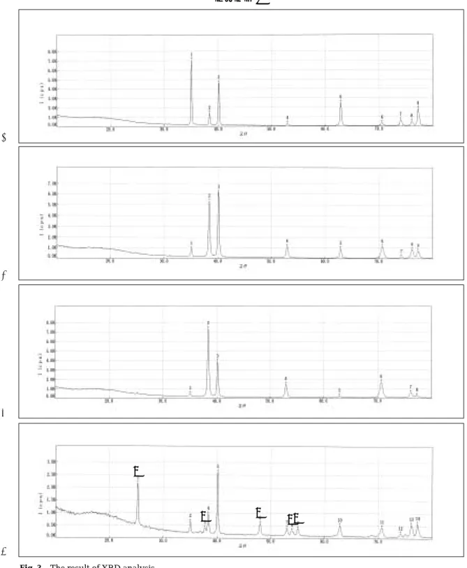
Surface composition and structure
Fig. 3 showed the result of XRD analysis.
In the electropolished, machined and anodized (low roughness) group, the X-ray diffraction spectroscopy expressed only Ti peaks. The titanium surface in air conditions has a naturally-formed very thin oxide lay- er. It is basically amorphous in crystal structure.
Hence, there was no TiO2peak in the XRD pat- tern of 1, 2, 3 groups. But anodized (high roughness) group showed TiO2peak in the XRD pattern. Only anodized (high roughness) group has crystallinity.
Looking at the TiO2crystallinity, the position of TiO2peak showed that crystal structure is anatase.
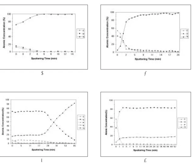
Surface composition and oxide thickness
The relative concentrations of the elements detected in AES survey spectra were represented graphi- cally in Fig. 4. The spectra were in all cases dominated by strong Ti, O and C signals, and in anodized groups trace amount of P and S were detected.
Especially high roughness anodized group had
high phosphate contents.
The depth profiles for Ti, O and C showed a dif- ferent qualitative behavior for all samples. In elec- tropolished and machined groups, O and C sig- nals rapidly disappeared. This decrease was ac- companied by an increase in Ti signals. The sputtering time required for the O signal to decrease halfway from its intensity in the oxide to that in the bulk was taken as a measure of the oxide thickness. The elec- tropolished group and machined group had similar oxide thrcknesses, and the anodized groups had ex- tremly thick oxide films. High roughness anodized group showed specific AES depth profile. Over 52min sputtering time the O and Ti signal maintained constant amount and constant Ti/O ratio value.
The oxide thicknesses were approximately 26nm in electropolished group, 16nm in machined group, 450nm in low roughness anodized group and over 550nm in high roughness anodized group.
Cell morphology
Cell morphologies were observed at 18 hour, 3rd day, 14thday.
Cells on electropolished and machined surfaces de- veloped a flattened cell shape and cells on two an- odized surfaces showed combined morphology.
Some cells on the anodized discs were flat and others were spherical. Especially on rougher anodized discs, lamelopodiae of cells dipped into the micro- pores. Generally it seemed like that the cells on electropolished and machined discs were spreading extensively and showed intimate adaptation, but the cells on the anodized surfaces showed less spread-
Table Ⅱ. Result of two dimension measurement
1 2 3 4
Ra(㎛) 0.121 0.35 0.623 1.123
Rt(㎛) 1.397 2.921 5.01 7.745
Rsk -1.736 -0.064 0.084 0.383
Table Ⅰ. Result of three dimension measurement
1 2 3 4
Sa(㎛) 0.098 0.234 0.29 0.535
St(㎛) 1.255 2.321 2.346 4.175
Ssk -0.887 -0.307 0.173 0.526
ing tendencies.
The filopodiae of cells on the anodized surfaces were thicker than those of electropolished and ma- chined surfaces. There were differences according to time. The cells on the anodized surfaces showing combined morphology at 18 hour and 3 day had ap- peared similar shapes of those of electropolished and machined surfaces at 14 day cultures (Fig. 5, 6).
Cell count
Table III showed the result of cell count.
Low roughness anodized group was signifi- cantly higher cell number than other groups.
There was no significant difference between 1, 2, 4 groups.
Total protein measurement
All four groups showed similar total protein amount.
There was no significant difference among four groups (Table IV and Fig. 10).
Alkaline phosphatase activity.
Table V showed ALP activities of four groups. High roughness anodized group was significantly high- er ALP activities than any other group. Low rough- ness anodized group was significantly higher ALP activities than electropolished and machined groups.
Electropolished group was higher ALP activities than machined group (P<.05).
DISCUSSION
The success of implant means formation and maintenance of osseointegration at bone-implant in- terface.
At an initial stage when the implant was in- stalled, implant and bone tissues were not inti- mate contact at microscopic level. At this time, the cells became active on implant-bone interface.
Several conditions of implant surface can directly in- fluence cellular responses. Recent in vitro and in vi- vo studies provided strong indications that biolog- ical responses to titanium are influenced both by sur- face structure and chemical composition. Surface which showed high levels of cell attachment, pro- liferation, differentiation, production of extracel- lular matrix and mineralization is good for os- seointegration. This study confirms those obser- vations that osteoblast-like cells respond in a different manner to surface roughness, oxide thickness, ox- ide crystallinity and microtopography.
Studies about cellular behaviors on the implant sur- faces were performed in a wide range of aspects and a large number of ways. One of the most often Table Ⅳ. The result of total protein measure-
ment(mg/ml)
Mean S.D.
1 0.37 0.024
2 0.35 0.012
3 0.42 0.008
4 0.32 0.009
Table Ⅲ. The result of cell count (X104)
Mean S.D.
1 4.8 4.0
2 8.8 5.2
3 19.8* 7.2
4 6.3 2.4
Significant diifference between values marked with asterisk (P<.05).
Table Ⅴ. Alkaline phosphatase activity(absorbance)
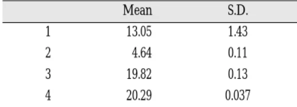
Mean S.D.
1 13.05 1.43
2 4.64 0.11
3 19.82 0.13
4 20.29 0.037
concerned subject is relationship of the surface roughness and cell attachment. For the osseointe- gration to occur at a biomaterial surface implanted in tissue, osteoblasts must first attach to the im- plant surface. In vitro studies using osteoblast-like cell culture have examined the effects of surface roughness on cell attachment.12,17,26,27They conclud- ed that rougher surface is more favorable than smoother one for cell attachment. In addition, some in vivo studies supported these observations.
Several studies suggested that surface rough- ness is not as important as other surface properties in biologic responses.9,16,19
Cell count can be measured by various meth- ods.24Direct and indirect methods could be used.
Attached or detached cells were counted using Coulter counter or hemocytometer. Some investigators used radioactively labeled cells directly from the sur- face giving more accurate count of cell adherence or captured image.38The cell count assay used in this study determined the amount of cells attached to the material surface directly. It is a relatively simple and easy procedure. However, the assay presented some variables and limitations. If some cells were not removed from the surface and remained on the surface, the result was not precise. It is more often at the rougher surface. Furthermore, this approach was unable to measure the strength of attachment to the test material. The result of the present study showed that rough anodized surface demonstrated lower level of cell count than smooth anodized one. But, the cell count of the rough anodized sur- face is similar to those of electropolished and ma- chined surfaces. It is possible that decreased pro- liferation precedes the expression of the differenti- ated osteoblast phenotype in culture. 25, 40, 41The rel- atively weak proliferative response, in conjunction with increased alkaline phosphatase activity on the rougher surfaces, suggesting that osteoblast- like cells grown on rough surfaces are at a more ad- vanced stage of development than their counterparts on the smooth surfaces. Second possible explanation
is such that the sharp angularity of this type of surface hinders anchorage of cell processes on ad- jacent regions and thus slows down the process of attachment.38And another possibility is the lim- itation of methods. It is probable that cells on rougher surface were more strongly adhered to the surface, therefore some of the cells on the rough surface might be remained on the surface after trypsination.
As noted above, other surface properties may be as or more important than surface roughness alone.16Chemical composition, oxide thickness, surface microtopography also might play roles.
Several studies have shown that even subtle dif- ferences in surface composition, including Ti oxide crystallinity can modify cell responses, even when surface roughness is held constant.42In this study, rough anodized surface had been found thicker oxide thickness and only this type of surface had TiO2 crystal structure. Titanium naturally forms a thin ox- ide layer in air. But the thickness, morphology, topography, chemical composition may be varied over a relatively wide range. The biocompatibility of ti- tanium is largely related to the characteristics of its oxide layer, as studies have suggested.43,44It is also likely that the four groups of discs used in this study were different with respect to the crystallinity of their surface oxides. These oxides in titanium having thin oxide film are essentially non-crys- talline (or quasiamorphous) while thicker oxide become increasingly more crystalline with in- creasing thickness having the anatase structure.
Consequently the discs anodized with rough surface measurement have surface oxides with a high degree of crystallinity (with the anatase structure), while the other three types of discs have non-crystalline sur- face oxides. The effect of difference of crystal struc- ture is not clear. Which structure is beneficial for cel- lular responses still remains unknown.
When discussing the AES results, it is important to point out that the compositions presented here should be seen as relative, rather than absolute
concentrations in the analyzed volume. The surface consisted of an oxide, mainly TiO2. The depth pro- files for Ti, O and C showed a different qualitative behavior for all samples. In electropolished and machined groups, O and C signals rapidly disap- peared. This decrease was accompanied by an in- crease in Ti signals. Another main difference in surface composition observed between the different types of discs was oxide thickness. The electropol- ished and machined discs had thin oxides of simi- lar thickness, while the two groups of anodized discs had much thicker oxides. High roughness anodized group showed specific AES depth profile.
Over 52min sputtering time the O and Ti signal maintained constant amount and Ti/O ratio value.
Therefore it was supposed that high roughness an- odized group had very thick oxide film. There were controversies about the relationship of oxide thickness and cellular responses. Ellingen and Videm showed that there was no significant corre- lation between oxide thickness on titanium im- plants and the bony response. These authors ex- plained the reason why the outermost molecular lay- ers of the biomaterial are the important part for the bone response, because the surface chemistry of these layer is exposed to the tissue.34But Hazan et al have shown recently that heat treatment of titanium alloy implants, leading to increased oxide-thick- ness, leads to higher removal torque and higher degree of calcification of the bone around implants in rats.45It is not known whether or not the oxide thickness alone affect the cellular responses. In this study thicker oxide film groups showed higher al- kaline phosphatase activities. It could be thought that thicker oxide thickness groups showed higher ini- tial cell differentiation.
SEM and three-dimension roughness measur- ing system identified significant differences also in the surface microtopography and roughness of the different groups of discs. The SEM observations of anodized discs showed that anodic oxides are con- taining regions of varying porosity. Depending on
the preparation conditions, thicker anodic oxides are more distinct grain (crater-like structure) structure with large micropores.
This paper described a series of experiments de- signed to examine cellular responses and the rela- tionships of interaction between a well characterized line of neonatal rat calvarial osteoblast and variously treated implant surfaces used in dental implant.
As far as cell morphology is concerned, the SEM ex- periment showed that two different cellular mor- phologies, spherical vs. flat, were present on the sub- strate surfaces. The spherical cells appear to be present in areas of calcifications.46Generally cells on the smooth surfaces are flattened shape and cells on the rough surfaces are spherical shape. Some authors reported that no obvious differences were among the attachment morphology on different surfaces.16,24 In contrasts, others presented that the differences of surface roughness influence cell attachment mor- phology.25,46,47In this study presented, the cells on elec- tropolished discs and machined discs were almost flattened shape and well spreaded on the discs.
On two anodized discs, some of the cells were flat- tened and the other cells were spherical. It could be supposed that the anodized surface was more fa- vorable for secreting extracellular matrix. But it is somewhat time-dependent. As time goes on, more amounts of flattened cells were observed on the anodized surfaces. SEM examination of osteoblast- like cells on materials with various surface rough- nesses generally demonstrated that cell spreading and continuous cell layer formation was better on smooth surfaces compared to rough ones. In this study, peculiar form of cell attachment was shown on rongh anodized surface. Some filopodiae dipped into the micropores on the surface.
Alkaline phosphatase (ALP) is a glycoprotein associated with the formation of calcified tissues.
Some authors presented the relationship of ALP and surface properties. Ong et al have found that rougher surface showed prolonged ALP specific activity and a more rapid osteocalcin production as
compared with the polished titanium surface.26But Martin et al reported somewhat different results.7ALP specific activity was lowest on the rougher sur- face. In this study alkaline phosphatase activity was significantly higher at anodized surface. This means that anodized surface was more favorable than other surfaces in osteoblast differentiation.
Although this study showed some similarities with prescribed investigations, some differences existed among other studies. Various explanations can be given for these observations. Differences in the origin of the cells and in the experimental meth- ods used in obtaining them make direct comparisons of results difficult. A significant difference is that in most of the reported studies, different cell types are used.
According to the Jones and Boyde, osteoblasts are the only cells that will migrate from the bone and, subsequently, colonize the biomaterial.51The ex- plant model has been used successfully in investi- gating migratory cell morphology and extracellular matrix formation by osteoblasts on ceramics, glass- es, and polymers. Since proper phenotype is es- sential in the correct interpretation of results obtained with cellular model, special care was taken in this study to characterize the rat osteoblast used.
Osteoblasts attached to and synthesized proteins on all the biomaterials examined. However, there were differences in the extent of certain cellular responses depending upon the substrate.
There were also methodological differences between the various studies. It can not be excluded that the time point chosen may not have been appropriate to reveal differences in cellular behavior. If cells were harvested too long, lack of surface area available oc- curred and cells were confluent. Therefore the cell morphologies were difficult to distinguish.
Preparation technique effect such as sterilization effects have been studied by several authors. They have demonstrated the crucial effect of sterilization methods of commercially pure titanium (cp Ti) on in vitro subsequent cell adhesion.19,52 These effect
may be related to sterilization-induced surface chemical modification.53Some clinical surface prepa- rations on titanium-based alloys are known to con- siderably modify the surface chemical characteris- tics. Improper glow discharge plasma treatment (a frequently used method for cleaning, preparation and modification of biomaterial) can produce un- wanted and irreproducible results. In this study ethylene dioxide gas was used for discs sterilization.
There are a few studies on ethylene dioxide gas sterilization method. It is necessary for more stud- ies on sterilization methods.
Ellingen and Williams54pointed out that ultra- structural, microstructural, and macrostructural levels of the surface topography influence the behavior of the adjacent tissue. There are many factors in ul- trasturucture: wettability, surface energy, zeta po- tential. As mentioned above, osteoblast adhesion on materials may also be considered in relation to the expression of the various adhesion proteins. Especially various biological molecules such as extracellular ma- trix proteins, cell membrane proteins, and cytoskele- ton proteins, which regulate cell attachment and dif- ferentiation, are worth noticing. Therefore further de- tailed studies on proteins and cellular behavior on controlled surface and on surface treatment and gene expression at transcription level are required.
CONCLUSIONS
The objective of this work was to investigate the influence of surface modifications on the cellular re- sponses. The different preparation methods produced discs with different surface roughness, microto- pography, chemical composition, oxide thickness and surface oxide crystallinity.
The findings of the present investigation were:
1. The four groups showed specific microtopography.
Anodized group showed grain structure with mi- cropores.
2. Surface roughness values were, from the lowest to the highest, electropolished group, machined
group, low roughness anodized group, and high roughness anodized group.
3. Highly roughened anodized group was found to have increased surface oxide thickness and sur- face crystallinity.
4. The morphology of cells, flattened or spherical, were different from each other. In the electrop- olished group and machined group, the cells were almost flattened. In two anodized groups, some cells were spherical and other cells were flat- tened. And the 14 day culture cells were nearly flattened due to confluency.
5. The number of cells was highest in less roughened anodized group. And the machined group had sig- nificantly lower cell count level than any other groups (P<.05).
6. Total protein contents showed no difference among all groups.
7. The level of alkaline phosphatase activities was higher in the anodized groups than electropolished and machined groups (P<.05).
REFERENCES
1. Albrektsson T, Bra�nemark PI, Hansson HA, Lundstro¨m J. Osseointerated titanim implants:
requirements for ensuring a longlasting, direct bone-to-implant anchorage in man. Acta Orthop Scand 1981;52:155-70.
2. Bra�nemark PI, Hansson BO, Adell R, Breine U, Lundstro¨m J, Hallen O, O¨hman A. Osseoietegrated implants in the treatment of the edentulous jaw: ex- perience from 10-year period. Scand J Plast Reconstr Surg 1977; 11:1-132.
3. Wyatt CCL, Zarb GA. Treatment outcomes of pa- tients with implant-supported fixed prostheses. Int J Oral Maxillofac implants 1998;13:204-11.
4. Lekholm U, Gunne J, Henry P, Higuchi K, Linden U, Bergstrom C, Steenberghe DV. Survival of the Bra�nemark implant of partially edentulous jaws:
a 10-year prospective multicenter study. Int J Oral Maxillofac implants 1999;14:639-45.
5. Albrektsson T, Jansson T, Lekholm U. Osseoint- egrated dental implants. Dent Clin N Am 1986;30:151-70.
6. Sullivan DY, Sherwood RL, Mai TN. Preliminary results of a multicenter study evaluating a chem- ically enhanced surface for machined commer- cially pure titanium implants. J Prosthet Dent 1997;78:379-86.
7. Martin JY, Dean DD, Cochran DL, Simpson J, Boyan BD, Schwartz Z. Proliferation, dif- ferenti- ation, and protein synthesis of human osteoblast like cells (MG63) cultured on previously used ti- tanium surfaces. Clin Oral Impl Res 1996;7:27- 37.
8. Ramires PA, Romito F, Cosentino F, Milella E.
The influence of titania/hydroxyapatite compos- ite coatings on in vitro osteoblasts behaviour.
Biomaterials 2001; 22:1467-74.
9. Lim YJ, Oshida Y, Andres CJ, Barco MT, Martin TB.
Surface characterizations of variously treated ti- tanium materials. Int J Oral Maxillofac implants 2001;16:333-42.
10. Ong JL, Carnes DL, Cardens HL, Cavin R. Surface roughness of titanium on bone morphogenetic protein-2 treated osteoblast cells in vitro. Implant Dent 1997;6:19-24.
11. Castellani R, de Ruijter JE, Renggli H, Jansen JA.
Response of rat bone marrow cells to differently roughened titanium discs. Clin Oral Impl Res 1999;10:369-78.
12. Larsson C, Thomsen P, Aronsson BO, Rodahl M, Lausmaa J, Kasemo B, Ericson LE. Bone response to surface-modified titanium implants: studies on the early tissue response to machined and electropolished implants with different oxide thicknesses. Biomaterials 1996;17:605-16.
13. Fini M, Cigada A, Rondelli G, Chiesa R, Giardino R, Giavaresi G, Aldini NN, Torricelli P, Vincentini B. In vitro and in vivo behaviour of Ca- and P- en- riched anodized titanium. Biomaterials. 1999;20:1587- 94.
14. Buser D, Schenk R, Steinemann S, Riorellini J, Fox C, Stich H. Influence of surface characteristics on bone integration of titanium implants. A his- tomorphomeric study in miniature pigs. J Biomed Mater Res 1991;25;889-902.
15. Piatelli A, Manzon L, Scarano A, Paolantonio M, Piatelli M. Histologic and histomorphometric analysis of the bone response to machined and sandblasted titanium implants: an experimental study in rabbits. Int J Oral Maxillofac Implants 1998;13;805-10.
16. Bowers KT, Keller JC, Randolph BA, Wick DG, Michaels CM. Optimization of surface micro- morphology for enhanced osteoblast responses in vitro. Int J Oral Maxillofac Implants 1992;7:302- 10.
17. Cooper LF, Masuda T, Whitson SW, Yliheik- kila¨ P, Felton DA. Formation of mineralizing os- teoblast cultures on machined, titanium oxide grit-blasted, and plasma-sprayed titanium sur- faces. Int J Oral Maxillofac Implants 1999;14:37-47.
18. Martin JY, Schwartz Z, Hummert TW, Schraub DM, Simpson J, Lankford Jr.J, Dean DD, Cochran DL, Boyan BD. Effect of titanium surface roughness on human osteoblsat-like cell(MG63) J Biomed Mater Res 1995;29:389-401.
19. Stanford CM, Keller JC, Solursh M. Bone cell ex-
pression on titanium surfaces is altered by steril- ization treatments. J Dent Res 1994;73:1061-71.
20. Kasemo B. Lausmaa J. Aspect of surface physics on titanium implants. Swed Dent J 1985; suppl 28:19- 36.
21. Albrecktsson T, Bra�nemark PI, Hansson HA, Kasemo B, Larsson K, Lundstro¨m I, McQueen D, Skalak R. The interface zone of inorganic im- plants in vivo: titanium implants in bone. Ann Biomed Eng 1983;11:1-27.
22. Sul Y-T, Johansson CB, Petronis S, Krozer A, Jeong Y, Wennerrberg A, Albrektsson T.
Characteristics of the surface oxides on turned and electrochemically oxidized pure titanium im- plants up to dielectric breakdown: the oxide thick- ness, micropore configurations, surface rough- ness, crystal structure and chemical composition.
Biomaterials 2002;23:491-501.
23. Ishizawa H, Oginno M. Formation and chatac- terization of anodic titanium oxide films contain- ing Ca and P. J Biomed Mat Res 1995;29:65-72.
24. Chang Y, Stanford CM, Wefel JS, Keller JC.
Osteoblastic cell attachment to hydroxyapatite- coated implant surfaces in vitro. Int J Oral Maxillofac Implants 1999;14:239-247.
25. Lincks J, Boyan BD, Blanchard CR, Lohmann CH, Liu Y, Cochran DL, Dean DD, Schwartz Z. Response of MG-63 osteoblast-like cells to titanium and ti- tanium alloy is dependent on surface roughness and composition. Biomaterials 1998;19:2219-32.
26. Ong JL, Cardens HL, Cavin R, Carnes DL.
Osteoblast responses to BMP-2-treated titanium in vitro. Int J Oral Maxillofac Implants 1997;12:649- 54.
27. Keller JC, Stanford CM, Wightman JP, Draughn RA, Zaharias R. Characterizations of titanium implant surfaces.III J Biomed Mater Res 1994;28:939-946.
28. Puleo DA, Holleran LA, Doremus RH, Bizios R.
Osteoblast responses to orthopedic implant materials in vitro. J Biomed Mater Res 1991;25:711-23.
29. Howlett CR, Evans MDM, Walsh WR, Johnson G, Steele JG. Mechanism of initial attachment of cells derived from human bone to commonly used prosthetic materials during cell culture. Biomaterials 1994;15:213-22.
30. Anselme K. Oseoblast adhesion on biomaterials.
Biomaterials 2000;21:667-681.
31. Freshney RI. Culture of animal cells : A manual of basic technique. Third edition. NewYork, wiley-liss inc. 1994 :4-5.
32. Larsson C, Thomsen P, Lausmaa J, Rodahl M, Kasemo B, Ericsson LE. Bone reponse to surface modified titanium implants: studies on electrop- olished implants with different oxide thicknesses and morphology. Biomaterials 1994;15:1062-1074.
33. Lowry O, Rosebrough N, Farr A, Randall R.
Protein measurement with the folin phenol reagent.
J Bioloc Chem 1951;193:265-75.
34. Ellingen JE. Surface configurations of dental im- plants. Periodontology 2000 1998;17:36-46.
35. Gofredsen K, Nimb L, Hjorting-Hansen E, Jenssen JS, Holmen A. Histomorphometric and removal torque analysis for TiO2-blasted titanium im- plants. An experimental study on dogs. Clin Oral Implants Res 1992;3:77-84.
36. Thomas K, Cook S. An evaluation of variables influencing implant fixation by direct bone ap- position J Biomed Mater Res 1985;19:875-901.
37. Cochran D, Simpson J, Weber H, Buser D.
Attachment and growth of periodontal cells on smooth and rough titanium. Int J Oral and Maxillofac Implants 1994;9:289-97.
38. Lumbikanonda N, Sammons R. Bone cell attach- ment to dental implants of different surface char- acteristics. Int J Oral Maxillifac Implants 2001;16:627- 36.
39. Nugiel DJ, Wood DJ, Paul Sung KL. Quantification of adhesiveness of osteoblasts to titnium surfaces in vitro by the micropipette aspiration technique.
Tissue Eng 1996;2:127-139.
40. Gichard DJ, Gowen M, MacDonald BR. Proliferative reponses to estradiol, IL-1alpha and TGF beta by cells expressing alkaline phosphatase in human osteoblast like cell cultures. Calcif Tissue Int 1993;52:227-33.
41. Owen TA, Aronow M, Shalhoub V, Barone LM, Wilming L, Tassinari MS, Kennedy MB, Pockwinse S, Lian JB, Stein GS. Progressive development of the rat osteoblast phenotype in vitro: Reciprocal re- lationships in expression of genes associated with osteoblast proliferation and differentiation using formation of the bone extracellular matrix. J Cell Physiol 1990;143:420-430.
42. Michaels CM, Keller JC, Stanford CM, Solursh M. In vitro cell atachment of osteoblast-like cells to titanium. J Dent Res 1989;68:276-81.
43. Oshida Y, Sachdeva R, Miyazaki S, Daly J. Effects of shotpeening on surface contact angles of bio- mateials. J Mater Sci 1993;4:443-47.
44. Oshida Y, Sachdeva R, Miyazaki S. Changes in con- tact angles as a function of time in some pre-oxi- dized biomateials. J Mater Sci Mater Med 1992;3:306- 12.
45. Hazan R, Brener R, Oron U. Bone growth to met- al implants is regulated by their surface chemical properties. Biomaterials 1993;14:570-574.
46. Lauer G, Wiedmann-Al-Ahmad M, Otten JE, Hu¨bner U, Schmelzeisen R, Schilli W. The titani- um surface texture effects adherence and growth of human gingival keratinocytes and human max- illar osteoblast-like cells in vitro. Biomaterials 2001;22:2799-2809.
47. Brunette DM. The effects of implant surface topog- raphy on the behavior of cells. Int J Oral Maxillofac Implants 1988;3:231-246.
48. Lampin M, Warocquier-Cle′rout R, Legris C, Degrange M, Sigot-Luizard MF. Corelation be- tween substratum roughness and wettability, cell adhesion, and cell migration. J Biomed Mater Res 1997;36:99-108.
49. Boyan BD, Hummert TW, Dean DD, Schwartz Z. Role of material surfaces in regulating bone and cartilage cell response. Biomaterials 1996;17:137- 46.
50. Yang Y, Tian J, Deng L, Ong JL. Morphologic be- havior of osteoblast-like cells on surface-modi- fied titanium in vitro. Biomaterials 2002:23;1383- 89.
51. Jones SJ, Boyde A. Colonization of various natural substrates by osteoblasts in vitro. SEM 1979;II:529- 538.
52. Vezeau PJ, Koorbusch GF, Draughn RA, Keller JC.
Effects of multiple sterilization on surface char- acteristics and in vitro biologic responses to titanium.
J Oral maxillofac Surg 1996;54:738-46.
53. Baier RE, Meyer AE, Akers CK, Natiella JR, Meenaghan M, Carter JM. Degradative effects of conventional steam sterilization on biomaterial surfaces. Biomaterials 1982;3:241-5.
54. Williams DF. Biocompatibility:performance in the surgical reconstruction of man. Interdisplinary Sci Rev 1990;15:20-33.
Reprint request to:
DR.YUNG-SOOKIM, D.D.S., M.S.D., Ph.D., M.Sc.(O.S.U.)
DEPT. OFPROSTHODONTICS,COLLEGE OF DENTISITY, SEOUL NATIONAL UNIV.
28-1 YEONGUN-DONG, CHONGNO-GU, 110-749, SEOUL KOREA [email protected]
사진부도 ①
A B
C D
Fig. 1. SEM photographs of each specimens.
A, Electropolished surface (×2000).
B, Machined surface (×2000).
C, Anodized surface (Low roughness)(×2000).
D, Anodized surface (High roughness)(×2000).
사진부도 ②
A B
C D
Fig. 2. The result of surface roughness measurement.
A, Electropolished surface.
B, Machined surface.
C, Anodized surface (Low roughness).
D, Anodized surface (High roughness).
사진부도 ③
A
B
C
D
Fig. 3. The result of XRD analysis.
A, Electropolished surface. B, Machined surface.
C, Anodized surface (Low roughness). D, Anodized surface (High roughness).
� TiO2(anatase)
Every peak in the A, B, C is Ti.
The 1, 3, 6, 8, 9 peaks in the D is TiO2(anatase) and the other peaks are Ti.
�
� �
��
사진부도 ④
A B
C D
Fig. 4. The result of auger electron scanning microscopy.
A, Electropolished surface. B, Machined surface.
C, Anodized surface (Low roughness). D, Anodized surface (High roughness).
Fig. 5. SEM photographs of cells cultured for 18 hours on the various substrates.
A, Electropolished surface(×1000, ×1000). B, Machined surface(×1000, ×500).
C, Anodized surface (Low roughness)(×1000, ×1000). D, Anodized surface (High roughness) (×1000, ×1000).
A,B, the cells were flattened shape and well spreaded on the discs,
C,D, some of the cells were flattened and the other cells were spherical shape.
A
B
C
D
사진부도 ⑤
사진부도 ⑥
Fig. 6. SEM photographs of the cells and matrix cultured for 14 days on the various substrates.
A, Electropolished surface (×1000). B, Machined surface (×1000).
C, Anodized surface (Low roughness)(×1000). D, Anodized surface (High roughness) (×1000).
The cells on the discs were flattened shape.
A B
C D