Ⅰ. Introduction
With regards to their biological and physiochemical proper- ties, titanium-based biomaterials have been successfully used in orthopaedic, dental and maxillo-facial surgery mainly as endosseous implants. The early osseointegration of titanium dental implants is an important factor for their clinical suc- cess1,2. The implant loading to bone is influenced by various factors, including surface chemistry, energetics, and surface topography3-6. These surface parameters are decisive during the bone healing period in the peri-implant region. After implanta- tion, implant surfaces are in contact with body fluids and inter- act with a number of proteins and different cell types. Cell
adhesion is one of the initial stages for subsequent prolifera- tion, and differentiation of osteoblastic cells producing bony tissue and extracellular matrix, which will ensure a high bone- implant contact. It has been shown that osteoblastic cell adhe- sion, growth, and differentiation are related to surface chem- istry, energetics, and roughness6-8.
Although many in vitro experiments have studied osteoblas- tic cell behavior on titanium surfaces, the optimal surface para- meters for adhesion, proliferation, and differentiation of osteoblastic cells have not yet been completely understood7,9. But among them, surface roughness is one of the most acknowledged parameters improving the bone anchorage of titanium implants10-12. A different evidence indicate that surface roughness of titanium implants strongly affects the behavior of osteoblastic cells. First of all, rough surfaces encourage the entrapment of fibrin protein, adhesion of osteogenic cells, and mechanical stability of implants in host bone13-15. On the other hand, numerous reports indicate that increased surface rough- ness stimulates osteoblastic cell differentiation and bone for- mation16-18.
Seung-Il Song
Department of Dentistry, School of Medicine, Ajou University San 5, Wonchon-dong, Youngtong-gu, Suwon, 443-721, Korea TEL: +82-31-219-5328 FAX: +82-31-219-5329
E-mail: [email protected]
Effect of implant surface microtopography by hydroxyapatite grit-blasting on adhesion, proliferation, and differentiation of
osteoblast-like cell line, MG-63
Sung-Jae Park1, Sang-Bum Bae1, Su-Kyoung Kim2, Tae-Gwan Eom2, Seung-Il Song1
1Department of Dentistry, School of Medicine, Ajou University, Suwon, Korea
2Department of Implant Research, Implant R&D Center, Osstem Co., Ltd., Busan, Korea
Objective: This study examined the potential of the in vitro osteogenesis of microtopographically modified surfaces, RBM (resorbable blasting media) surfaces, which generate hydroxyapatite grit-blasting.
Methods:RBM surfaces were modified hydroxyapatite grit-blasting to produce microtopographically modified surfaces and the surface morphology, roughness or elements were examined. To investigate the potential of the in vitro osteogenesis, the osteoblastic cell adhesion, proliferation, and differ- entiation were examined using the human osteoblast-like cell line, MG-63 cells. Osteoblastic cell proliferation was examined as a function of time. In addition, osteoblastic cell differentiation was verified using four different methods of an ALP activity assay, a mineralization assay using alizarin red-s staining, and gene expression of osteoblastic differentiation marker using RT-PCR or ELISA.
Results: Osteoblastic cell adhesion, proliferation and ALP activity was elevated on the RBM surfaces compared to the machined group. The cells exhibited a high level of gene expression of the osteoblastic differentiation makers (osteonectin, type I collagen, Runx-2, osterix). imilar data was rep- resented in the ELISA produced similar results in that the RBM surface increased the level of osteocalcin, osteopontin, TGF-beta1 and PGE2 secre- tion, which was known to stimulate the osteogenesis. Moreover, alizarin red-s staining revealed significantly more mineralized nodules on the RBM surfaces than the machined discs.
Conclusion: RBM surfaces modified with hydroxyapatite grit-blasting stimulate the in vitro osteogenesis of MG-63 cells and may accelerate bone formation and increase bone-implant contact.
Key words: Osseointegration, Osteogenesis, Microtopography, Surface roughness, RBM (resorbable blasting media)
[paper submitted 2011. 2. 8 / revised 2011. 5. 31 / accepted 2011. 6. 7]
Abstract (J Korean Assoc Oral Maxillofac Surg 2011;37:214-24)
*This study was supported by Osstem Implant R&D Center.
There are various methods have been developed to create rough implant surfaces in order to improve the clinical perfor- mance of implants and to guarantee a stable mechanical load- ing. Among them, the popular methods for roughening titani- um implants uses a biocompatible and resorbable blasting media, such as hydroxyapatite and β-tricalcium phosphate (β- TCP) ceramic particles or other calcium phosphate ceramic particles14,19-21. These calcium phosphate biomaterials have been contemplated as a potential abrasive materials. Indeed hydrox- yapatite and β-TCP are biocompatible and well known to form a direct bond with surrounding tissue after bone implantation.
As these calcium phosphate materials are soluble in acids, they can be easily removed from the implant surface after blast-
ing22,23. As these treatments generate surfaces with random
topography and various chemical compositions, the optimal surface properties that result in rapid osteoblastic cell adhe- sion, proliferation, and differentiation.
In view of the above-mentioned data, the main objectives of this work were (i) to develop a new technique of blasting using hydroxyapatite particles to provide a roughened surface which improve the osteoblastic cell adhesion, proliferation, and dif- ferentiation and (ii) to assess the effects of such a roughened surface with respect to osteoblastic cells. In this attempt, we examined the roughness of pure titanium discs prepared by hydroxyapatite grit-blasting technique. Two groups were investigated: polished (machined) and hydroxyapatite grit- blasted resorbable blasting media (RBM) titanium surfaces.
After analyzing the properties of the implant surfaces, osteoblastic cell adhesion, proliferation, and differentiation were studied.
Ⅱ. Materials and Methods 1. Materials
Commercially pure titanium (cpTi) discs with a diameter of 12 mm and 1 mm in thickness (Carpenter, Washington, USA) was used. The samples were cut from a cpTi bar and used as machined discs. Abrasive hydroxyapatite powders (40-80 mesh) were obtained from Himed (NY, USA). Cell culture plastic ware was purchased from Falcon (Becton-Dickinson, Franklin Lakes, NJ, USA). Fetal bovine serum (FBS), DMEM, penicillin/streptomycin, trypsin/EDTA were purchased from HyClone (Salt Lake City, Utah, USA). Phosphate puffered saline (PBS) were purchased from Invitrogen Corporation (Paisley, UK). Triton X-100, Alizarin red-s, p-nitro- phenylphosphate (p-NPP), cresyl violet, MgCl2were purchased from Sigma (St. Louis, MO, USA). 3-(4,5-Dimethylthiazol-2-
gl)-5-(3-carboxymethoxylphenyl)-2-(4-sulphophenyl-2H) tetrasolium inner salt (MTS) and hTGF-beta1 ELISA kit were purchased from Promega (Madison, WI, USA). hOsteocalcin ELISA kit was purchased from Biosource (Nivelles, Belgium).
hOsteopontin and hPGE2 ELISA kit were purchased form Assay Designs (MI, USA). QuantiTech Reverse Transcription kit was obtained from Qiagen (CA, USA). PCR premix was purchased from Bioneer (Daejeon, Korea). All other chemicals were from standard laboratory suppliers and were of the high- est purity available.
2. Methods
1) Preparation of titanium discs
cpTi discs were cleaned with acetone and ethanol. Discs were thereafter randomly divided into two groups. The polish group was obtained by polishing with silicon carbide abrasive paper (Jinan Abrasive Cloth Factory, Jinan, China). Decreased grind paper P400, P600, P1200, P2000 were successfully used on a rotative polisher at 250 rotation per minute (rpm) for 1 min. The hydroxyapatite grit-blasted surface (RBM group) was prepared by blasting using hydroxyapatite powders (Himed, NY, USA) of 40-80 mesh with a microhardness. The hydrox- yapatite powders were blasted at air pressure of 5 bars for 10 sec. Hydroxyapatite grit-blasted (RBM) discs were then passi- vated by immersion for 10 min in 15% nitric acid at room tem- perature and rinsed with deionized water. Then the samples were ultrasonically cleaned for 30 min in Et-OH, washed 3 times in DW. Finally they were dried at 80℃ dry oven. Before use in the cell-based assays, all discs were packaged and steril- ized using autoclave machined for 20 min at 120℃.
2) Surface roughness analysis
Three cpTi discs of each group were randomly selected and roughness measurements were carried out with a SV-3000 S4 digital profilometer (Mitutoyo, Tokyo, Japan) linked to an acquisition software Surfpak-SV. This profilometer system consists of a probe which scanned the surface 2.4 mm in length. Results were expressed 2D parameter, as a Ra (average roughness), Rz (average maximum height of the profile) and Rt (maximum height of the profile). These parameters expressed in μm, gave a general description of surface roughness.
3) Scanning electron microscopy (SEM) and energy-disper- sive spectroscopy (EDS)
The different surface morphologies of cpTi and RBM surface were examined using a SEM (JSM-6480LV, Tokyo, Japan) at 20Kv. An EDS attached with the JSM-6480LV. SEM has also
been used to characterize the elemental atomic composition of samples. Using the INCA-sight system of Oxford Instrument, elemental analysis of a defined area on the cpTi and RBM sur- face can easily be determined to a high degree of precision (~5 wt %).
4) Cell culture
The MG-63 cells (ATCC, VA, USA) is a non-transformed cell line established from human osteosarcoma. This cell were routinely grown in DMEM supplemented with 10% FBS (HyClone, Salt Lake City, Utah) and 0.1 mg/mL penicillin and streptomycin (P/S) (HyClone, Salt Lake City, Utah). Cells were subcultured twice a week using trypsin/EDTA and main- tained at 37℃ in a humidified atmosphere of 5% CO2in air.
Medium was completely renewed every two days.
5) Cell adhesion
The cell adhesion assay of the MG-63 cells was performed using previous report24,25. Briefly, the cells were grown to approximately 80% confluence prior to cell adhesion assay.
Mg-63 cells were cultured either onto the various titanium disc or in the culture plastic in 24 multi-well plate at a density of 1
×105cells. After 1hr, culture media were removed and rinsed three times by Dulbecco’s phosphate-buffered saline (DPBS).
And the cells were fixed using 4% formaldehyde solution for 1 hr at 4℃ and were rinsed three times by DPBS. Then the viable cells were stained using cresyl violet which stained ribonucleic acid and were eluted using citric acid. Finally col- orimetric measurement of cresyl violet was performed on a DTX 880 (Beckman Coulter, CA, USA) with optical density reading at 590 nm.
6) Cell Proliferation
The cell proliferation assay of the MG-63 cells was per- formed using CellTiter 96 Aqueous Non-Radioactive Cell Proliferation Assay kit (Promega, Madison, USA) according to the manufacturer’s protocol. MG-63 cells were cultured either onto the various titanium disc or in the culture plastic in 24 multi-well plate at a density of 1×10⁵cells. After indicated times, culture media was removed and 500 μL MTS/PMS/
Media mixture was added in each well for 1 hr. Finally colori- metric measurement of formazan dye was performed on a DTX 880 with optical density reading at 490 nm. Results were expressed as optical density and relative MTS activity as com- pared to control.
7) Alkaline phasphatase (ALP) activity
ALP activity was examined as previously described26-29, in
MG-63 cells cultured either onto the various titanium disc or culture plastic in 24 multi-well plates (1×10⁵cells/well).
Osteogenic medium (0.5 μM dexamethasone, 50 μg/mL L- ascorbic acid, 10 mM b-glycerolphosphate)30,31, was completely renewed every two days and after indicated times, cells were washed twice with DPBS and collected in 0.1% ALP lysis buffer. In order to extract the dissolved proteins from the crude cell lysates, the supernatants were centrifuged at 13,000 rpm for 10 min. The protein concentrations were determined using a BCA reagent (Pierce, Rockford, USA). For ALP activity measurement with isolated cell supernatants, 100 μg total pro- tein were incubated with 2 mg/mL p-NPP (as a chromogenic substrate, Sigma) using ALP assay buffer.(pH 9.75 in 0.1 M Glycine containing 1 mM MgCl2) at 37℃ for 2 hrs. The degree of colorimetic reaction was measured by DTX 880 for an opti- cal density measurements at 405 nm.
8) Alizarin red-s staining
The alizarin red-s staining with experimental samples was performed as reported elsewhere32-34 with a minor modifica- tions. Briefly, MG-63 cells were cultured either onto the vari- ous titanium disc or culture plastic in 24 multi-well plates (1×
105cells/well). Osteogenic medium was completely renewed every two days and after indicated times. Cells were washed twice with DPBS and fixed in 10% (v/v) formaldehyde (Junsei, Tokyo, Japan) for 20 min at room temperature and rinsed with triple distilled H2O. Cells were stained with 2%
alizarin red-s (Sigma, St. Louis, MO, USA), pH4.2, for 20 min with gentle agitation. After aspiration of the unstained dye, the wells were washed five times with distilled H2O while shaking for 5 min. For quantification of staining, 500 μL 10% (v/v) acetic acid was added to each well, and the plate was heated to 70-80℃ for 1hr. The cells were then scraped from the plate and centrifuged at 13,000 rpm for 10 min. The supernatant was transferred to a new e-tube and the 0.4 volume of 10% (v/v) ammonium hydroxide added to neutralize the acid.
Absorbance of extracted alizarin red-s in acetic acid solution (200 μL) was measured at 405 nm. Amount of alizarin red-s was determined according to an alizarin red-s standard curve and normalized to the total protein of the cell lysates using BCA method (Pierce, Rockford, USA).
9) ELISA
MG-63 cells were cultured either onto the various titanium disc or culture plastic in 24 multi-well plates (1×10⁵ cells/well). Osteogenic medium was completely renewed every two days and after indicated times, supernatants were collected using centrifuge (at 13,000 rpm for 2 min) to eliminate a cell
debris. The ELISA of the MG-63cells was performed using each ELISA kits (hTGF-beta1 ELISA kit; Promega, hOsteocalcin ELISA kit; Biosource, hPGE2 ELISA kit: Assay Design, hOsteopontin ELISA kit; Assay Design) according to the manufacturer’s protocol.
10) Semi-quantitative RT-PCR
Changes in osteoblast gene expression for osteonectin (OSN), Type I collagen, osteoprotegrin, BSP-2, Runx-2 and Osterix (OSX) were analyzed using semi-quantitative RT- PCR. MG-63 cells cultured either onto the various titanium disc or culture plastic in 24 multi-well plates (1×10⁵ cells/well). Osteogenic medium was completely renewed every two days. After 72 hrs, total RNA was extracted with Trizol (Invitrogen, CA, USA), according to the manufacturer’s instructions. Using the extracted RNA as a template, reverse transcription were carried out with QuantiTech Reverse Transcription kit (Qiagen, CA, USA) and RT reactions were performed in a Mastercycler (Eppendorf, NY, USA). RT reac- tion mixture was incubated at 42℃ for 20 min heated at 94℃
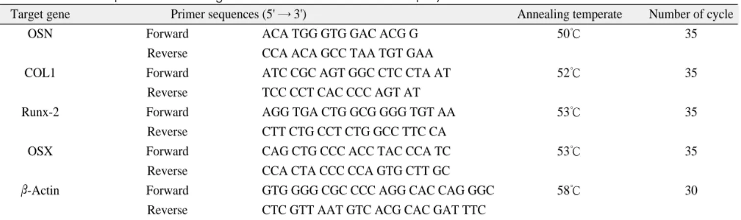
for 10 min, and subsequentially chilled to 4℃. Then OSN, type I collagen (COL1), runt-related transcription factor 2 (Runx-2) and OSX were amplified by Mastercycler using PCR premix (Bioneer, Daejeon, Korea).(Table 1) The captured image of amplification of osteogenic marker gene expression products from 2% agarose gel stained with ethidium bromide was analyzed using a Doc-It system (UVP, CA, USA). Semi- quantitative analysis was achieved by calibration the level of specific osteogenic marker bands was normalized for the housekeeping β-actin cDNA content.
11) Statistical analysis
Each experiment was repeated at least twice with similar results. Results are expressed as mean±SD of triplicate deter-
minations. For statistical analysis of experiments (N=6, three groups repeated two times), unpaired t-tests and ANOVA were performed with SPSS (SPSS INC., Chicago, IL, USA).
(*; P<0.05, **; P<0.01, ***; P<0.001)
Ⅲ. Results
1. Surface Parameter Tuning for Optimization of RBM
The purpose of this investigation was to find an optimal RBM surface that show the appropriated cell response. To decide the optimal RBM surface, we established surface roughness as a key parameter that influence to RBM surface characteristics. Briefly, cell adhesion, proliferation, and osteoblastic differentiation displays tendency that increase as RBM surface roughness grows (data not shown). Through this approach, we select the optimal RBM surface that has a 1.5 μm range surface roughness, and a following experiments used selected RBM condition.
2. Surface Characterization of Optimized RBM
SEM indicates a quite discrepancy between each of the two surfaces. RBM surfaces exhibited a highly rugged and irregu- lar surface.(Figs. 1. A, 1. B) The roughness parameters as mea- sured by optical profilometer are represent in Table 2. The val- ues refer to the mean of ten measurements for each of the two types of surfaces. The surfaces scored from rougher to more smoother as follows: culture plate < machined < RBM.
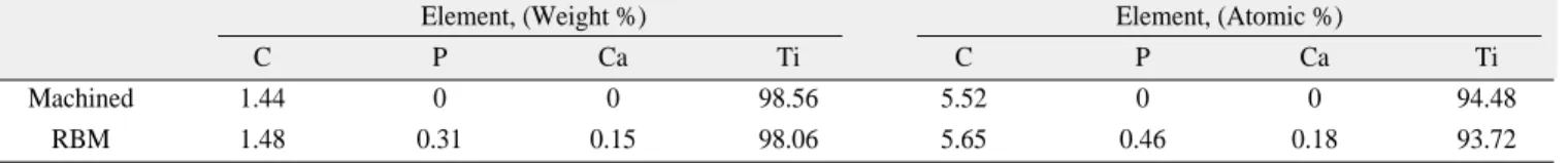
According to the EDS data(Figs. 1. C, 1. D), machined or RBM surface was basically similar elemental composition.
The RBM surfaces possesses the phosphorous and calcium of infinitesimal quantity.(Fig. 1. D, Table 3) This phenomenon is due to hydroxyapatite powder that remain behind after blasting.
Table 1.Rrimer sequences of osteogenic marker and conditions for polymerase chain reaction
Target gene Primer sequences (5' → 3') Annealing temperate Number of cycle
OSN Forward ACA TGG GTG GAC ACG G 50℃ 35
Reverse CCA ACA GCC TAA TGT GAA
COL1 Forward ATC CGC AGT GGC CTC CTA AT 52℃ 35
Reverse TCC CCT CAC CCC AGT AT
Runx-2 Forward AGG TGA CTG GCG GGG TGT AA 53℃ 35
Reverse CTT CTG CCT CTG GCC TTC CA
OSX Forward CAG CTG CCC ACC TAC CCA TC 53℃ 35
Reverse CCA CTA CCC CCA GTG CTT GC
β-Actin Forward GTG GGG CGC CCC AGG CAC CAG GGC 58℃ 30
Reverse CTC GTT AAT GTC ACG CAC GAT TTC
OSN, Osteonectin; COL1, Type I Collagen; Runx-2, Runt-Related Transcription Factor-2; and OSX, Osterix
3. Cell Adhesion
The cell adhesion data of the MG-63 cells on the different surfaces are presented in Fig. 2. Statistical testing of the data, using a two-way analysis of variance (ANOVA) and a multiple comparison test (Turkey), revealed that after 1hr of incubation in the RBM surfaces, the adherent cells were significantly higher than the machined surface and the culture plate.
(*P<0.05; Fig. 2)
Fig. 1.Surface morphologhy of titanium discs for osteoblastic cell culture. SEM images of machined titanium (A) and RBM (B). Bar = 10 μm. EDA Data of machined titanium (C) and RBM (D). Surfaces were examined using the JSM-6480LV SEM at working distance of 10 mm and accelerating voltage of 20 kV.
A B
C D
Table 2. Roughbess values of a machined Ti and a RBM surfaces measured by profilometer
Machied RBM
Ra, [um] 0.222 (±0.039) 1.523 (±0.156) Ra2max, [um] 0.244 (±0.032) 1.663 (±0.163) Rz, [um] 1.566 (±0.199) 10.514 (±1.177) Rt, [um] 1.727 (±0.278) 11.648 (±1.378) Ra, average roughness; Ra2max, maximum value of roughness; Rz, average maximum height of the profile; and Rt, maximum height of the height
RBM, resorbable blasting media
Table 3. EDS data of element contents on machined Ti and RBM surfaces
Element, (Weight %) Element, (Atomic %)
C P Ca Ti C P Ca Ti
Machined 1.44 0 0 98.56 5.52 0 0 94.48
RBM1.48 0.31 0.15 98.06 5.65 0.46 0.18 93.72
C, Carbon; P, Phosphorous; Ca, Calcium and Ti, Titanium
EDS, energy-dispersive spectroscopy, RBM, resorbable blasting media
4. Cell Proliferation
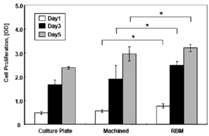
To examine the proliferation of osteoblastic cells in RBM surface, we carried out a measurement of MTS activity on MG-63 cells after 1, 3, 5 days in culture. Results indicated that cell proliferation on RBM surface was increased after 1 days of culture than the machined surfaces.(*P<0.05; Fig. 3) This phenomenon was observed similar at 3 days and 5 days. Also gradient of cell proliferation curve was more steep in RBM surfaces.(Table 4) Therefore these results showed that prolifer- ation of MG-63 cells was increased as early as 1 day in culture on RBM surface as compared to the machined surface.
5. Cell Differentiation
1) ALP activity
Generally, ALP activity was affected by the surface microto- pography. In addition osteoblastic cell differentiation was assessed by measuring the ALP activity normalized to total
protein content. On RBM surfaces, we observed a significant enhancement of ALP activity (10.8±2.6%) as compared with machined surface after 7 days.(***P<0.001; Fig. 4. A) These results indicate that osteoblastic cells cultured in direct contact with RBM surface increased their capability to express ALP.
2) Mineralization (Quantification of alizarin red-s staining) We used the alizarin red-s staining for quantification of min- eralization induced by osteoblastic cells. As shown Fig. 4. B, similar pattern in ALP activity data, MG-63 cells cultured on RBM surfaces had increased mineralization dramatically.
(***P<0.001)
3) ELISA
According to the acceleration of the differentiation, various osteoblastic differentiation-related proteins, such as extracellu- lar matrix proteins or other signaling factors, were secreted. So we checked the expression levels of osteoblastic differentia- tion-related proteins using ELISA methods. Osteocalcin secre- tion by MG-63 cells was slightly greater (**P<0.01) on RBM surface than machined surface.(Fig. 4. C) Moreover, osteopon- tin (***P<0.001), PGE2 (***P<0.001) and TGF-beta1 (*P<0.05) secretion was greater on RBM surface than the machined surface.(Figs. 4. D, 4. E, and F separately) As a result ELISA data showed that the RBM surface enhances the secre- tion of various osteoblastic differentiation-related proteins.
Fig. 2.Effect of surface roughness on the adhesion of MG- 63 cells. MG-63 cells were stained with cresyl violet dye and counted using spectrophotometer on surfaces of dif- fering roughness. Data are represented as the average ± standard deviation. RBM surface, the rough surface, were promoted the cell adhesion compared to smooth machined surface. *: Statistically significant compared with cells cul- tured on machined surfaces.(P<0.05)
Fig. 3.Results of MG-63 cell proliferation experiments. Data are represented as the average±standard deviation. The differences between the number of cells on RBM and machined surfaces during 5 days is statistically significant.
*: Statistically significant compared with cells cultured on machined surfaces.(P<0.05)
Table 4. Gradient of MG-63 cell proliferation on a machined Ti and a RBM surfaces during 5 days
Culture plate Machine RBM Gradient of
0.9493 1.1937 1.2196
cell proliferation
RBM, resorbable blasting media
4) Semi-quantitative RT-PCR
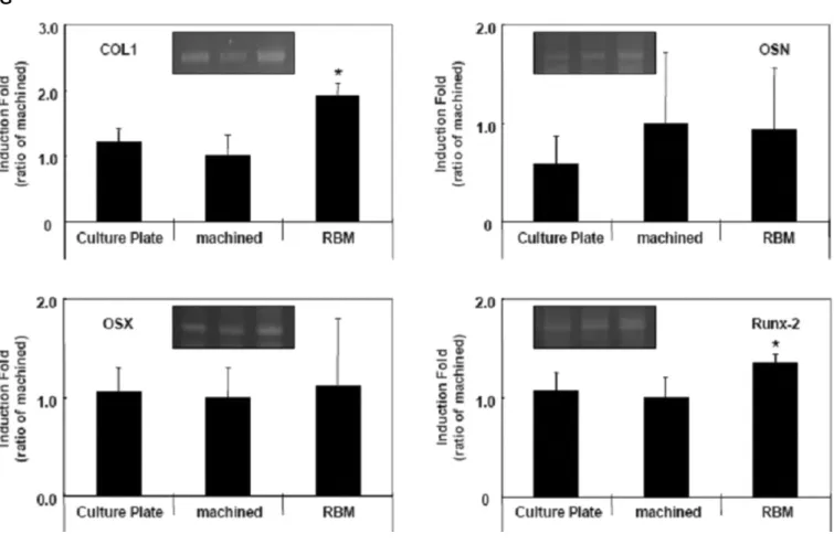
Semi-quantitative RT-PCR showed significantly (*P<0.05) increased COL1 and Runx-2 (cbfa-1, OSF-2) gene expression in MG-63 grown on RBM, relative to machined surface or cul- ture plate.(Fig. 4. G) There was a slight, but insignificant, increase in OSN and OSX gene expression on the RBM sur- face relative to culture plate or machined surface.(Fig. 4. G) These results strongly suggested that RBM surface increases
the expression level of osteoblastic differentiation marker gene associated with osteogenesis and osseointegration.
Ⅳ. Discussion
Osteoblastic cells play a critical role in the early stages of ossteointegration. Several reports have assessed that osteoblas- tic cell response to smooth surfaces more rapidly than rough
A B
C D
E F
surfaces while their osteoblastic differentiation was enhanced by rough topography35-39. Little is known about the regulatory mechanism of the osteoblastic cell adhesion, proliferation, and differentiation on the titanium discs. This study provided the evidence that the roughness and topography generated by hydroxyapatite-grit blasting (RBM) is an important regulating factor of the osteoblastic cell adhesion, proliferation, and dif- ferentiation. This was examined in six ways, using the cresyl
violet staining, MTS assay, ALP activity assay, alizarin red-s staining, ELISA, semi-quantitative RT-PCR. In all six cases, the osteoblastic cell adhesion, proliferation, and differentiation was dramatically enhanced in RBM surfaces. These results provided unequivocal evidence that surface roughness and topography generated by RBM surface is an important factor that enhances the osteoblastic cell adhesion, proliferation, and differentiation.
G
Fig. 4. Effect of surface roughness on the osteoblastic differentiation of MG-63 cells. Data are represented as the average±
standard deviation. (A) ALP (alkaline phosphatase) activity of MG-63 cells cultured either on machined Ti and RBM surfaces.
Culture plate without osteogenic differentiation was served as a internal negative control. And a culture plate with osteogenic differentiation was a internal positive control. On RBM surface, the ALP activity enhanced about 10% than machined surface (B). Mineralization properties of the machined Ti and RBM surface. MG-63 cells were cultured as before for 14 days with osteogenic differentiation medium. Total calcium deposition measured by alizarin red-s stain via extraction with acetic acid.
(C-F) Effect of surface roughness on osteogenesis-related local factor levels determined by using an ELISA kit. Osteocalcin levels were measure in the conditioned media using an ELISA kit specific for human osteoclacin (C). Osteopontin levels were measured in the conditioned media using an ELISA kit specific for human osteopontin (D). PGE2 levels were measured in the conditioned media using an ELISA kit specific for humans PGE2 (E). And Active of latent TGF-β1 (transforming growth factor-β1) levels were measure in the conditioned media using an ELISA kit specific for human TGF-β1 (F). (G) RT-PCR analysis of roughness-influenced osteogenic marker gene. Type I collagen, osteonectin, osterix and Runx-2 RNA levels in MG-63 cells exposure for 72hrs on a machined Ti or RBM surface. RT-PCR products were subjected to electrophoresis on 2% agarose gel and visualized UV exposure. A representative analysis is shown inside of the each figure. And the level of specific bands was normalized for the β-actin cDNA content. Statistically significant compared with cells cultured on machined surfaces by unpaired t-tests.(*P<0.01, **P<0.05, ***P<0.001)
While the effects of surface roughness and topography remains controversial with respect to osteoblastic phenotype expression40,41, we aimed at testing whether MG-63 preserve their phenotype in direct contact with hydroxyapatite grit- blasted (RBM) surface. Therefore, this study further investigat- ed the phenomenon underlying the surface roughness and topography modulate the osteoblastic cell behavior. Briefly, MG-63 cells cultured on very irregular surfaces, such as RBM, exhibited the increased cell adhesion, proliferation, and differ- entiation. MTS activity was increased more steeply on RBM surfaces. ALP is a marker of osteogenic differentiation, bone formation and mineralization. Previous reports have demon- strated the influence of surface roughness on ALP activity37,39. Like a report of Kim et al.36, ALP activity was enhanced in parallel with the roughness parameters in our study. Moreover this osteoblastic phenotypic characterization can be based on the analysis of various osteobalstic markers including COL1, OSN, OSX and Runx-2 (cbfa-1/OSF-2). Hydroxyapatite grit- blasted (RBM) surface induced a slight increase in COL1 and Runx-2 expression. Also secretion of osteoblastic differentia- tion-related protein and deposition of mineral was enhanced in RBM surface.
Nevertheless, the biological effects of residual alumina parti- cles on titanium surfaces are controversial in the literature42-45. Bone healing around residual alumina in alumina grit-blasted titanium implants is impaired. On the contrary, Sader et al.19 did not observe any detrimental defect of residual blasting alu- mina particles in vivo and in vitro experiment. Furthermore, Wennerberg et al.46did not find any significant differences in bone-implant contact for alumina-blasted and machined titani- um implants.
Calcium phosphate ceramics were used as the blasting mate- rials in order to avoid the possible negative effects of residual alumina on the osseointegration between titanium and implant.
Our results demonstrate the biocompatible and resorbable hydroxyapatite abrasive particles can be used to create titani- um surface roughness. This grit-blasting process increased sur- face roughness of titanium implants and offered an osteoblastic cell favorable surfaces. As shown as EDS data (Table 2), even if infitesibal calcium and phosphorous were found on the RBM surfaces after blasting and cleaning, they should not be detri- mental to the osseointegration periods.
Although both alumina and hydroxyapatite grit-blasting media had comparable roughness, their surface energetics were different. The alumina-blasted surface was more hydrophobic than the hydroxyapatite grit-blasted surface. Hydrophilic sur- face should be more favorable to the osteoblastic cell adhesion, proliferation, and differentiation47. These difference may be
related to the presence of residual alumina particles on the sur- faces. Therefore, grit-blasting titanium surfaces with a biocom- patible, resorbable, and osteoconductive materials like hydrox- yapatite and β-TCP ceramic particles are alternative method for avoiding the presence of residual alumina particles.
In this study, osteoblastic cell adhesion, proliferation, and differentiation was greater for the RBM surfaces than for the machined surfaces. This suggests that the RBM surfaces allowed more great osteoblastic cell phenotype than the machined surfaces48. Moreover we showed that rough implant surface can alter the expression of bone associated regulatory transcription factors and key osseointegration-related proteins.
Maybe this occurred as a result of differences in cell adhesion, as a result of integrin-mediated adhesion and regulation of downstream signaling pathways as previously reported49-53. This early osteoblastic cell differentiation in contact with roughened RBM surfaces may be favorable during the early phases of bone healing. Further investigations on the relation- ship between surface roughness, topography, and chemistry should improve the understanding of the osseointegration- related osteoblastic cell behavior.
Ⅴ. Conclusion
In summary, this study examined the effect of microtopo- graphically modified rough surfaces by hydroxyapatite grit- blasting on osteoblastic cell adhesion, proliferation, and differ- entiation. Human osteoblast-like cell line, MG-63 cells, cul- tured on the RBM surfaces exhibited more osteoblastic cell adhesion, proliferation, differentiated osteoblastic phenotype and produced more local factors that stimulate the osteoblastic differentiation. Our results demonstrated the RBM surfaces stimulate the in vitro osteogenesis in MG-63 cells and raise the potential that RBM surfaces accelerate the bone formation and finally increase bone-implant contact.
References
1. Davies JE. Mechanisms of endosseous integration. Int J Prosthodont 1998;11:391-401.
2. Berglundh T, Abrahamsson I, Lang NP, Lindhe J. De novo alve- olar bone formation adjacent to endosseous implants. Clin Oral Implants Res 2003;14:251-62.
3. Albrektsson T, Wennerberg A. Oral implant surfaces: Part 2.
Review focusing on clinical knowledge of different surfaces. Int J Prosthodont 2004;17:544-64.
4. Esposito M, Coulthard P, Thomsen P, Worthington HV. The role of implant surface modifications, shape and material on the suc- cess of osseointegrated dental implants: a Cochrane systematic review. Eur J Prosthodont Restor Dent 2005;13:15-31.
5. Puleo DA, Thomas MV. Implant surfaces. Dent Clin North Am 2006;50:323-38.
6. Zhao G, Schwartz Z, Wieland M, Rupp F, Geis-Gerstorfer J,
Cochran DL, et al. High surface energy enhances cell response to titanium substrate microstructure. J Biomed Mater Res A 2005;
74:49-58.
7. Cooper LF, Masuda T, Yliheikkila¨ PK, Felton DA.
Generalizations regarding the process and phenomenon of os- seointegration: Part II. In vitro studies. Int J Oral Maxillofac Implants 1998;13:163-74.
8. Anselme K. Osteoblast adhesion on biomaterials. Biomaterials 2000;21:667-81.
9. Kieswetter K, Schwarts Z, Dean DD, Boyan BD. The role of im- plant surface characteristic in the healing of bone. Crit Rev Oral Biol Med 1996;7:329-45.
10. Thomas KA, Cook SD. An evaluation of variables influencing implant fixation by direct bone apposition. J Biomed Mater Res 1985;19:875-901.
11. Predecki P, Stephan JE, Auslaender BA, Mooney VL, Kirkland K. Kinetics of bone growth into cylindrical channels in aluminum oxide and titanium. J Biomed Mater Res 1972;6:375-400.
12. Carlsson L, Ro¨stlund T, Albrektsson B, Albrektsson T. Removal torques for polished and rough titanium implants. Int J Oral Maxillofac Implants 1988;3:21-4.
13. Lauer G, Wiedmann-Al-Ahmad M, Otten JE, Hu¨bner U, Schmelzeisen R, Schilli W. The titanium surface texture effects adherence and growth of human gingival keratinocytes and hu- man maxillar osteoblast-like cells in vitro. Biomaterials 2001;22:
2799-809.
14. Mustafa K, Wennerberg A, Wroblewski J, Hultenby K, Lopez BS, Arvidson K. Determining optimal surface roughness of TiO2
blasted titanium implant material for attachment, proliferation, and differentiation of cells derived from human mandibular alve- olar bone. Clin Oral Implants Res 2001;12:515-25.
15. Ellingsen JE, Johansson CB, Wennerberg A, Holme′n A.
Improved retention and bone-to implant contact with fluoride- modified titanium implants. Int J Oral Maxillofac Implants 2004;
19:659-66.
16. Anselme K, Bigerelle M, Noel B, Dufresne E, Judas D, Iost A, et al. Qualitative and quantitative study of human osteoblast adhe- sion on materials with various surface roughnesses. J Biomed Mater Res 2000;49:155-66.
17. Anselme K. Osteoblast adhesion on biomaterials. Biomaterials 2000;21:667-81.
18. Wennerberg A, Albrektsson T, Andersson B. Bone tissue re- sponse to commercially pure titanium implants blasted with fine and coarse particles of aluminum oxide. Int J Oral Maxillofac Implants 1996;11:38-45.
19. Sader MS, Balduino A, Soares Gde A, Borojevic R. Effect of three distinct treatments of titanium surface on osteoblast attach- ment, proliferation, and differentiation. Clin Oral Implants Res 2005;16:667-75.
20. Le Guehennec L, Lopez-Heredia MA, Enkel B, Weiss P, Amouriq Y, Layrolle P. Osteoblastic cell behaviour on different titanium implant surfaces. Acta Biomater 2008;4:535-43.
21. Citeau A, Guicheux J, Vinatier C, Layrolle P, Nguyen TP, Pilet P, et al. In vitro biological effects of titanium rough surface ob- tained by calcium phosphate grit blasting. Biomaterials 2005;26:
157-65.
22. Sanz A, Oyarzu′n A, Farias D, Diaz I. Experimental study of bone response to a new surface treatment of endosseous titanium im- plants. Implant Dent 2001;10:126-31.
23. Novaes AB Jr, Souza SL, de Oliveira PT, Souza AM. Histomor- phometric analysis of the bone-implant contact obtained with 4 different implant surface treatments placed side by side in the dog mandible. Int J Oral Maxillofac Implants 2002;17:377-83.
24. Ginsberg SD, Che S. Combined histochemical staining, RNA amplification, regional, and single cell cDNA analysis within the hippocampus. Lab Invest 2004;84:952-62.
25. Hutton LC, Castillo-Melendes M, Smythe GA, Walker DW.
Microglial activation, macrophage infiltration, and evidence of
cell death in the fetal brain after uteroplacental administration of lipopolysaccharide in sheep in late gestation. Am J Obstet Gynecol 2008;198:e1-11.
26. Engvall E. Enzyme immunoassay ELISA and EMIT. Methods Enzymol 1980;70(A):419-39.
27. Jones JV, Mansour M, James H, Sadi D, Carr RI. A substrate am- plification system for enzyme-linked immunoassays: II.
Demonstration of its applicability for measuring anti-DNA anti- bodies. J Immunol Methods 1989;118:79-84.
28. Harada M, Hiraoka BY, Fukasawa K, Fukasawa KM.
Purification and properties of bovine dental-pulp alkaline-phos- phatase. Arch Oral Biol 1982;27:69-74.
29. Jung K, Pergande M. Influence of inorganic phosphate on the ac- tivity determination of isoenzymes of alkaline phosphatase in various buffer systems. Clin Chim Acta 1980;102:215-9.
30. Valarmathi MT, Yost MJ, Goodwin RL, Potts JD. The influence of proepicardial cells on the osteogenic potential of marrow stro- mal cells in a three-dimensional tubular scaffold. Biomaterials 2008;29:2203-16.
31. Rausch-fan X, Qu Z, Wieland M, Matejka M, Schedle A.
Differentiation and cytokine synthesis of human alveolar os- teoblast compared to osteoblast-like cells (MG63) in response to titanium surfaces. Dent Mater 2008;24:102-10.
32. Gregory CA, Gunn WG, Peister A, Prockop DJ. An Alizarin red- based assay of mineralization by adherent cells in culture: com- parison with cetylpyridinium chloride extraction. Anal Biochem 2004;329:77-84.
33. Malladi P, Xu Y, Chiou M, Giaccia AJ, Longaker MT. Effect of reduced oxygen tension on chondrogenesis and osteogenesis in adipose-derived mesenchymal cells. Am J Physiol Cell Physiol 2006;290:C1139-46.
34. Sudo H, Kodama HA, Amagai Y, Yamamoto S, Kasai S. In vitro differentiation and calcification in a new clonal osteogenic cell line derived from newborn mouse calvaria. J Cell Biol 1983;96:
191-8.
35. Ba¨chle M, Kohal RJ. A systematic review of the influence of dif- ferent titanium surfaces on proliferation, differentiation and pro- tein synthesis of osteoblast-like MG63 cells. Clin Oral Implants Res 2004;15:683-92.
36. Kim MJ, Choi MU, Kim CW. Activation of phospholipase D1 by surface roughness of titanium in MG63 osteoblast-like cell.
Biomaterials 2006;27:5502-11.
37. Schwartz Z, Lohmann CH, Oefinger J, Bonewald LF, Dean DD, Boyan BD. Implant surface characteristics modulate differentia- tion behavior of cells in the osteoblastic lineage. Adv Dent Res 1999;13:38-48.
38. Anselme K, Bigerelle M. Topography effects of pure titanium substrates on human osteoblast long-term adhesion. Acta Biomater 2005;1:211-22.
39. Kim MJ, Kim CW, Lim YJ, Heo SJ. Microrough titanium surface affects biologic response in MG63 osteoblast-like cells. J Biomed Mater Res A 2006;79:1023-32.
40. Wennerberg A, Albrektsson T, Andersson B. Bone tissue re- sponse to commercially pure titanium implants blasted with fine and coarse particles of aluminum oxide. Int J Oral Maxillofac Implants 1996;11:38-45.
41. Deligianni DD, Katsala ND, Koutsoukos PG, Missirlis YF.
Effect of surface roughness of hydroxyapatite on human bone marrow cell adhesion, proliferation, differentiation and detach- ment strength. Biomaterials 2001;22:87-96.
42. Diniz MG, Pinheiro MA, Andrade Junior AC, Fischer RG.
Characterization of titanium surfaces for dental implants with in- organic contaminant. Braz Oral Res 2005;19:106-11.
43. Piattelli A, Degidi M, Paolantonio M, Mangano C, Scarano A.
Residual aluminum oxide on the surface of titanium implants has no effect on osseointegration. Biomaterials 2003;24:4081-9.
44. Canabarro A, Diniz MG, Paciornik S, Carvalho L, Sampaio EM, Beloti MM, et al. High concentration of residual aluminum oxide
on titanium surface inhibits extracellular matrix mineralization. J Biomed Mater Res A 2008;87A:588-97.
45. Rodrigo A, Valle′s G, Saldaña L, Rodriguez M, Martinez ME, Munuera L, et al. Alumina particles influence the interactions of cocultured osteoblasts and macrophages. J Orthop Res 2006;24:
46-54.
46. Wennerberg A, Albrektsson T, Johansson C, Andersson B.
Experimental study of turned and grit-blasted screw-shaped im- plants with special emphasis on effects of blasting material and surface topography. Biomaterials 1996;17:15-22.
47. Li LH, Kong YM, Kim HW, Kim YW, Kim HE, Heo SJ, Koak YK. Improved biological performance of Ti implants due to sur- face modification by micro-arc oxidation. Biomaterials 2004;
25:2867-75.
48. Giordano C, Sandrini E, Busini V, Chiesa R, Fumagalli G, Giavaresi G, et al. A new chemical etching process to improve endosseous implant osseointegration: in vitro evaluation on hu-
man osteoblast-like cells. Int J Artif Organs 2006;29:772-80.
49. Xavier SP, Carvalho PS, Beloti MM, Rosa AL. Response of rat bone marrow cells to commercially pure titanium submitted to different surface treatments. J Dent. 2003;31:173-80.
50. Schneider GB, Perinpanayagam H, Clegg M, Zaharias R, Seabold D, Keller J, et al. Implant surface roughness affects os- teoblast gene expression. J Dent Res 2003;82:372-6.
51. Schneider GB, Whitson SW, Cooper LF. Restricted and coordi- nated expression of beta3-integrin and bone sialoprotein during cultured osteoblast differentiation. Bone 1999;24:321-7.
52. Schneider GB, Zaharias R, Seabold D, Keller J, Stanford C.
Differentiation of preosteoblasts is affected by implant surface microtopographies. J Biomed Mater Res A 2004;69:462-8.
53. Schneider GB, Zaharias R, Stanford C. Osteoblast integrin adhe- sion and signaling regulate mineralization. J Dent Res 2001;
80:1540-4.