S
ince the introduction of the concept of osseoin- tegration, implants of various composition and design have been studied and developed.Currently, commercially pure titanium (Ti) is
the material of choice for uncoated implants because of its biologic acceptance in bone.1, 2)This high degree of biocompatibility is thought to result, in part, from the oxide layer that resides over the titanium surface and facilitates the bonding of
EFFECT OF TITANIUM SURFACE ROUGHNESS ON CELL ADHESION OF HUMAN OSTEOBLAST- LIKE CELLS (MG63)
Soon-Ho Yim, D.D.S., Ph.D.
Department of Prosthodontics, Samsung Medical Center, College of Medicine, Sungkyunkwan University
Statement of problem.The effects of surface roughness have not or insufficiently been ana- lyzed on earlier events such as cell adhesion though cell behavior most germane to implant per- formance is cell adhesion.
Purpose.The purpose of this study was to evaluate cell adhesion of osteoblast-like cells (MG63) onto three types of titanium disks with varying roughness using the Elisa assay.
Materials and methods. Representative disks from each group (SLA, HA, machined) were subjected to surface analysis and surface roughness was measured by the optical interferometer (Accura 2000, Intekplus Co., Seoul, Korea). Following this, MG63 cells were cultured on the titanium disks and released. Cell adhesion measurements using the Elisa assay were performed specifically at three points: after 24, 48, and 72 hours of culture.
Results. Among the 3 types of surface analyzed, the SLA surface was the roughest with a Ra value of 1.114 μm followed by HA coated surface and machined surface, consecutively. The optical density values for the SLA surface group was significantly higher than that of the machined and HA coated suface groups following 24 and 48 hours of culture. The cell culture on HA coat- ed surface showed significantly higher values compared to the machined surface following 24, 48 and 72 hours of culture.
Conclusion. The results suggest that surface treatment of titanium surfaces enhanced cell adhe- sion of human osteoblast-like cells (MG63).
Key Words
Cell adhesion, Osteoblast-like cells (MG63), Elisa assay, Optical density values
J Korean Acad Prosthodont : Volume 42, Number 3, 2004
the extracellular matrix at the implant-tissue interface.3, 4)
The characteristic compositon and structure of the oxide layer may be altered when different preparation techniques are applied to the surface of the titanium. It has been shown that meth- ods of implant surface preparation can significantly affect the resultant properties of the surface and subsequently the biological responses that occur at the surface. Windeler et al.5)demonstrated that osteoblast-like cells adhered more strongly to Titanium surfaces whereas osteoclasts adhered more strongly to hydroxyapatite surfaces. In a study comparing cell adhesion to different surfaces, Martin et al.6) have found that osteoblast-like cells adhered more strongly to Titanium sur- faces than they do to smooth surfaces. Moreover, it was also shown that surface roughness affects proliferation; as roughness increases, proliferation decreases.
MG63 cells, an osteoblast-like human cell line, have phenotypic and genetic characteristics typ- ical of a relatively immature osteoblast.7)Although MG63 cells do not calcify their extracellular matrix in culture, the cell line is sufficiently dif- ferentiated far along the osteogenic lineage to serve as an excellent system for examining early events in the response of bone cells to surfaces.
Studies using these cells have demonstrated pos- itive effects of increased surface roughness of titanium discs on cellular differentiation and matrix production.6)
However, these effects of surface roughness have not or insufficiently been analyzed on earlier events such as cell adhesion though cell behavior most germane to implant per- formance is cell adhesion. The objective of this study was to evaluate cell adhesion of osteoblast- like cells (MG63) onto three types of Titanium disks with varying roughness using the Elisa assay.
MATERIALS AND METHODS
Surface preparation
The Titanium disks subjected for cell culture were processed to produce three types of surfaces of varying roughness as follows.
1. Machined
2. SLA surface: Disks were blasted with 220um corundum grit at 3 bar until the surface reached a uniform gray tone. They were then acid-etched in hydrochloric acid/sulfuric acid at room temperature for 4 minutes fol- lowed by rinsing in deionized water, neu- tralization in 5 % sodium bicarbonate solution, and three 5-minute rinses in deionized water contained in the ultrasonic bath.
3. HA: Thin HA layers were deposited on Titanium-substrates by an electron beam deposition method. After evacuating the chamber down to 10-7torr using a cryop- ump (OB-10, Helix Technology, Mansfield, MA, USA), an electron beam (Telemark, Fremont, CA, USA) of 8.5 kV and ~0.1 A was directed onto the source target. Prior to deposition, the substrates were sputter- cleaned with an ion beam (Mark II, Commonwealth Scientific, Alexandria VA, USA) of 120 V and 0.6 A for 20 minutes. In order to increase the uniformity of the coat- ing layer during the deposition process, sub- strates were rotated at a speed of 8 rpm. The targets were made using a commercially available HA (Ca10(PO4)6(OH)2) powder (Alfa Aesar Co., Ward Hill, MA, USA). Extra weight % of CaO powder (Cerac Co., Milwaukee, WI, USA) was added to the HA and mixed by ball milling in ethyl alcohol for 24 hours with Al2O3balls as media. The powder mixtures were sintered in air at 1200�C for 2 hours. The deposited coating lay- er was heat treated in air at temperatures between 300�C and 500�C for 1 hour.
Surface analyses
Representative 3 disks from each group were sub- jected to surface analysis. 5 different areas of each samples were measured. Surface rough- ness was measured by optical interferometer (Accura 2000, Intekplus Co., Seoul, Korea).
(Fig. 1, 2 & 3) This system provides visual images as well as numerical values for the different surface roughness parameters.
Cell cultures
MG63 osteoblast-like cells, originally isolated from a human osteosarcoma, were used in this study.
This osteoblast-like cell line has been well-char- acterized and contains numerous osteoblastic traits that are typical of a relatively immature osteoblast, including high levels of 1,25-(OH)2D3- responsive alkaline phosphatase and osteocalcin synthesis inhibition of proliferation when treat- ed with 1,25-(OH)2D3. As a result, they are a good model for examining the early stages of osteoblastic differentiation. MG63 cells were obtained from the American Type Culture Collection (Rockvill, MD). Cells were plated at a concentration of 2.5x10⁵/ml in Dulbecco’s mod- ified Eagel’s medium (DMEM) containing 10%
fetal bovine serum (FBS) and 1 % antibiotics and cultured at 37�in 5% CO2.
Cell adhesion assay (ELISA ASSAY) Cell adhesion onto each titanium disk was measured using the Elisa assay. 8 disks were
used for each groups. After cell culture, cells were washed in PBS (Phosphated buffered saline) and fixed with 10% formalin for 15 minutes.
Cells were then stained overnight with 1% crys- tal violet 1ml. After staning, disks containing the cultured cells were washed 3 times with DW. With the addition of 1% SDS (sodium dode- sil sulfate) 0.2ml and vortexing for 5 minutes, cells from the disk surface were released. Lysed cells were transferred to 96-well plate and the absorp- tion optical density was calculated at 570nm using the Elisa assay.
RESULTS
Surface analysis
The results of the optical interferometer analy- sis are shown in Table I. Among the 3 types of sur- face analyzed, the SLA surface was the roughest with a Ra value of 1.114 μm followed by HA coated surface and machined surface, consecutively.
(Fig. 1, 2 & 3)
Cell adhesion assay
Cell adhesion was measured using the Elisa assay following 24, 48, and 72 hours of culture. The control sample was calculated at the beginning of each cell culture and showed no significant dif- ference in absorption optical density values between the samples. The optical density val- ues for the SLA surface group was significantly higher than that of the machined and HA coated suface groups following 24 hours of culture.
This was also true following 48 hours of culture as the SLA surface displayed significantly high- er values. The cell culture on HA coated sur- face showed significantly higher values com- pared to the machined surface following 24, 48 and 72 hours of culture. All three groups showed decreased optical density values following 72 hours of culture. The results of the Elisa test are listed in Table II.
Table I. Surface roughness (Ra) of the titanium implants (Mean±SD, n=15)
Ra (arithmetic mean of the absolute values of the surface)
Machined 0.45±0.04 μma
HA coated 0.49±0.06 μma
SLA 1.11±0.08 μmb
* The same letter denotes groups that were not signif- icantly different from each other (P>0.05)
DISCUSSION
The objective of this study was to evaluate cell adhesion of osteoblast-like cells (MG63) onto three types of Titanium disks with varying rough- ness using the Elisa assay. Representative disks from each group was first subjected to surface roughness analysis using the optical interfer- ometer. (Accura 2000, Intekplus Co., Seoul, Korea). The results revealed that the Ra values of SLA surface were higher than that of HA coated and machined surfaces. Also, the Ra values of HA coated implants when compared to machined
implants, showed only a marginal difference.
This was interesting in respect to earlier findings where HA coated implants consistently showed rougher surfaces than the machined implants. Not well understood, one may only speculate the cause of these results because direct compar- isons to earlier studies are difficult to make since different methodology and techniques have been applied during processing of these surfaces.
Different preparation techniques may have con- tributed to this phenomenon.
As the second part of this study, cell adhesion was measured following 24, 48 and 72 hours of culture. The control sample was calculated at the beginning of each cell culture. The results of the cell adhesion assay suggest enhanced adhesion of osteoblastic cells on surface treated Table II. Results of the Elisa test (n=8)
Control 24 hours 48 hours 72 hours machined 0.16±0.04a 0.49±0.03a 0.49±0.05a 0.35±0.04c HA coated 0.09±0.03a 0.66±0.02b 0.6±0.02b 0.08±0.02a SLA 0.1±0.03a 1.1±0.05c 1.43±0.06c 0.19±0.03b
* The same letter denotes groups that were not signif- icantly different from each other (P>0.05)
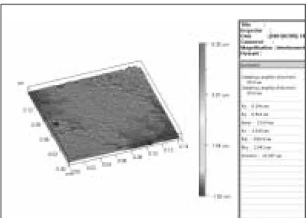
Fig. 2.Three-dimensional image analysis of the SLA sur- face measured by the optical interferometer.
Fig. 1.Three-dimensional image analysis of the machined surface measured by the optical interfer- ometer.
Fig. 3.Three-dimensional image analysis of the HA coat- ed surface measured by the optical interferometer.
titanium disks. Also, as roughness increased, so did adhesion of osteoblastic cells. This is in accor- dance with a number of studies that evaluated the effect of surface roughness on early cellular activ- ities of human osteoblasts or osteoblastic cells.
Bowers et al.8) have demonstrated that rough titanium surfaces which measured in the microm- eter range due to sandblasting or coating by plasma spray significantly enhanced cellular attachment and the production of extracellular matrix and subsequent mineralization in vitro. Also, it was shown by Michaels et al.16)that osteoblast- like cells exhibit greater initial attachment to rough Titanium surfaces.
Results following 72 hours of culture suggest decrease in optical density values in all three types of surfaces. This may be due to the fact that cell adhesion properties of osteoblastic cells occur early, presumably within 24 hours of culture.
Further evaluation is needed to confirm this phenomenon.
The present study possessed interesting aspects as it utilized (MG63) osteoblast-like cells and Elisa assay for evaluation. In a previous study using human osteoblast-like cell line (MG63), Martin et al.6)showed that increasing surface roughness of titanium discs reduced proliferation but induced cellular differentiation and matrix pro- duction. However, earlier events such as cell adhesion was not evaluated. This study con- firmed that increasing surface roughness con- tributes to adhesion of osteoblast-like cells (MG63).
Although further studies seem necessary to strengthen this hypothesis, this finding presents a significant leap in understanding the early events of osteoblast-like cells on Titanium surfaces.
CONCLUSION
The effects of surface roughness have not or insuf- ficiently been analyzed on earlier events such as cell adhesion though cell behavior most germane
to implant performance is cell adhesion. The purpose of this study was to evaluate cell adhe- sion of osteoblast-like cells (MG63) onto three types of titanium disks with varying roughness.
Representative disks from each group (SLA, HA, machined) were subjected to surface analysis and surface roughness was measured by the optical interferometer (Accura 2000, Intekplus Co., Seoul, Korea). Following this, MG63 cells were cultured on the titanium disks and released. Cell adhesion measurements using the Elisa assay were performed specifically at three points: after 24, 48 and 72 hours of culture. It can be concluded that :
1. The surface roughness of machind group was not different from that of HA coated group.
(P>0.05) The SLA group showed the roughest surface among three groups.
2. The SLA group showed the highest cell adhe- sion following the HA group and the machined group. (P<0.05) After 24 hours and 48 hours.
3. Cell attachment of all three groups decreased following 72 hours of culture.
4. Surface modifications using HAcoating and SLA can promote cellular attachments and enhance bone formation.
REFERENCES
1. Branemark P-I. Osseointegration and its experi- mental background. Journal of Prosthetic Dentistry 1983;50:399-410.
2. Branemark P-I. Introduction to osseointegration, in Branemark P-I, Zarb GA, Albrektsson T (eds):
Tissue-Integrated Prostheses: Osseointegration in Clinical Dentistry. Chicago, Quintessence Publishing Co, 1985, pp 11-76.
3. Kasemo B, Lausamm J. Biomaterial and implant sur- faces: A surface science approach. International Journal of Oral Maxillofacial Implants 1988;3:247- 259.
4. Stanford CM, Keller JC. Osseointegration and matrix production at the implant surface. Crit Rev Oral Biol Med 1991;2:83-101.
5. Windeler AS, Bonewald LF, Khare AG, Boyan BD, Mundy GR. The influence of sputtered bone substitutes on cell growth and phenotypic ex-
pression. In: The Bone-Biomaterial Interface, J.E.
Davies (Ed). Toronto: Toronto Press 1991:205-213.
6. Martin JY, Schwartz Z, Hummert TW, Schraub DL, Boyan BD. Effect of titanium surface roughness on proliferation, differentiation, and protein synthe- sis of human osteoblast-like cells (MG63). Journal of Biomedical Materials Research 1995;29:389- 401.
7. Boyan BD, Batzer R, Kieswetter K, Liu Y, Cochran DL, Szmuckler-Moncler S, Dean DD, Schwartz Z. Titanium surface roughness alters responsive- ness of MG63 osteoblast-like cells to 1α,25-(OH)2D3.
Journal of Biomedical Materials Research 1998;39:77- 85.
8. Bowers KT, Keller JC, Randolph BA, Wick DG, Michaels CM. Optimization of surface micro- morphology for enhanced osteoblast responses in vitro. International Journal of Oral and Maxillofacial Implants 1992; 7:302-310.
9. Michaels CM, Keller JC, Stanford CM, Solursh M, Mackenzie IC. In vitro connective tissue cell at- tachment to cpTi. Journal of Dental Research 1989;68:276.
10. Boyan BD, Hummert TW, Kieswtter K, Schraub D, Dean DD, Schwartz Z. Effect of titanium surface characteristics on chondrocytes and osteoblasts in vitro. Scanning Electron Microscopy (Cells &
Materials) 1995;5:323-335.
11. Brunette DM. The effects of implant surface topog- raphy on the behavior of cells. International Journal of Oral Maxillofacial Implants 1988;3:231- 246.
12. Buser D, Schenk R, Steinemann S, Fiorellini J, Fox C, Stich H. Influence of surface characteristics on bone integration of titanium implants. A histo- morphometric study in miniature pigs. Journal of Biomedical Materials Research 1991;25:889-902.
13. Davies JE. In vitro modeling of the bone/implant
interface. The Anatomical Record 1996;245:426-445.
14. Davies JE, Lowenberg B, Shiga A. The bone-titanium interface in vitro. Journal of Biomedical Materials Research 1990;24:1289-1306v
15. Gotfredsen K, Wennerberg A, Johansson C, Skovgaard LTT, Hjorting-Hansen E. Anchorage of TiO2-blasted, HA-coated, and machined implants:
An experimental study with rabbits. Journal of Biomedical Materials Research 1995;29:1223-1231.
16. Kieswetter K, Schwartz Z, Hummert TW, Cochran DL, Simpson J, Dean DD, Boyan BD. Surface roughness modulates the local production of growth factors and cytokines by osteoblast-like MG63 cells. Journal of Biomedical Materials Research 1996;32:55-63.
17. Larsson C, Thomsen P, Lausmaa J, Rodahl M, Kasemo B, Ericson LE. Bone response to surface modified titanium implants: studies on electrop- olished implants with different oxide thicknesses and morphology. Biomaterials 1994;15:1062-1074.
18. Larsson C, Thomsen P, Aronsson BO, Rodahl M, Lausmaa J, Kasemo B, Ericson LE. Bone response to surface-modified titanium implants: studies on the early tissue response to machines and elec- tropolished implants with different oxide thick- nesses. Biomaterials 1996;17:605-616.
19. Piatelli A, Scarano A, Piatelli M, Calabresse L.
Direct bone formation on sand-blasted titanium im- plants: an experimental study. Biomaterials 1996;17:1015-1018.
Reprint request to:
SOON-HOYIM
SUNGKYUNKWANUNIVERSITY,SCHOOL OFMEDICINE
741-1, HANNAM2-DONG, YONGSAN-GU, SEOUL,KOREA140-895 [email protected]