http://e-nrp.org
Inhibitory effect of Erythronium japonicum on the human breast cancer cell metastasis
Mi-Kyoung You
1, Min-Sook Kim
2, Jin Rhyu
1, Mi-Ae Bang
3and Hyeon-A Kim
1§1Department of Food & Nutrition / Research Institute of Human Ecology, Mokpo National University, 1666, Yeongsan-ro, Cheonggye-myeon, Muan-gun, Jeonnam 534-729, Korea
2Nature Pure Korea Inc. Jeonnam 517-803, Korea
3Jeonnam Biofood Technology Center, Jeonnam 520-330, Korea
BACKGROUND/OBJECTIVES: In this study, the inhibitory effect of Erythronium japonicum extracts on the metastasis of MDA-MB-231 human breast cancer cell line was determined.
MATERIALS/METHODS: Cells were cultured with DMSO or with 50, 75, 100 or 250 μg/ml of Erythronium japonicum methanol or ethanol extract.
RESULTS: Both methanol and ethanol extracts significantly inhibited the growth and induced apoptosis of MDA-MB-231 cells in a dose-dependent manner. Erythronium japonicum extracts inhibited the adhesion of MDA-MB-231 cells. The invasion of breast cancer cells was suppressed by Erythronium japonicum extracts in a dose-dependent manner. The motility and MMP-2 and MMP-9 activities were also inhibited by both methanol and ethanol extracts.
CONCLUSIONS: Our results collectively indicate that Erythronium japonicum extracts inhibit the growth, adhesion, migration and invasion as well as induce the apoptosis of human breast cancer cells. Clinical application of Erythronium japonicum as a potent chemopreventive agent may be helpful in limiting breast cancer invasion and metastasis.
Nutrition Research and Practice 2015;9(1):17-21; doi:10.4162/nrp.2015.9.1.17; pISSN 1976-1457 eISSN 2005-6168
Keywords: Erythronium japonicum, cancer, proliferation, apoptosis, invasion
INTRODUCTION
3)The basic characteristics of cancer progression include the capability to invade the surrounding tissues and metastasize to regional and remote sites [1]. To invade the surrounding tissues, primary tumor cells must grow progressively in order to expand the tumor mass [2]. In most cancers, each of the pathways that impel the proliferative response in normal cells is disturbed [3]. Another factor that affect the tumor mass is apoptosis, a multi-step and multi-pathway cell-death program [4]. Every cell in a multicellular organism has the potential to die by apoptosis. Defects in the apoptosis can eventually lead to the expansion of a population of tumor cells [4]. For tumor invasion, detached tumor cells from the primary tumor adhere to the surrounding extracellular matrix (ECM). The adhered tumor cells produce proteolytic enzymes, which degrade ECM proteins. Tumor cells can migrate through proteolytically modified ECM and penetrate into the blood or lymph. Apoptosis resistance of tumor cells might lead to escape from immune- surveillance and consequently, carried to distant sites where they attach to the basement membrane. A secondary tumor is formed by tumor cell motility and invasion into the new target
tissue [4-6]. These events occur repeatedly during tumor invasion is blocking the adhesiveness, motility and invasiveness which are required to prevent cancer progression [6-8].
Erythronium japonicum is a perennial herb belonging to the fawn lily family, which inhabits the cool-temperate mesic deciduous-forest floor. Erythronium japonicum is distributed in Japan, Korea, northeast China, Sakhalin and the Kurile Islands.
After a long winter with deep snow, buds come into bloom immediately after the snow melts [9]. Erythronium japonicum has been used for a long time for stomachics, antidiarrheics detoxification and nourishment [10]. Fructan, one of the starches in the Erythronium japonicum bulb, was reported to have anticancer effects [11]. Another research reported that the administration of methanol extract prolonged the life of ICR mouse with induced abdominal cancer. Further, it increased SOD and GPx enzyme activities in L1210 cell death [10]. However, little research has been performed on the investigations of its pharmacological importance.
Therefore, we examined the anti-metastatic effects of Erythronium japonicum and demonstrated the inhibitory effect of Erythronium japonicum on the metastasis of MDA-MB-231 human breast cancer cells.
This research was supported by Bio-industry Technology Development Program 311013-03-2-HD110), Ministry of Agriculture, Food and Rural Affairs.
§Corresponding Author: Hyeon-A Kim, Tel. 82-61-450-2525, Fax. 82-61-450-2529, Email. [email protected] Received: July 25, 2014, Revised: September 3, 2014, Accepted: September 17, 2014
This is an Open Access article distributed under the terms of the Creative Commons Attribution Non-Commercial License (http://creativecommons.org/licenses/by-nc/3.0/) which permits unrestricted non-commercial use, distribution, and reproduction in any medium, provided the original work is properly cited.
MATERIALS AND METHODS Preparation of the extracts
Whole plants of Erythronium japonicum were collected from Jogye Mountain in Sooncheon, Korea; they were then freeze- dried and powdered. The methanol or ethanol extracts were obtained by macerating the powder with 80% methanol for 2 days at room temperature; this procedure was repeated twice.
The respective extract was filtered under reduced pressure and freeze-dried. The yields obtained were 38.47% for the methanol extract and 43.78% for the ethanol extract.
Cells and culture conditions
Human breast cancer cell line, MDA-MB-231, was purchased from the American Type Culture and Collection (Rockville, MD).
All cells were maintained in Dulbecco’s modified Eagle’s medium (DMEM; Gibco, Gaithersburg), supplemented with penicillin 100 U/ml, streptomycin 100 μg/ml and 10% fetal bovine serum. Cells were cultured at 37°C in a humidified incubator with 5% CO
2. Cultures used in subsequent experiments were at < 20 passages.
Cell proliferation
Cells were seeded in 6-well culture plates at a concentration of 6×10
4/well. A complete medium with DMSO or with 50, 75, 100 or 250 μg/ml of methanol or ethanol extracts was replaced every other day, beginning the day after seeding. Cells cultured in a complete medium were harvested with trypsin- EDTA on day 5 after treatment, and then counted with hemocy- tometer.
Apoptosis
For the assessments of apoptosis, the Muse
TMAnnexin V &
Dead Cell Kit was used. MDA-MB-231 cells were incubated with either DMSO or Erythronium japonicum extracts for 24 h.
Thereafter, the treatment medium was removed and washed with PBS, and collected using trypsin-EDTA. The harvested cells were centrifuged at 1,400 rpm for 5 m and re-suspended with a complete medium. Muse
TMAnnexin V & Dead Cell reagent (100 μl) was added to each of the re-suspended cell and incubated for 20 m. At the end of 20 m, the re-suspended cell was assayed by a Muse Cell Analyzer.
Cell adhesion
Twelve well culture plates were coated with 30 μg of Matrigel (Becton Dickinson, Bedford, MA) and air-dried in a laminar flow fume hood [12]. In order to block the nonspecific binding sites, Matrigel coated plates were re-hydrated with serum free DMEM containing 0.1% BSA for 90 m at 37°C, and then washed with the same medium. MDA-MB-231 cells were trypsinized and resuspended in serum free DMEM containing 0.1% BSA. Cells were seeded at a concentration of 2×10
5/ well in the presence of DMSO or of 50, 75, 100 or 250 μg/ml of methanol or ethanol extract; they were then incubated for 90 m at 37°C and 5%
CO
2. At the end of 90 m, the medium was removed and the cells were washed gently twice with PBS. The attached cells were harvested, resuspended and counted. Each assay was performed in triplicate and repeated in three independent experiments. The values were expressed as the average of the
triplicate experiments.
Invasion
The ability of cells to migrate across a Matrigel barrier (invasion) was determined using modified Boyden chamber [13].
In brief, Boyden chambers were assembled using 8 μm Falcon transwell inserts (Becton Dikinson, Bedford, MA) as the upper chamber and 24-well plates as the lower chamber. Matrigel applied insert was re-hydrated with serum free DMEM for 90 m at 37°C and 5% CO
2, and washed with serum free DMEM.
Cells resuspended in 0.1% BSA DMEM were seeded at a concentration of 2×10
5cells. DMSO or 50, 75, 100 or 250 μg/ml of methanol or ethanol extract was added to the upper chamber and fibronectin, a chemoattractant, was added to the lower chamber. The Boyden chamber was incubated for 24 h at 37°C and 5% CO
2. After incubation, the cells in the upper chamber were gently removed; then, the cells that transverse the Matrigel and attached to the lower surface of the insert were fixed with 10% formalin and stained with 0.5% crystal violet. The inserts were examined under 20× or 40× phase contrast field microscopy and photographed. The values for invasion were expressed as the number of migrated cells per microscopic field and four fields were counted. Each invasion assay was performed in triplicate and repeated in three independent experiments. The values were expressed as the average of the triplicate experiments.
Cell migration
Cell migration was assessed using a scratch wound assay [14].
Cells were seeded into 6-well culture plates at a concentration of 3×10
5cells and cultured in 10% FBS DMEM to near con- fluence. The confluent monolayer was carefully wounded using a sterile pipette tip and washed gently with PBS to remove cellular debris. The wounded monolayer was incubated in 10%
FBS DMEM containing 20 μg/ml fibronectin and DMSO or 50, 75, 100 or 250 μg/ml of methanol or ethanol extract for 24 h. Migrating cells were examined under 10× by phase contrast microscope and photographed.
Gelatin zymography
The activity of MMP-2 and MMP-9 in the conditioned media was assayed by gelatin zymography [15]. In brief, MDA-MB-231 cells were seeded at a concentration of 2×10
5/well in 6-well plates. After treatment with DMSO or 50, 75, 100 or 250 μg/ml of methanol or ethanol extracts for 24 h, the supernatant was collected and subjected to gel electrophoresis on 10% running gels containing 0.1% gelatin. The gels were washed in 2.5%
Triton X-100 for 30 min, followed by incubation for 20 h at 37°C in the incubation buffer containing 50 mM Tris-HCl (pH 7.5), 0.2 M NaCl, 1 mM CaCl
2and 0.2% NaN
3. The gels were stained for 10 h in 0.5% Coomassie brilliant blue, and then destained with a destaining solution (45% methanol and 10%
acetic acid); finally, they were photographed.
Statistical analysis
Statistics were analyzed using SPSS version 21.0 (Chicago, IL,
USA). The results were expressed as the mean ± S.D. of the three
independent experiments. Moreover, the comparisons were
Fig. 1. Effects of Erythronium japonicum on the proliferation of MDA-MB-231 cells. C: Control; M: Methanol extract of Erythronium japonicum; E: Ethanol extract of Erythronium japonicum. Data are mean ± SD. Means with the same letter are not significantly different by Duncan’s multiple range test (P< 0.05).
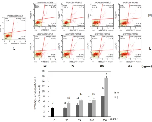
Fig. 2. Effects of Erythronium japonicum on the apoptosis of MDA-MB-231 cells. C: Control; M: Methanol extract of Erythronium japonicum; E: Ethanol extract of Erythronium japonicum. Data are mean ± SD. Means with the same letter are not significantly different by Duncan’s multiple range test (P< 0.05).
Fig. 3. Effects of Erythronium japonicum on the adhesion of MDA-MB-231 cells.
C: Control; M: Methanol extract of Erythronium japonicum; E: Ethanol extract of Erythronium japonicum. Data are mean ± SD. Means with the same letter are not significantly different by Duncan’s multiple range test (P< 0.05).
based on a one-way ANOVA, followed by Duncan's multiple range test. A P-value < 0.05 was considered to be statistically significant.
RESULTS
Effect of Erythronium japonicum on the proliferation and apoptosis of breast cancer cells
Erythronium japonicum extracts inhibited the growth of MDA-MB-231 breast cancer cells. Ethanol extract was more potently inhibited the cell growth than methanol extract (Fig.
1). Extracts at all concentrations, which we treated, inhibited cell growth and showed no cytotoxicity. Therefore, in this study, we treated methanol and ethanol extracts at the concentrations of 50, 75, 100 and 250 μg/ml for further investigation. Erythronium
japonicum extracts induced the apoptosis of MDA-MB-231 cells (Fig. 2). Methanol extract significantly induced apoptosis of tumor cells at the concentration of 250 μg/ml, whereas, ethanol extract induced apoptosis at the lower concentration, 75 μg/ml.
Effect of Erythronium japonicum on the adhesion of breast cancer cells
The attachment of MDA-MB-231 cells to the extracellular matrix (ECM) was determined using Matrigel binding assay.
Erythronium japonicum extracts repressed the attachment of
MDA-MB-231 cells to the ECM (Fig. 3). At a concentration of
250 μg/ml, both methanol and ethanol extracts reduced by 21.3
and 32.5%, respectively, compared to the control.
Fig. 4. Effects of Erythronium japonicum on the invasion of MDA-MB-231 cells.
C: Control; M: Methanol extract of Erythronium japonicum; E: Ethanol extract of Erythronium japonicum. Data are mean ± SD. Means with the same letter are not significantly different by Duncan’s multiple range test (P< 0.05).
(μg/mL)
Fig. 5. Effects of Erythronium japonicum on the migration of MDA-MB-231 cells.
C: Control; M: Methanol extract of Erythronium japonicum; E: Ethanol extract of Erythronium japonicum. 0 h & 24 h: hours after making wound
Fig. 6. Effects of Erythronium japonicum on the activities of MMP-2 and MMP-9 of MDA-MB-231 cells. C: Control; M: Methanol extract of Erythronium japonicum;E:
Ethanol extract of Erythronium japonicum
Effect of Erythronium japonicum on the invasion and migration of breast cancer cells
The invasion of MDA-MB-231 was determined using a Boyden chamber model; both methanol and ethanol extracts dose dependently suppressed the invasion of MDA-MB-231cells (Fig.
4). Furthermore, Erythronium japonicum ethanol extract inhibited the invasion of breast cancer cells more potently than the methanol extract. We also analyzed the effect of Erythronium japonicum on the migration capability of MDA-MB-231 cells by a wound healing assay. The MDA-MB-231 cell line was able to close the wound completely within 24 h and moreover, Erythronium japonicum extracts reduced the rate of the wound closure (Fig. 5).
Effect of Erythronium japonicum on MMP-2 and MMP-9 enzyme activities
The effect of Erythronium japonicum extract on the gelatinolytic MMP-2 and MMP-9 activities was analyzed in MDA-MB-231 breast cancer cells; both methanol and ethanol extracts had an inhibitory effect on the MMP activities (Fig. 6).
DISCUSSION
Malignant tumors are characterized by excessive growth, immortalization and metastatic spread [6]. We observed that Erythronium japonicum extracts suppressed the excessive growth of MDA-MB-231 breast cancer cells without cytotoxicity;
further, ethanol extract was more potent than methanol extract regarding the inhibition of cell growth (Fig. 1). Because progressive growth of tumor cells, and thereby expanding tumor mass, are crucial for the invasion of surrounding tissues [2], Erythronium japonicum extracts might exert to suppress cancer progression by inhibiting tumor cell growth. The research revealed that Erythronium japonicum contained anthocyanin in the petals [16]
and flavonoids, such as di-O-n-decyphthalate, kaempferol, quercetin, astragalin and isoquercitrin, in the leaves [17]. We determined caffeic acid using an HPLC analysis of ethanol extract (data not shown) and this phenolic compound might contribute to the inhibition of tumor cell growth. Caffeic acid and polymeric caffeic acids have been shown to suppress the proliferation of various types of cancer cells and induce apoptotic pathways [18-21]. We also observed the proapoptotic effect of Erythronium japonicum extracts (Fig. 2). Many physiolo- gical growth-regulate mechanisms that control cell proliferation and tissue homeostasis are linked to apoptosis [4]. Therefore, sensitizing tumor cells to apoptosis is one of the strategies of tumor therapy. Deregulated cell proliferation and suppressed cell death together provide the underlying basis for cancer progression [3]. Our results revealed that Erythronium japonicum extracts might suppress the progression of breast cancer by inhibiting proliferation and inducing apoptosis of tumor cells.
The attachment of tumor cells to ECM is the initial invasive action of metastatic cells, which is necessary for cell motility and invasion [22,23]. Erythronium japonicum extracts suppressed the attachment of MDA-MB-231 cells to the ECM (Fig. 3). The next step of metastasis involves local invasion and migration through stromal tissues by degrading the ECM [5,6]. In the present study, both methanol and ethanol extracts dose depen- dently suppressed the invasion of MDA-MB-231cells (Fig. 4).
Furthermore, Erythronium japonicum ethanol extract more potently inhibited the invasion of breast cancer cells than the methanol extract. Cell migration was also inhibited by Erythronium japonicum extracts (Fig. 5). Cell motility is crucial for cancer cell invasion and metastasis [24,25]. A previous study reported that antiproliferative and proapoptotic properties can contribute to the inhibitory effect on migration and invasion [26]. In the present study, Erythronium japonicum extracts demonstrated antiproliferative and proapoptotic properties. In fact, the ethanol extract of Erythronium japonicum showed more antiproliferative and proapoptotic properties as well as also more effectively inhibited the invasion of MDA-MB-231 cells.
Matrix metalloproteinases (MMPs) produced by the adhered
tumor cells are crucial for degrading and modifying the ECM component [27,28]. MMPs have been known to promote tumor cellular invasion in vitro and in vivo [27]. We analyzed the effect of Erythronium japonicum extract on the MMP activities; both methanol and ethanol extracts inhibited MMP activities (Fig. 6).
The previous researches demonstrated that MMP-2 and -9 (gelatinase A and B) are connected to tumor growth, invasion and metastasis by their capacities for tissue remodeling and angiogenesis induction [19,27,28]. Contrary to our expectation, the difference of MMP activity between ethanol and methanol extracts were not very obvious, even though the ethanol extract more potently inhibited tumor cell growth, adhesion and invasion compared to the methanol extract. Therefore, other factors, which can affect cell motility, need to be investigated in order to account for the discordance between the effect of Erythronium japonicum extracts on cell proliferation to apoptosis ratio, invasion and MMP activities.
Our study clearly identified that Erythronium japonicum extracts inhibited MDA-MB-231 cell metastasis by altering the proliferation to the apoptosis ratio and the suppression of adhesion, migration and invasion; further, the ethanol extract was much potent than the methanol extract. Further study would be necessary to determine the possible mechanisms of Erythronium japonicum.
REFERENCES
1. Yokota J. Tumor progression and metastasis. Carcinogenesis 2000;21:497-503.
2. Fidler IJ. The pathogenesis of cancer metastasis: the 'seed and soil' hypothesis revisited. Nat Rev Cancer 2003;3:453-8.
3. Evan GI, Vousden KH. Proliferation, cell cycle and apoptosis in cancer. Nature 2001;411:342-8.
4. Igney FH, Krammer PH. Death and anti-death: tumour resistance to apoptosis. Nat Rev Cancer 2002;2:277-88.
5. Song G, Ohashi T, Sakamoto N, Sato M. Adhesive force of human hepatoma HepG2 cells to endothelial cells and expression of E-selectin. Mol Cell Biomech 2006;3:61-8.
6. Weber GF. Molecular mechanisms of metastasis. Cancer Lett 2008;270:181-90.
7. Festuccia C, Guerra F, D'Ascenzo S, Giunciuglio D, Albini A, Bologna M. In vitro regulation of pericellular proteolysis in prostatic tumor cells treated with bombesin. Int J Cancer 1998;75:418-31.
8. Festuccia C, Bologna M, Gravina GL, Guerra F, Angelucci A, Villanova I, Millimaggi D, Teti A. Osteoblast conditioned media contain TGF-beta1 and modulate the migration of prostate tumor cells and their interactions with extracellular matrix components. Int J Cancer 1999;81:395-403.
9. Kondo T, Okubo N, Miura T, Honda K, Ishikawa Y. Ecophysiology of seed germination in Erythronium japonicum (Liliaceae) with underdeveloped embryos. Am J Bot 2002;89:1779-84.
10. Shin YJ, Jung DY, Ha HK, Park SW. Anticancer effect of Erythronium japonicum extract on ICR mouse and L1210 cells with alteration of antioxidant enzyme activities. Korean J Food Sci Technol 2004;36:968-73.
11. Ritsema T, Smeekens S. Fructans: beneficial for plants and humans.
Curr Opin Plant Biol 2003;6:223-30.
12. Virtanen SS, Väänänen HK, Härkönen PL, Lakkakorpi PT. Alendronate
inhibits invasion of PC-3 prostate cancer cells by affecting the mevalonate pathway. Cancer Res 2002;62:2708-14.
13. Chen PN, Hsieh YS, Chiang CL, Chiou HL, Yang SF, Chu SC. Silibinin inhibits invasion of oral cancer cells by suppressing the MAPK pathway. J Dent Res 2006;85:220-5.
14. Goodman SL, Vollmers HP, Birchmeier W. Control of cell locomotion:
perturbation with an antibody directed against specific glycoproteins.
Cell 1985;41:1029-38.
15. Albini A, D'Agostini F, Giunciuglio D, Paglieri I, Balansky R, De Flora S. Inhibition of invasion, gelatinase activity, tumor take and metastasis of malignant cells by N-acetylcysteine. Int J Cancer 1995;61:121-9.
16. Lee MS, Lim SC, Park HJ. The anthocyanin contents of the petals of Erythronium japonicum collected from several stocks. Korean J Pharmacogn 1993;24:251-4.
17. Lee MS, Lim SC, Park HJ. Phthalate ester and flavonoids isolated from leaves of Erythronium japonicum. Korean J Med Crop Sci 1994;2:67-72.
18. Chang WC, Hsieh CH, Hsiao MW, Lin WC, Hung YC, Ye JC. Caffeic acid induces apoptosis in human cervical cancer cells through the mitochondrial pathway. Taiwan J Obstet Gynecol 2010;49:419-24.
19. Hwang HJ, Park HJ, Chung HJ, Min HY, Park EJ, Hong JY, Lee SK.
Inhibitory effects of caffeic acid phenethyl ester on cancer cell metastasis mediated by the down-regulation of matrix metallopro- teinase expression in human HT1080 fibrosarcoma cells. J Nutr Biochem 2006;17:356-62.
20. Sanderson JT, Clabault H, Patton C, Lassalle-Claux G, Jean-François J, Paré AF, Hébert MJ, Surette ME, Touaibia M. Antiproliferative, antiandrogenic and cytotoxic effects of novel caffeic acid deriva- tives in LNCaP human androgen-dependent prostate cancer cells.
Bioorg Med Chem 2013;21:7182-93.
21. Yamanaka D, Tajima K, Adachi Y, Ishibashi KI, Miura NN, Motoi M, Ohno N. Effect of polymeric caffeic acid on antitumour activity and natural killer cell activity in mice. J Funct Foods 2014;6:513-22.
22. Hazan RB, Phillips GR, Qiao RF, Norton L, Aaronson SA. Exogenous expression of N-cadherin in breast cancer cells induces cell migration, invasion, and metastasis. J Cell Biol 2000;148:779-90.
23. Zheng DQ, Woodard AS, Fornaro M, Tallini G, Languino LR. Prostatic carcinoma cell migration via alpha(v)beta3 integrin is modulated by a focal adhesion kinase pathway. Cancer Res 1999;59:1655-64.
24. Hwang SL, Kim HN, Jung HH, Kim JE, Choi DK, Hur JM, Lee JY, Song H, Song KS, Huh TL. Beneficial effects of β-sitosterol on glucose and lipid metabolism in L6 myotube cells are mediated by AMP-activated protein kinase. Biochem Biophys Res Commun 2008;377:1253-8.
25. Kawasaki G, Yanamoto S, Yoshitomi I, Yamada S, Mizuno A.
Overexpression of metastasis-associated MTA1 in oral squamous cell carcinomas: correlation with metastasis and invasion. Int J Oral Maxillofac Surg 2008;37:1039-46.
26. Azios NG, Dharmawardhane SF. Resveratrol and estradiol exert disparate effects on cell migration, cell surface actin structures, and focal adhesion assembly in MDA-MB-231 human breast cancer cells.
Neoplasia 2005;7:128-40.
27. Johansson N, Ahonen M, Kähäri VM. Matrix metalloproteinases in tumor invasion. Cell Mol Life Sci 2000;57:5-15.
28. Radisky ES, Radisky DC. Matrix metalloproteinase-induced epithelial- mesenchymal transition in breast cancer. J Mammary Gland Biol Neoplasia 2010;15:201-12.