• 검색 결과가 없습니다.

Anti-cancer Effect of Apigenin on Human Breast Carcinoma MDA-MB-231 through Cell Cycle Arrest and Apoptosis

N/A
N/A
Protected

Academic year: 2021

Share "Anti-cancer Effect of Apigenin on Human Breast Carcinoma MDA-MB-231 through Cell Cycle Arrest and Apoptosis"

Copied!
9
0
0

로드 중.... (전체 텍스트 보기)

전체 글

(1)

Anti-cancer Effect of Apigenin on Human Breast

Carcinoma MDA-MB-231 through Cell Cycle Arrest and Apoptosis

Hwan Hee Lee

1,2

and Hyosun Cho

1,2

*

1

College of Pharmacy, Duksung Women’s University, Seoul 01369, Republic of Korea

2

Duksung Innovative Drug Center, Duksung Women’s University, Seoul 01369, Republic of Korea

Received: September 19, 2018 / Revised: October 9, 2018 / Accepted: October 10, 2018

Introduction

Breast cancer is the most common cancer among woman worldwide with a high mortality rate. Breast cancers are classified according to different schemes cri- teria and the major categories include the histopatholog- ical type, the grade, the stage, and the various receptor expressions of cancer. The three major receptors are estrogen receptor (ER), progesterone receptor (PR), and human epidermal growth factor receptor (HER2/neu).

Breast cancer with or without these receptors are called ER positive (ER+), ER negative (ER-), PR positive (PR+), PR negative (PR-), HER2 positive (HER2+), and HER2

negative (HER2-) and breast cancer with none of these receptors is called triple negative (TN). The status of these receptors is reported to highly associate with dis- ease progress and clinical outcomes under certain treat- ments [1, 2]. Triple-negative breast cancer (TNBC) lacking ER, PR, and Her2/neu is known to be about 15 − 20% of all breast cancer [3]. TNBC is a very aggressive tumor with higher histological grade and is more likely to migrate to distant sites, resulting in poor prognosis [4].

Apigenin (4 ,5,7-trugtdrixyflavone) is a natural flavo- noid found in many plants including vegetables, fruits and Chinese herb. The biological activity of apigenin includes antioxidant, anti-inflammatory [5, 6], antibac- terial, antiviral and anti-cancer effects [7 −9]. Birt et al.

reported the tumor inhibition-related effect of apigenin in 1986 for the first time [10]. Apigenin potently inhib- Apigenin, a common natural product that is found in many plants and vegetables, has been reported to have many biological activities, including antioxidative, anti-inflammatory, and anticancer effects. The tri- ple-negative breast carcinoma cell line MDA-MB-231 is known to be highly invasive and resistant to chemo- therapy. In this study, we investigated the anticancer effect of apigenin on human MDA-MB-231 cells. First, the cytotoxicity of apigenin toward MDA-MB-231 cells was analyzed by MTT assay. Then, the cell cycle and apoptotic effects of apigenin were examined, and the molecular mechanism underlying its anticancer activity was explored. Apigenin inhibited the growth of the cells in a dose-dependent manner, correlating with the cell cycle arrest at the G2-M phase as well as an increase of early apoptosis. The cell-cycle inhibi- tory effect was highly associated with the increased expression of p21 and decreased expression of CDK6, cyclin D1, and cyclin B1. The induction of apoptosis by apigenin was associated with the upregulated expression of cleaved PARP and cleaved caspase-3, -7, and -9.

Keywords: Apigenin, human breast carcinoma, MDA-MB-231, cell cycle arrest, apoptosis

*Corresponding author

Tel: +82-2-901-8678, Fax: +82-2-901-8386 E-mail: [email protected]

© 2019, The Korean Society for Microbiology and Biotechnology

(2)

ited the epidermal ornithine decarboxylase induction, which highly connected with skin tumor promotion in mice [10]. Since then, anti-tumor activity of apigenin on a variety of cancers such as colorectal cancer, liver can- cer, lung cancer, prostate cancer, breast cancer and osteosarcoma has been observed [11 −14]. The molecular mechanism of anti-cancer activity of apigenin was found to be related with an inhibition of cell proliferation by arresting cell cycle or induction of cell apoptosis [13, 15].

Also, apigenin seemed to inhibit cell motility and migra- tion [10, 11]. Recently, apigenin showed a strong anti- cancer activity through modulating immune responses against cancer. Cardenas reported that apigenin sup- pressed leukocyte infiltration by reducing the activation of NF-kB [16]. And, Nelson group showed that apigenin recovered T cell homeostasis and function by increasing the expression of Ikaros in murine pancreatic cancer [17].

The proliferation of cancer cell is known to be regu- lated by many complicated molecular processes includ- ing cell cycle and cell apoptosis. p53 is a representative tumor suppressive protein that plays a critical role in cell apoptosis [18]. And p53 is also one of cell cycle con- trol related proteins along with p21, which is a potent cyclin-dependent kinase inhibitor. The p21 protein is known to directly bind to cyclin-CDKs complexes and inhibits the activity of CDKs [19]. Cell apoptosis associ- ates with not only p53 protein but also caspases and poly ADP-ribose polymerase (Parp) [20]. Caspases are essen- tial proteases that are activated in the process of cell apoptosis. They can be categorized as initiators (caspase -2, -8, -9 and -10) or executioner (caspase -3, -6 and -7) and involves two different apoptotic pathways, the extrinsic and intrinsic pathways [21].

A few studies reported the anti-cancer activities of api- genin on human breast carcinoma MDA-MB-231 [22, 23]. Vrhovac group found that apigenin induced apopto- sis, DNA damage and oxidative stress in MDA-MB-231 [22]. Tseng group presented that apigenin inhibited cell proliferation of MDA-MB-231 through up-regulation of p21, which resulted in cell cycle arrest at G2/M phase [23]. However, the further molecular basis of the anti- cancer effect by apigenin still remains to be clarified. In this study, we investigated the cellular anti-cancer mechanisms of apigenin underlying the induction of apoptosis and cell cycle arrest in MDA-MB-231.

Materials and Methods

Specimen preparation

Apigenin (≥ 95% pure) was purchased from Sigma- Aldrich, Inc. (USA). Apigenin was dissolved using dimethyl sulfoxide (DMSO; Sigma-Aldrich, USA) and diluted with medium for human breast carcinoma MDA- MB-231.

Cell culture

Human breast carcinoma MDA-MB-231 was obtained from American Type Cell Culture (ATCC-HTB26).

MDA-MB-231 was cultured in RPMI1640 (Corning, USA) supplemented with 10% heat-inactivated fetal bovine serum (FBS; Yong-In Frontier, Korea), 100 U/ml penicillin and streptomycin (Gibco, USA) at 37 ℃ in a humidified atmosphere with 5% CO2.

MTT assay

Cell cytotoxicity of apigenin on MDA-MB-231 was assessed using thiazolyl blue tetrazolium bromide (MTT;

Sigma-Aldrich, USA). Cells were seeded at a density of 1 × 10

4

cells per each well in a 96-well flat bottom plate and serially diluted apigenin (0, 10, 20, 30, 40, 50, 60, 70, 80, 90, or 100 μM) was added for 24, 48 or 72 h. Then, MTT solution was added to each well, and the cells incu- bated in accordance with the reactive time of the solu- tion. The absorbance was measured using a microplate reader (BMG Labtech, Ortenberg, Germany) at 570 nm.

Cell cycle analysis

Cell cycle was assessed using propidium iodide (Invit- rogen, USA) staining assay. MDA-MB-231 was plated with a density of 5 × 10

5

per each well of 6-well bottom plate overnight and treated with different concentra- tions of apigenin (0, 10, 20 and 40 μM) for 24 h. After the treatment, cells were fixed 70% ice-ethanol and stained with propidium iodide (PI) for 15 min at room tempera- ture and then analyzed by flow cytometry (Novocyte

®

Flow Cytometer, ACEA Biosciences, USA).

Cell apoptosis analysis

Cell apoptosis was analyzed by FITC Annexin V Apop-

tosis Detection Kit I (BD Biosciences, USA). Briefly,

MDA-MB-231 was plated in 6-well bottom plate and

treated with different concentrations of apigenin (0, 10,

(3)

20 and 40 μM) for 24 h. And then, cells were stained with FITC Annexin-V and propidium iodide for 30 min at room temperature. Cell apoptosis was measured by flow cytometry (Novocyte

®

Flow Cytometer, ACEA Bio- sciences).

Western blot analysis

MDA-MB-231 was plated with a density of 5 × 10

5

per each well in 6-well bottom plate and treated with differ- ent concentrations of apigenin (0, 10, 20 and 40 μM) for 24 h. Cells were lysed by protein extraction buffer (Intron Biotechnology, Korea). Proteins in cell lysates were quantified by the Bradford assay, separated by electrophoresis, and transferred to nitrocellulose mem- branes, which was then blotted with 1

st

and 2

nd

antibod- ies. Anti-PARP (#9542) (and anti-cleaved PARP, #5625), anti-Caspase 3 (#9665) (and anti-cleaved caspase 3,

#9664), anti-Caspase 7 (#12827) (and anti-cleaved caspase 7, #8438) and anti-Caspase 9 (#9508) (and anti- cleaved caspase 9, #7237) antibodies and Cell cycle regu- lation sampler kit (#9932) were obtained from Cell Sig- naling Technology (USA). Anti- β-actin (#A5441) and anti-p53 (#05-224) antibodies were purchased from

sigma Aldrich (USA) and Millipore (USA), respectively.

The blots were visualized by enhanced chemilumines- cent (ECL) detection solution (Intron Biotechnology, Korea).

Statistical analysis

Data were processed using Microsoft Excel and results are presented as means ± SDs. Comparisons of several means were performed by one-way or two-way analysis of variance followed by Fisher’s exact test to identify sig- nificant differences between groups with p-values of less than 0.05 being considered significant.

Results and Discussion

Cytotoxic effect of apigenin on human breast carcinoma MDA-MB-231

To identify the cell cytotoxic effect of apigenin on human breast carcinoma MDA-MB-231, cells were treated with serially diluted concentrations (0, 10, 20, 30, 40, 50, 60, 70, 80, 90, and 100 μM) of apigenin for 24, 48 and 72 h. Cytotoxicity was determined by MTT assay.

Apigenin showed a great cytotoxicity on MDA-MB-231

Fig. 1. Cytotoxic effect of apigenin on human breast carcinoma MDA-MB-231. MDA-MB-231 cells were seeded in 96-well bottom

plate with a density of 1 × 10

4

cells per well and incubated with a serially diluted apigenin (0 to 100 µM) for 24, 48, and 72 h. And

then, the cell cytotoxicity was measured by MTT assay. (A) Cell viability (%) and (B) morphological changes.

(4)

at 48 or 72 h (IC

50

= 54 μM at 72 h) in a dose dependent manner (Fig. 1A). Fig. 1B shows the morphological changes of MDA-MB-231 under 24 h of treatment with apigenin (0, 10, 20 and 40 μM). As the concentrations of apigenin increased, the proliferation of cells seems to be

inhibited even though the IC

50

value at 24 h was not determined within the range of apigenin concentrations used in this experiment (Figs. 1A and 1B). In fact, the IC

50

value of apigenin on MDA-MB-231 at 72 h is very close to the IC

50

value (54.64 μM at 72 h) reported by

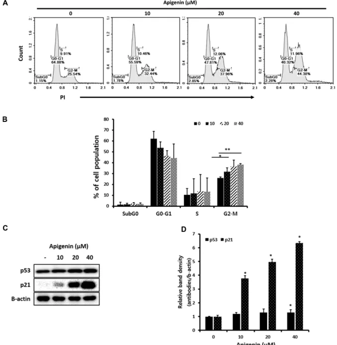
Fig. 2. Effect of apigenin on cell cycle regulation, p53 and p21 in MDA-MB-231. MDA-MB-231 cells were plated in 6-well bottom

plate with a density of 5 × 10

5

cells per well and cultured with or without apigenin (0, 10, 20, and 40 µM) for 24 h. For cell cycle

assay, cells were stained with propidium iodide (PI), and analyzed using flow cytometry. And, cells were also lysed, and western blot

analysis for proteins expression of p53 and p21 is performed. (A) Representative plots of cell cycle progress in MDA-MB-231, (B) the

percentage of individual cell cycle distribution from three independent experiments, (C) Expression of p53 and p21 proteins, and

(D) relative band intensity of p53 and p21 compared to the loading control.

(5)

Vrhovac group [22]. Both studies indicates that apigenin significantly suppresses the cell proliferation of MDA- MB-231 with dose-dependent and time-dependent manner.

Effect of apigenin on cell cycle regulation, p53 and p21 in MDA-MB-231

To investigate whether apigenin affects the cell cycle of MDA-MB-231, cells were treated with or without three different concentrations of apigenin (0, 10, 20 and 40 μM). After 24 h, cells were stained with propidium iodide (PI) and analyzed using flow cytometry. As shown in Fig. 2A and 2B, apigenin considerably increased the cell cycle arrest at G2/M phase in a dose-dependent manner (untreated, 25.74%; 10 μM, 31.63%; 20 μM, 36.48%; 40 μM, 38.19%). Apigenin also decreased the G1/S transition although it was not statistically signifi- cant (Fig. 2B). Recently, Tseng group also reported that apigenin induced G2/M phase arrest in MDA-MB-231 through upregulation of histone H3 acetylation-medi- ated p21 expression, which agrees with our result to a certain extent [23]. Furthermore, we examined if api- genin affected the expression of p53 or p21 proteins because they are known as cell cycle control related pro- teins as mentioned earlier.

Fig. 2C and 2D shows that the expression of p21 pro- tein is significantly upregulated as the concentration of apigenin increases (Figs. 2C and 2D). Also, the expres- sion of p53 protein seems to be induced by 40 uM of api-

genin treatment, which suggests that apigenin induces cell cycle arrest at G2/M phase in p53-dependent manner.

Effect of apigenin on the expression of CDKs (CDK1,2,4) and Cyclins (Cyclin B1, D1) in MDA-MB-231

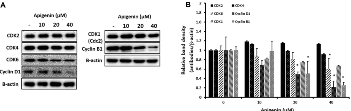
Since apigenin dramatically stimulated the expression of p21 (Figs. 2C and 2D), we determined to examine other cell cycle regulation proteins such as CDK1,2,4,6, Cyclin B1 and D1, which associate with p21. As shown in Fig. 3A and 3B, the expressions of CDK6, Cyclin D1 and Cyclin B1 proteins are decreased in apigenin treated MDA-MB-231 compared with that from untreated control. Of note, the expressions of Cyclin D1 and Cyclin B1 was almost blocked by 40 μM of apigenin treatment (Figs. 3A and 3B). Interestingly, Cyclin D1 and CDK6 proteins are reported to be overexpressed in several cancers including breast cancer, colorectal can- cer, oral squamous carcinoma and brain cancer [24 −27].

In detail, Cyclin D1, as a key regulator in cell cycle con- trol, stimulates G1 progression, leading an increase of cell proliferation. Thus, overexpression of cyclin D1 could induce cell proliferation, which results in the development of cancer [24]. CDK6 is also involved in promoting G1 progression and G1/S transition [27].

Cyclin B1 is linked with inducing G2 progression and G2/M transition [28]. And, overexpression of Cyclin B1 has been reported in pancreatic cancer and colorectal cancer [29, 30]. Therefore, our study suggests that api- genin may target both G1/S and G2/M check points by

Fig. 3. Effect of apigenin on the expression of CDKs (CDK1,2,4) and Cyclins (Cyclin B1, D1) in MDA-MB-231. MDA-MB-231 cells

were plated in 6-well bottom plate with a density of 5 × 10

5

cells per well and cultured with or without apigenin (0, 10, 20, and

40 µM) for 24 h, and western blot analysis for proteins expression of CDKs and Cyclins is performed. (A) Expression of CDK2, CDK4,

CDK6, cyclinD1, CDK1 and cyclinB1 proteins, and (B) Relative band intensity of CDK2, CDK4, CDK6, cyclinD1, CDK1 and cyclinB1 pro-

teins compared to the loading control.

(6)

inhibiting the expressions of Cyclin D1 and CDK6 for G1/S phase arrest and the expression of Cyclin B1 for G2/M phase arrest in MDA-MB-231.

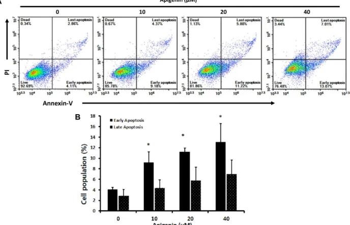
Effect of apigenin on cell apoptosis in MDA-MB-231 To investigate whether apigenin affects cell apoptosis in MDA-MB-231, cells were treated with three different concentrations of apigenin (0, 10, 20 and 40 μM) for 24 h and stained with FITC annexin-V and propidium iodide (PI) for apoptotic cells or dead cells, respectively. Fig. 4A and 4B shows that both early and late apoptosis seem to be increased in a treatment with apigenin. Especially, the early apoptosis is significantly enhanced by apigenin treatment in a dose dependent manner compared with that from untreated control (untreated, 4.11%; 10 μM, 9.18%; 20 μM, 11.22%; 40 μM, 13.07%). Of note, 40 μM of apigenin generated up to 20% of apoptosis totally (Fig.

4). In fact, many recent studies reported that apigenin- related flavonoids strongly stimulated cell apoptosis in

several cancer cells including various human breast car- cinomas, hepatocellular carcinoma and colon cancer [31 − 33]. Chen group demonstrated that apigenin induced cell apoptosis by inhibiting proteasome activity in MDA- MB-231 and the molecular mechanism of apoptosis was associated with activation of caspase-3 and caspase-7 [34]. Ding found that upregulation of TRAIL pathway is closely connected to the induction of apoptosis by api- genin in MDA-MB-231 [35].

Effect of apigenin on the expression of (cleaved) parp and caspase-3, 7, 9 in MDA-MB-231

Since apigenin stimulated cell apoptosis of MDA-MB- 231 dramatically with dose dependency (Figs. 4A and 4B). We further analyzed how apoptosis-related cellular molecules are affected by apigenin in MDA-MB-231. We measured the expressions of parp, caspase-3, -7 and -9 as well as the individual cleaved forms because the increased expression of cleaved forms of parp or

Fig. 4. Effect of apigenin on cell apoptosis in MDA-MB-231. MDA-MB-231 cells were plated in 6-well bottom plate with a density

of 5 × 10

5

cells per well and cultured with or without apigenin (0, 10, 20, and 40 µM) for 24 h. For apoptosis assay, cells were stained

with FITC annexin-V and propidum iodide (PI), and then assessed by flow cytometry. (A) Representative plots of stained cell for apop-

tosis in MDA-MB-231, and (B) the percentage of apoptotic cell (early and late apoptotic) from three independent experiments.

(7)

caspases directly reflects the activation of cell apoptosis.

Fig. 5A and 5B shows that there is a clear decrease in the expression of parp, caspase-3 and -9 whereas an increase in the expression of cleaved forms of parp, caspase-3, -7 and -9 by apigenin treatment in MDA-MB- 231, which indicates the significant cleavage of parp and caspase-3,-7, and -9. Of note, we observed a strong dose

dependency of apigenin in the expression of parp, caspase-3, and -9 (Fig. 5A and 5B). Cao group reported that 40 μM or 80 μM of apigenin induced a considerable cleavage of caspase-3 and parp in MDA-MB-231 [36].

And Chen et al. presented that dose dependent increase of caspase-3/caspase-7 activity by apigenin is positively linked with cell apoptosis of MDA-MB-231 [34]. Both Fig. 5. Effect of apigenin on the expression of (cleaved) parp and cleaved caspase-3,-7,-9 in MDA-MB-231. MDA-MB-231 cells were plated in 6-well bottom plate with a density of 5 × 10

5

cells per well and cultured with or without apigenin (0, 10, 20, and 40 µM) for 24 h, and western blot analysis for proteins expression of (cleaved) parp and (cleaved) caspase-3,-7, and -9 is performed.

(A) Expression of (cleaved) parp and (cleaved) caspase-3,-7, and -9 proteins, and (B) Relative band intensity of (cleaved) parp and (cleaved) caspase-3,-7, and -9 proteins compared to the loading control.

Fig. 6. Anti-cancer mechanisms of apigenin in MDA-MB-231.

(8)

studies agree with our study and we additionally reported for the first time that caspase-9 is also involved in cell apoptosis of MDA-MB-231.

In this study, we investigated the cell cycle regulation and apoptosis related cellular mechanisms underlying the anti-cancer effect of apigenin in human breast carci- noma MDA-MB-231. Collectively, our data indicates that apigenin has a potent cytotoxic effect on human breast carcinoma MDA-MB-231through cell cycle arrest at G2/M phase and induction of apoptosis (Fig. 6). In case of cell cycle regulation, apigenin specifically inhibits Cyclin B1, Cyclin D1 and CDK6 proteins along with upregulation of p53 and p21. Apigenin also induces a strong cell apoptosis through cleavege of parp, caspase- 3, -7 and -9 in MDA-MB-231. Our study provides a com- prehensive understanding of anti-cancer mechanism of apigenin in human breast carcinoma cells, which sug- gests that apigenin could be a good food supplement for a prevention and treatment against human breast cancer.

Acknowledgments

This research was supported by Duksung Women’s University Research Grants 2018-3000003028.

Conflict of Interest

The authors have no financial conflicts of interest to declare.

References

1. Sorlie T, Perou CM, Fan C, Geisler S, Aas T, Nobel A, et al. 2006.

Gene expression profiles do not consistently predict the clinical treatment response in locally advanced breast cancer. Mol. Cancer Ther. 5: 2914-2918.

2. van de Vijver MJ, He YD, van't Veer LJ, Dai H, Hart AA, Voskuil DW, et al. 2009. A gene-expression signature as a predictor of survival in breast cancer. N. Engl. J. Med. 347: 1999-2009.

3. Hudis CA, Gianni L. 2011. Triple-negative breast cancer: an unmet medical need. Oncologist. 16 Suppl 1: 1-11.

4. Dent R, Trudeau M, Pritchard KI, Hanna WM, Kahn HK, Sawka CA, et al. 2007. Triple-negative breast cancer: clinical features and patterns of recurrence. Clin. Cancer Res. 13: 4429-4434.

5. Papay ZE, Kosa A, Boddi B, Merchant Z, Saleem IY, Zariwala MG, et al. 2017. Study on the pulmonary delivery system of apigenin- loaded albumin nanocarriers with antioxidant activity. J. Aerosol.

Med. Pulm. Drug Deliv. 30: 274-288.

6. Wang YC, Huang KM. 2013. In vitro anti-inflammatory effect of apigenin in the Helicobacter pylori-infected gastric adenocarci-

noma cells. Food Chem. Toxicol. 53: 376-383.

7. Yan X, Qi M, Li P, Zhan Y, Shao H. 2017. Apigenin in cancer ther- apy: anti-cancer effects and mechanisms of action. Cell Biosci. 7:

50.

8. Ozcelik B, Kartal M, Orhan I. 2011. Cytotoxicity, antiviral and anti- microbial activities of alkaloids, flavonoids, and phenolic acids.

Pharm. Biol. 49: 396-402.

9. Zhang W, Qiao H, Lv Y, Wang J, Chen X, Hou Y, et al. 2014. Api- genin inhibits enterovirus-71 infection by disrupting viral RNA association with trans-acting factors. PLoS One. 9: e110429.

10. Birt DF, Walker B, Tibbels MG, Bresnick E. 1986. Anti-mutagenesis and anti-promotion by apigenin, robinetin and indole-3-carbinol.

Carcinogenesis 7: 959-963.

11. Huang C, Wei YX, Shen MC, Tu YH, Wang CC, Huang HC. 2016.

Chrysin, abundant in morinda citrifolia fruit water-EtOAc extracts, combined with apigenin synergistically induced apop- tosis and inhibited migration in human breast and liver cancer cells. J. Agric. Food Chem. 64: 4235-4245.

12. Lee YM, Lee G, Oh TI, Kim BM, Shim DW, Lee KH, et al. 2016. Inhi- bition of glutamine utilization sensitizes lung cancer cells to api- genin-induced apoptosis resulting from metabolic and oxidative stress. Int. J. Oncol. 48: 399-408.

13. Gupta S, Afaq F, Mukhtar H. 2002. Involvement of nuclear factor- kappa B, Bax and Bcl-2 in induction of cell cycle arrest and apop- tosis by apigenin in human prostate carcinoma cells. Oncogene 21: 3727-3738.

14. Angulo P, Kaushik G, Subramaniam D, Dandawate P, Neville K, Chastain K, et al. 2017. Natural compounds targeting major cell signaling pathways: a novel paradigm for osteosarcoma therapy.

J. Hematol. Oncol. 10: 10.

15. Zhao G, Han X, Cheng W, Ni J, Zhang Y, Lin J, et al. 2017. Apigenin inhibits proliferation and invasion, and induces apoptosis and cell cycle arrest in human melanoma cells. Oncol. Rep. 37: 2277- 2285.

16. Cardenas H, Arango D, Nicholas C, Duarte S, Nuovo GJ, He W, et al. 2016. Dietary apigenin exerts immune-regulatory activity in vivo by reducing NF- κB activity, halting leukocyte infiltration and restoring normal metabolic function. Int. J. Mol. Sci. 17: 323.

17. Nelson N, Szekeres K, Iclozan C, Rivera IO, McGill A, Johnson G, et al. 2017. Apigenin: selective CK2 inhibitor increases Ikaros expression and improves T cell homeostasis and function in murine pancreatic cancer. PLoS One. 12: e0170197.

18. Lakin ND, Jackson SP. 1999. Regulation of p53 in response to DNA damage. Oncogene 18: 7644-7655.

19. Ballestar E, Esteller M. 2008. Epigenetic gene regulation in cancer.

Adv. Genet. 61: 247-267.

20. Koh DW, Dawson TM, Dawson VL. 2005. Mediation of cell death by poly(ADP-ribose) polymerase-1. Pharmacol. Res. 52: 5-14.

21. Matt S, Hofmann TG. 2016. The DNA damage-induced cell death response: a roadmap to kill cancer cells. Cell Mol. Life Sci. 73:

2829-2850.

22. Vrhovac Madunic I, Madunic J, Antunovic M, Paradzik M, Garaj-

Vrhovac V, Breljak D, et al. 2018. Apigenin, a dietary flavonoid,

(9)

induces apoptosis, DNA damage, and oxidative stress in human breast cancer MCF-7 and MDA MB-231 cells. Naunyn. Schmiede- bergs Arch. Pharmacol. 391: 537-550.

23. Tseng TH, Chien MH, Lin WL, Wen YC, Chow JM, Chen CK, et al.

2017. Inhibition of MDA-MB-231 breast cancer cell proliferation and tumor growth by apigenin through induction of G2/M arrest and histone H3 acetylation-mediated p21(WAF1/CIP1) expres- sion. Environ. Toxicol. 32: 434-444.

24. Li Z, Cui J, Yu Q, Wu X, Pan A, Li L. 2016. Evaluation of CCND1 amplification and CyclinD1 expression: diffuse and strong stain- ing of CyclinD1 could have same predictive roles as CCND1 amplification in ER positive breast cancers. Am. J. Transl. Res. 8:

142-153.

25. Liu L, Yan X, Wu D, Yang Y, Li M, Su Y, et al. 2018. High expression of Ras-related protein 1A promotes an aggressive phenotype in colorectal cancer via PTEN/FOXO3/CCND1 pathway. J. Exp. Clin.

Cancer Res. 37: 178.

26. Andisheh-Tadbir A, Ashraf MJ, Jeiroodi N. 2012. Expression of CDK6 in oral squamous cell carcinomas. Asian Pac. J. Cancer Prev.

19: 1013-1016.

27. Li B, He H, Tao BB, Zhao ZY, Hu GH, Luo C, et al. 2012. Knockdown of CDK6 enhances glioma sensitivity to chemotherapy. Oncol.

Rep. 28: 909-914.

28. Wang Z, Fan M, Candas D, Zhang TQ, Qin L, Eldridge A, et al. 2014.

Cyclin B1/Cdk1 coordinates mitochondrial respiration for cell- cycle G2/M progression. Dev. Cell 29: 217-232.

29. Zhou L, Li J, Zhao YP, Cui QC, Zhou WX, Guo JC, et al. 2014. The prognostic value of Cyclin B1 in pancreatic cancer. Med. Oncol.

31: 107.

30. Fang Y, Liang X, Jiang W, Li J, Xu J, Cai X. 2015. Cyclin b1 sup-

presses colorectal cancer invasion and metastasis by regulating E-cadherin. PLoS One. 10: e0126875.

31. Kabala-Dzik A, Rzepecka-Stojko A, Kubina R, Iriti M, Wojtyczka RD, Buszman E, et al. 2018. Flavonoids, bioactive components of propolis, exhibit cytotoxic activity and induce cell cycle arrest and apoptosis in human breast cancer cells MDA-MB-231 and MCF-7 - a comparative study. Cell Mol. Biol. (Noisy-le-grand). 64: 1- 10.

32. Yang J, Pi C, Wang G. 2018. Inhibition of PI3K/Akt/mTOR pathway by apigenin induces apoptosis and autophagy in hepatocellular carcinoma cells. Biomed. Pharmacother. 103: 699-707.

33. Maeda Y, Takahashi H, Nakai N, Yanagita T, Ando N, Okubo T, et al.

2018. Apigenin induces apoptosis by suppressing Bcl-xl and Mcl- 1 simultaneously via signal transducer and activator of transcrip- tion 3 signaling in colon cancer. Int. J. Oncol. doi: 10.3892/

ijo.2018.4308.

34. Chen D, Landis-Piwowar KR, Chen MS, Dou QP. 2007. Inhibition of proteasome activity by the dietary flavonoid apigenin is associ- ated with growth inhibition in cultured breast cancer cells and xenografts. Breast Cancer Res. 9: R80.

35. Ding J, Polier G, Kohler R, Giaisi M, Krammer PH, Li-Weber M.

2012. Wogonin and related natural flavones overcome tumor necrosis factor-related apoptosis-inducing ligand (TRAIL) protein resistance of tumors by down-regulation of c-FLIP protein and up-regulation of TRAIL receptor 2 expression. J. Biol. Chem. 287:

641-649.

36. Cao X, Liu B, Cao W, Zhang W, Zhang F, Zhao H, et al. 2013. Auto-

phagy inhibition enhances apigenin-induced apoptosis in

human breast cancer cells. Chin. J. Cancer Res. 25: 212-222.

수치

Fig. 1. Cytotoxic effect of apigenin on human breast carcinoma MDA-MB-231. MDA-MB-231 cells were seeded in 96-well bottom plate with a density of 1 × 10 4  cells per well and incubated with a serially diluted apigenin (0 to 100  µM) for 24, 48, and 72 h
Fig. 2. Effect of apigenin on cell cycle regulation, p53 and p21 in MDA-MB-231. MDA-MB-231 cells were plated in 6-well bottom plate with a density of 5 × 10 5  cells per well and cultured with or without apigenin (0, 10, 20, and 40  µM) for 24 h
Fig. 2C and 2D shows that the expression of p21 pro- pro-tein is significantly upregulated as the concentration of apigenin increases (Figs
Fig. 4. Effect of apigenin on cell apoptosis in MDA-MB-231. MDA-MB-231 cells were plated in 6-well bottom plate with a density of 5 × 10 5  cells per well and cultured with or without apigenin (0, 10, 20, and 40  µM) for 24 h
+2

참조

관련 문서

Abstract — The objective of this study is to evaluate the anticancer effects of the isoflavone extract from Chungkukjang in human breast cancer, MDA-MB-453 cells.

Induction of G2/M Cell Cycle Arrest by Glutamine Deprivation in Human Prostate Carcinoma PC3 Cells.. Dong Yeok Shin 1 , Sung Hyun Choi 2 , Dong Il Park 3 and Yung Hyun Choi

(A) MDA-MB-231 cells treated with resveratrol 0, 200 µM for 24, 48 h and apoptotic bodies were stained with DAPI (n=4, Mean±standard deviation from triplicate separated experiments

The Effect of Red Cabbage ( Brassica oleracea L. rubra ) Extract on the Apoptosis in Human Breast Cancer MDA-MB-231 Cells.. Mi Kyung Nam and Keum Jee

Induction apoptosis and G2/M phase cell cycle arrest by non-ther- mal plasma in human osteosarcoma MG-63 cell line.. Byul Bo Ra Choi and Gyoo

It was found that METO treatment showed cytotoxic activity in a dose-dependent manner, and induced G2/M cell cycle arrest and apoptosis in HT29 cells.. The induction of G2/M arrest

Induction of cancer cell apoptosis by α -lipoic acid treatment When treated with α-lipoic acid at concentrations of 500 μ mol/L and higher, the protein expression of Bcl-2

Deoxypodophyllotoxin Induces G2/M cell cycle arrest and apoptosis in SGC-7901 cells and inhibits tumor growth in vivo. Anthricin Isolated from Anthriscus sylvestris