41(5), 584~590(2012) http://dx.doi.org/10.3746/jkfn.2012.41.5.584
국화과 추출물의 암세포 증식 억제 효과
안인정1․권중기1․이진석1․박하승2․김동찬2․최병준2․이규민3․박용진4․정지윤1†
1공주대학교 특수동물학과, 2예산국화시험장
3상명대학교 식물산업공학과, 4공주대학교 두과녹비자원연구센터
Induction of Apoptosis in Human Cancer Cells with Compositae Extracts
In-Jung An1, Jung-Ki Kwon1, Jin-Seok Lee1, Ha-Seung Park2, Dong-Chan Kim2, Byung-Jun Choi2, Kyu-Min Lee3, Youg-Jin Park4, and Ji-Youn Jung1†
1Dept. of Companion and Laboratory Animal Science, Kongju National University, Chungnam 340-702, Korea
2Yesan Chrysanthemum Experiment Station, Chungnam 340-910, Korea
3Dept. of Plant Science and Techonology, Sangmyung University, Chungnam 330-720, Korea
4Legume Bio-Resource Center of Green Manure (LBRCGM), Kongju National University, Chungnam 340-702, Korea
Abstract
DriedCompositae flowers have traditionally been used for the treatment of anti-inflammatory and anti-oxida- tive stress in Korea. This paper investigates the effects ofCompositae extracts on the inhibition of proliferation and apoptosis of human gastric cancer AGS cells, human breast cancer MDA-MB-231 cells, and SK-BR-3 cells. The proliferation of AGS cells, MDA-MB-231 cells, and SK-BR-3 cells were determined by MTT assay.
Several Compositae extracts inhibited proliferation of AGS cells, MDA-MB-231 cells, and SK-BR-3 cells in a dose-dependent manner. To assess the apoptosis of Compositae extracts, the nuclei of MDA-MB-231 cells were stained with DAPI. The presence of chromatin condensation in the Compositae extract-treated cells was detected on a fluorescent microscope (×200). We conducted Western blot analysis of changes in Bcl-2, Bax, and p53 protein expression levels. Apoptosis by Chrysanthemum zawadskii subsp. coreanum, Chrysanthemum zawadskii var. tenuisectum and Rudbeckia laciniata var. hortensis treatment created a decrease in Bcl-2 ex- pression, whereas the expression of Bax and p53 were increased. These results indicate that Chrysanthemum zawadskii subsp. coreanum, Chrysanthemum zawadskii var. tenuisectum and Rudbeckia laciniata var. hortensis inhibit breast cancer cell growth through the induction of apoptosis.
Key words: Compositae, anticancer, breast cancer, apoptosis
†Corresponding author. E-mail: [email protected]
†Phone: 82-41-330-1526, Fax: 82-41-330-1529
서 론
최근 생활수준의 향상으로 평균수명의 연장과 건강에 대 한 관심이 높아지고 식습관에 기인하는 여러 가지 만성질환 의 증가로 인해 항산화, 항암, 면역증진 및 혈행 개선 등의 생리활성을 갖는 약용식물에 대한 연구가 진행되게 되었다 (1,2).
국화과(Compositae) 식물은 세계에 널리 분포하고 있으 며 약 920속 20,000종이 알려져 있고, 지금까지 우리나라에 서 자생 또는 귀화되어있는 것으로 밝혀진 국화과 식물의 수는 약 300여 종이 있다(3,4). 전통식품의 기능성 소재로서 위장, 감기, 두통, 현기증, 고혈압 등에 유효하고(5) 바이러스 억제효과(6), 항균활성 및 항암활성 등에 효과가 알려져 약 용소재로 다양하게 사용되어 왔다(7). 그중 삼잎국화(Rud-
beckia laciniatavar.hortensis)는 우리나라 전국에 널리 분 포하며 어린 순은 식이하며 7~8월에 황색을 꽃을 핀다. 구 절초(Chrysanthemum zawadskii var. latilobum(Maxim.) Kitam)는 국화과에 속하는 식물로 꽃이 단아하고 아름다워 경관 조성용으로 널리 알려져 있으며 예로부터 월경불순, 자궁냉증, 불임증 등의 효능이 있는 생약으로 사용되어왔다 (8). 또한 최근에는 구절초의 ethyl acetate 및 water 분획물 에서 우수한 항산화 효과가 보고되고 있다(9). 기존에 보고 된 국화과 식물의 생리활성과 항균활성은 많은 연구가 보고 되고 있으나 항암효능에 대해서는 연구가 미흡하며, 국화과 식물의 항암효능에 관한 연구는 찾아보기 어려운 실정이다.
우리나라에서 현재 사망원인 1위는 암이 차지하고 있으 며, 최근에는 대장암, 전립선암, 유방암, 자궁암의 발병이 증 가하고 있는 추세이다. 특히 여성에게서 유방암은 가장 많이
발생하는 암이다(10). 최근 연구에 따르면 많은 종류의 항암 제와 항암화학요법에 사용되는 물질들은 많은 암에서 암세 포 apoptosis를 유도함으로써 암의 발생과 진행을 억제하는 것으로 알려져 있다(11). 세포가 삶과 죽음의 길을 결정하는 데 가장 중요한 역할을 하는 Bcl-2 family는 folicullar B cell lympoma의 원인이 되는 암유전자로서 발견되었다(12). Bcl- 2 family는 크게 apoptosis를 억제하는 유전자(대표적으로 Bcl-2)와 apoptosis를 유도하는 유전자(대표적으로 Bax)로 나누어진다(13). 따라서 apoptosis를 유도하는 식품 성분들 은 향후 항암제, 암 예방제 및 항암보조제로 개발할 수 있는 가능성이 크다.
따라서 본 연구에서는 14종 국화과 식물을 대상으로 위암 세포인 AGS, 유방암세포인 MDA-MB-231과 SK-BR-3의 세포 증식에 미치는 영향을 조사하고 구절초와 삼잎국화가 유방암세포에서 apoptosis에 영향을 미치는지 알아보았다.
재료 및 방법 실험 재료
본 실험에 사용한 위암세포 AGS와 유방암세포 MDA- MB-231, SK-BR-3 세포주(KCLB, Seoul, Korea)로서 한국 세포주은행에서 분양 받았다. 세포배양에 사용한 RPMI- 1640(Hyclone Laboratoris Inc, Logen, UT, USA), 5% fetal bovine serum(Gibco BRL, Grand Island, NY, USA)과 1%
streptomycin/penicillin(Gibco BRL)에서 구입하였다. 본 연 구에 사용한 일반적인 시약은 Sigma Chemical Co.(St.
Louis, MO, USA)에서 구입하였다. Goat anti-mouse IgG, anti-rabbit IgG와 Bcl-2 antibody, Bax antibody, p53 anti- body, β-actin antibody는 Cell signaling Technology(Dan- vers, MA, USA)에서 구입하였다.
시료 추출
본 실험에 사용한 14종의 국화과 추출물은 식물재료를 부 위별로 구분, 건조(음지, 실온 5일)한 후 분쇄하고 메탄올로 50oC에서 HPLC 추출(시료와 3차 증류수를 1:10 비율로 3시 간 정도 약탕기에서 추출한 후 filtering하여 Freeze dryer로 물을 제거)하여 45oC에서 감압하여 농축시킨 후 저온에 보관 한 시료를 한국식물추출물은행에서 분양 받아 실험에 사용 하였다.
세포 배양
위암세포 AGS, 유방암세포 MDA-MB-231과 SK-BR-3 세포는 RPMI-1640 배지에 5% fetal bovine serum, 1%
streptomycin/penicillin을 첨가하여 사용하여 37oC, 5% CO2
배양기에서 배양하였다. 세포가 80% 정도 배양접시를 덮으 면 PBS(pH 7.4)로 세포의 단층을 씻어낸 후 trypsin-EDTA 로 처리하여 계대배양 하였고 배지는 2~3일마다 교환하였 다.
MTT assay
AGS, MDA-MB-231, SK-BR-3 세포를 96 well plate에 2×10⁴ cells/mL로 분주한 후, 24시간 동안 배양시킨 다음 세 포주에 14종의 국화과 추출물을 50 μg/mL, 100 μg/mL, 200 μg/mL의 농도로 24시간 처리하였다. 이후 각각의 well에 MTT[3-(4,5-dimethylthiazol-2-yl)-2,5-diphenyltetra- zolium bromide]용액(5 mg/mL)을 20 μL 첨가하여 2시간 동안 37oC에서 배양하고, 이후 상층액을 제거한 후 dime- thylsulfoxid(DMSO)를 100 μL/well로 첨가하여 shaker에서 5분간 흔들어 준 후 ELISA-reader(Bio-Rad Laboratories Inc, Hercules, CA, USA)로 595 nm에서 흡광도를 측정하 였다.
DAPI staning
MDA-MB-231 유방암세포주를 12 well plate에 4×10⁴ cells/mL로 분주한 후, 24시간 동안 배양시킨 다음 세포주에 한라구절초(CZS.C) 전초, 포천구절초(CZV.T) 전초, 삼잎국 화(RLV.H of SP) 지하부, 낙동구절초(CZV.N) 전초, 산국 (CB) 전초, 해국(AS of F) 꽃 추출물을 25 μg/mL, 50 μg/mL 농도로 처리하여 24시간 배양한 후 PBS로 조심스럽게 세척 한 후에 4% paraformaldehyde를 1 mL/well 첨가하여 10~
20분간 배양하였다. PBS로 다시 세척 후 PBS로 10배 희석 한 DAPI stain 용액을 500 μL/well 처치하였다. 처치 후 암 실에서 형광현미경으로 관찰하였다.
Western blot analysis
MDA-MB-231 유방암세포주를 60-mm dish에서 한라구 절초 전초, 포천구절초 전초 및 삼잎국화 지하부 추출물을 처리하고 48시간 배양한 후 trypsin-EDTA를 처리하여 세 포를 떼어낸 후 원심분리(1,000 rpm, 5 min, 4oC)하였다. Cell lysate를 만들기 위해 세포를 차가운 PBS에 세척하고, cell lysis buffer(Invitrogen, Carlsbad, CA, USA)를 첨가하여 4oC에서 30분간 반응시켰다. Lysate는 13,000 rpm에서 5분 간 원심분리 하여 상층액을 취해 cell lysate로 사용하였다.
Cell lysate의 단백질 농도는 Bradford assay를 사용하여 측 정하였다. 단백질(30 μg)를 12% sodium dodecyl sulfate polyacrylamide gel electrophoresis(SDS-PAGE)로 분리한 후 nitrocellulose membranes(Bio-Rad Laboratories)에 이 동시켰다. Membrane은 5% skim milk-TBST(20 mM Tris·HCl, pH 7.5, 150 mM NaCl, 0.1% Tween 20)에서 1시 간 동안 blocking하고, anti-Bax(1:1000 희석), anti-Bcl-2 (1:1000 희석), anti-p53(1:1000 희석), anti-β-actin(1:1000 희석) 등 측정하고자 하는 antibody를 각각 첨가하여 4oC에 서 overnight을 하였다. 그 후 goat anti-mouse IgG 또는 anti-rabbit IgG를 첨가하여 1시간 반응시켰다. 각 protein band는 SuperSignal West Dura Extended Duration Sub- strate(Pierce, Rockford, IL, USA)를 사용하여 enhanced chemiluminescence 방법으로 가시화하였다(14,15). 각 밴드
는 imaging program인 Image J Launcher(provided by NCBI)를 이용하여 밀도를 측정하였다.
통계처리
모든 실험결과는 평균치와 표준오차를 사용하여 나타내 고 각 군간 비교는 one-way ANOVA에 이은 t-test 분석을 실시하였다. 대조군과 비교하여 p값이 0.05 미만일 때를 통 계학적으로 유의성이 있다고 판정하였다.
결과 및 고찰 암세포의 성장에 미치는 영향
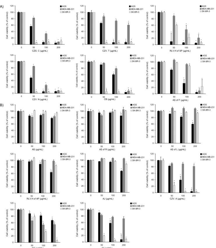
국화과 식물의 대표적인 약리효과로는 항에이즈, 항균, 항 진균 및 항산화효과 등이 있으며(16-18) 주로 산국과 감국에 서 기능성 생리활성 연구가 보고되었다(19). 최근 연구에 사 용된 산국과 감국 이외에 14종의 국화과 추출물에서 항암효 과를 확인하였다. 위암세포인 AGS와 유방암세포인 MDA- MB-231과 SK-BR-3의 성장에 영향을 주는지 확인하기 위 해 MTT assay를 시행하였다. 암세포주를 seeding 후 24시 간 동안 배양시킨 다음 14종의 국화과 추출물을 50, 100, 200 μg/mL의 농도로 24시간 처리하였다. Fig. 1에 나타난 바와 같이 대조군은 지속적으로 세포성장이 일어나는데 비해 14 종의 국화과 추출물 중 6종의 국화과 추출물인 한라구절초 (CZS.C) 전초, 포천구절초(CZV.T) 전초, 삼잎국화(RLV.H of SP) 지하부, 낙동구절초(CZV.N) 전초, 산국(CB) 전초 그 리고 해국(AS of F) 꽃에서 농도 의존적으로 암세포의 성장 억제효과를 나타내었다. 국화과 식물 중 하나인 감국에서 인간의 전립선암세포인 PC3, 백혈병세포인 HL 60 또한 자 궁경부암세포인 HeLa 세포에서 농도와 시간 의존적으로 항 암효과가 있다고 보고하였다(20-22). 이러한 결과를 통해 6 종의 국화과 추출물은 위암세포인 AGS와 유방암세포인 MDA-MB-231과 SK-BR-3의 세포증식 억제효과 탁월하 며 암예방제 및 항암보조제로 개발할 수 있는 가능성이 있다 고 사료된다.
유방암세포의 apoptosis에 미치는 영향
암세포의 성장 억제효과를 나타낸 6종의 국화과 추출물이 유방암세포인 MDA-MB-231 세포의 apoptosis에 미치는 영향을 살펴보았다. Apoptosis는 암세포의 증식을 억제하는 기전 중의 하나이며 apoptosis의 형태학적 특성을 확인하기 위해 DAPI 염색을 하였다(Fig. 2). MDA-MB-231 유방암세 포에 한라구절초(CZS.C) 전초, 포천구절초(CZV.T) 전초, 삼잎국화(RLV.H of SP) 지하부, 낙동구절초(CZV.N) 전초, 산국(CB) 전초 그리고 해국(AS of F) 꽃을 25, 50 μg/mL의 농도로 24시간 동안 처리하였다. 6개 중 3개의 추출물인 CZS.C, CZV.T 그리고 RLV.H of SP에서는 농도 의존적으 로 세포의 성장이 감소한 것과 동일하게 apoptotic body와 세포질 위축, 염색사의 응축 등을 포함하는 전형적인 apop-
tosis 형태가 관찰되었다. 이는 nucleosome의 linker DNA 부분의 절단에 의한 DNA 단편화의 결과(23)이므로 apopto- sis 유발과 밀접한 관련이 있음을 알 수 있다. 국화과에 속하 는 목향을 전립선암에 4 mg/L의 농도로 처리하였을 때 apoptotic cell이 관찰된다는 연구결과(24)와 일치하게 국화 과 추출물인 CZS.C, CZV.T 그리고 RLV.H of SP의 유방암 세포 성장 억제효과는 암세포의 apoptosis를 통해 유발시키 는 것으로 사료된다.
p53 단백질 발현에 미치는 영향
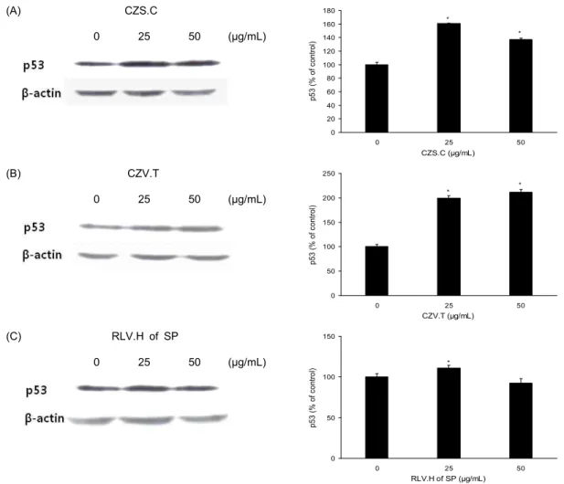
p53은 정상적인 세포의 세포질에 소량 존재하는 암억제 단백질로 DNA가 손상을 받는 경우에 그 양과 기능이 증가 되고 세포질에서 핵으로 이동된다(25). 인체 암의 50% 이상 에서 p53 유전자는 돌연변이 되어 있거나 단백질이 기능적 으로 소실되어 있다(26). 따라서 DAPI 염색에서 apoptosis 형태가 관찰된 한라구절초(CZS.C) 전초, 포천구절초(CZV.T) 전초와 삼잎국화(RLV.H of SP) 지하부에서 p53 단백질의 발현에 미치는 영향을 살펴보았다. 그 결과 Fig. 3A와 3B에 서 보는 것과 같이 CZS.C와 CZV.T에서는 p53 단백질의 발 현이 증가하였지만, RLV.H of SP에서는 25 μg/mL에서 대 조군보다 증가하였다(Fig. 3C). CZS.C, CZV.T, RLV.H of SP 추출물을 MDA-MB-231 유방암세포에 처리했을 때 암 억제 단백질인 p53 발현의 증가로 apoptosis가 유발시키는 것으로 사료된다.
Bcl-2 family 단백질 발현에 미치는 영향
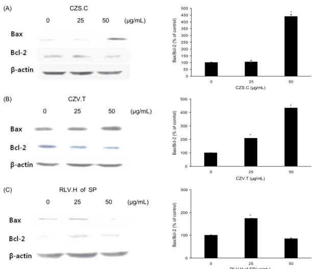
Bcl-2 family 단백질들은 미토콘드리아 막 투과성을 조절 하는 단백질로 미토콘드리아를 통한 apoptosis의 경로를 조 절하는데 중요한 역할을 담당한다(27). Bcl-2 family 단백질 들은 미토콘드리아 막에 존재하거나 또는 apoptotic 자극에 의하여 미토콘드리아 막으로 이동한다. Bcl-2 family에 속하 는 몇 가지 인자들은 apoptosis 유발 조절에 가장 대표적인 유전자로 알려져 있으며 그중 Bcl-2는 anti-apoptotic 분자 로서 apoptosis의 유발을 억제하는 기능을 가지며 Bax는 대 표적인 pro-apoptotic 분자로 apoptosis의 유발과 관계가 있 다(28). 미토콘드리아 막 투과성의 변화에 의한 것인지 확인 하기 위하여 western blot을 통해 Bcl-2와 Bax 단백질들의 변화를 확인하였다. Fig. 4A와 4B에서 보는 것과 같이 CZS.C와 CZV.T에서는 anti-apoptotic 분자인 Bcl-2 단백질 이 감소하고 pro-apoptotic 분자인 Bax 단백질은 농도 의존 적으로 증가하였지만, RLV.H of SP는 25 μg/mL에서 Bax/
Bcl-2 비율이 증가하였다(Fig 4C). 국화과인 목향 헥산추출 물이 전립선암에서 Bax의 발현에서 유의적인 변화가 없었 으며 Bcl-2의 발현은 감소시켰고(4) 대장암세포에서는 Bax 의 발현을 증가시켰다(29). 하지만 그 밖의 국화과 추출물인 CZS.C, CZV.T, RLV.H of SP 추출물은 Bax와 Bcl-2의 단백 질 발현 수준을 변화시켜 apoptosis를 유도시키는 것으로 사료된다. CZS.C, CZV.T, RLV.H of SP 추출물을 암 예방제
A)
0 20 40 60 80 100 120
0 50 100 200
CZS. C (μg/mL)
Cell viability (% of control) .
AGS MDA-MB-231 SK-BR-3
*
* *
* *
*
*
*
*
0 20 40 60 80 100 120
0 50 100 200
CZV. T (μg/mL)
Cell viability (% of control) .
AGS MDA-MB-231 SK-BR-3
*
*
*
* *
*
* *
*
0 20 40 60 80 100 120
0 50 100 200
RLV.H of SP (μg/mL)
Cell viability (% of control) .
AGS MDA-MB-231 SK-BR-3
*
*
*
*
*
*
*
*
*
0 20 40 60 80 100 120
0 50 100 200
CZV. N (μg/mL)
Cell viability (% of control) .
AGS MDA-MB-231 SK-BR-3
*
**
*
*
*
* *
*
0 20 40 60 80 100 120
0 50 100 200
CB (μg/mL)
Cell viability (% of control) .
AGS MDA-MB-231 SK-BR-3
*
* *
* *
*
*
*
0 20 40 60 80 100 120
0 50 100 200
AS of F (μg/mL)
Cell viability (% of control) .
AGS MDA-MB-231 SK-BR-3
*
*
*
*
*
*
B)
0 20 40 60 80 100 120
0 50 100 200
AS (μg/mL)
Cell viability (% of control) .
AGS MDA-MB-231 SK-BR-3
* *
0 20 40 60 80 100 120
0 50 100 200
AS of R (μg/mL)
Cell viability (% of control) .
AGS MDA-MB-231 SK-BR-3
*
*
0 20 40 60 80 100 120
0 50 100 200
AS of L (μg/mL)
Cell viability (% of control) .
AGS MDA-MB-231 SK-BR-3
* *
*
*
0 20 40 60 80 100 120
0 50 100 200
RLV.H of AP (μg/mL)
Cell viability (% of control) .
AGS MDA-MB-231 SK-BR-3
*
*
*
0 20 40 60 80 100 120
0 50 100 200
AJ (μg/mL)
Cell viability (% of control) .
AGS MDA-MB-231 SK-BR-3
*
*
0 20 40 60 80 100 120
0 50 100 200
CZV. A (μg/mL)
Cell viability (% of control) .
AGS MDA-MB-231 SK-BR-3
*
*
*
* *
*
*
0 20 40 60 80 100 120
0 50 100 200
CC (μg/mL)
Cell viability (% of control) .
AGS MDA-MB-231 SK-BR-3
*
*
*
*
*
*
* *
*
0 20 40 60 80 100 120
0 50 100 200
CI (μg/mL)
Cell viability (% of control) .
AGS MDA-MB-231 SK-BR-3
*
*
*
*
*
*
Fig. 1. Growth inhibitory effect of Compositae extracts on AGS, MDA-MB-231 and SK-BR-3 cells. A: Growth inhibitory effect ofCompositae extracts on cancer cells. B: No show growth inhibitory effect of Compositae extracts on cancer cells. For studies accessing the effect of Compositae extracts on exponentially growing AGS, MDA-MB-231 and SK-BR-3 cells growth, 2×10⁴cells were plated in 96 well plate and after 24 hr treated with vehicle (DMSO) alone orCompositae extracts (50, 100, 200 μg/mL) in medium.
The growth inhibition was measured by the MTT assay. Data are mean±standard deviation (SD) for three samples. The significance was determined by Student’s t-test (*p<0.05 compared with untreated control). CZS.C:Chrysanthemum zawadskii subsp. coreanum, CZV.T: Chrysanthemum zawadskii var. tenuisectum, RLV.H of SP: Rudbeckia laciniata var. hortensis of subterranean part, CZV.N:
Chrysanthemum zawadskii var. naktongense, CB: Chrysanthemum boreale, AS of F: Aster spathulifolius of flower, AS: Aster spathulifo- lius, AS of R: Aster spathulifolius of root, AS of L: Aster spathulifolius of leaf, RLV.H of AP: Rudbeckia laciniata var. hortensis of aerial part, AJ:Artemisia japonica, CZV.A: Chrysanthemum zawadskii var. alpinum, CC: Centaurea cyanus, CI: Chrysanthemum indicum.
CZV.N
Fig. 2.Compositae extracts induces apoptosis of MDA-MB-231 cells. A: Apoptosis effect of Compositae extracts on MDA-MB-231 cells. B: No show apoptosis effect ofCompositae extracts on MDA-MB-231 cells. MDA-MB-231 cells were treated with 25, 50 μg/mL Compositae extracts or vehicle in medium containing 5% FBS for 24 hr and stained with DAPI. Chromatin condensation, representing apoptotic cell death (arrow), was examined using a fluorescence microscope (×200).
(A) CZS.C
0 25 50 (μg/mL)
0 20 40 60 80 100 120 140 160 180
0 25 50
CZS.C (µg/mL)
p53 (% of control) .
*
*
(B) CZV.T
0 25 50 (μg/mL)
0 50 100 150 200 250
0 25 50
CZV.T (µg/mL)
p53 (% of control) . *
*
(C) RLV.H of SP
0 25 50 (μg/mL)
0 50 100 150
0 25 50
RLV.H of SP (µg/mL)
p53 (% of control) . *
Fig. 3. Compositae extracts change the levels of p53 protein in MDA-MB-231 cells. A: Cells were treated with CZS.C for 24 hr. Cell lysates were prepared as described in the Materials and Methods and analyzed by 12% SDA-PAGE followed by Western blotting. The membranes were incubated with anti-p53 antibodies. B: Cells were treated with CZV.T for 24 hr. The membranes were incubated with anti-p53 antibodies. C: Cells were treated with RLV.H of SP for 24 hr. The membranes were incubated with anti-p53 antibodies. The blots were also probed with anti-β-actin antibodies to confirm equal sample loading. Each bar represents the mean±SD calculated from three independent experiments.
(A) CZS.C
0 25 50 (μg/mL)
0 50 100 150 200 250 300 350 400 450 500
0 25 50
CZS.C (µg/mL)
Bax/Bcl-2 (% of control) . *
(B) CZV.T
0 25 50 (μg/mL)
0 100 200 300 400 500
0 25 50
CZV.T (µg/mL)
Bax/Bcl-2 (% of conrtol) .
*
*
(C) RLV.H of SP
0 25 50 (μg/mL)
0 100 200 300
0 25 50
RLV.H of SP(µg/mL)
Bax/Bcl-2 (% of control) .
*
Fig. 4. Compositae extracts change the levels of Bax and Bcl-2 protein in MDA-MB-231 cells. A: Cell were treated with CZS.C for 24 hr. Cell lysates were prepared as described in the Materials and Methods and analyzed by 12% SDA-PAGE followed by Western blotting. The membranes were incubated with anti-Bax antibodies and anti-Bcl-2 antibodies. B: Cell were treated with CZV.T for 24 hr. The membranes were incubated with anti-Bax antibodies and anti-Bcl-2 antibodies. C: Cell were treated with RLV.H of SP for 24 hr. The membranes were incubated with anti-Bax antibodies and anti-Bcl-2 antibodies. The blots were also probed with anti-β-actin antibodies to confirm equal sample loading. Each bar represents the mean±SD calculated from three in- dependent experiments.
로 사용하기 위해서는 좀 더 세밀한 기전연구와 후속 연구가 계속적으로 수행되어야 할 것이다.
요 약
국화과 꽃은 우리나라에서 전통적으로 항염증과 항산화 치료에 사용되었다. 본 연구에서는 국화과 추출물이 인간 위암세포 AGS, 인간 유방암세포 MDA-MB-231과 SK- BR-3 암세포에서 성장을 억제하고 세포자멸사를 유발하는 지 확인하였다. AGS, MDA-MB-231 그리고 SK-BR-3 암 세포의 성장을 MTT로 측정하였다. 14종의 국화과 추출물 을 24시간 동안 50, 100, 200 μg/mL의 농도로 처치하였다.
한라구절초 전초, 포천구절초 전초, 삼잎국화 지하부, 낙동 구절초 전초, 산국 전초 그리고 해국 꽃 추출물에서 암세포 의 성장을 농도 의존적으로 억제시켰다. 우리나라 여성에서 가장 많이 발생하는 유방암세포인 MDA-MB-231 암세포에
서 세포자멸사를 확인하기 위해 DAPI 염색을 수행하였다.
MTT assay에서 암세포를 억제시킨 6종의 국화과 추출물중 3종인 한라구절초 전초, 포천구절초 전초, 삼잎국화 지하부 추출물을 처치한 세포에서 핵의 응축이 농도 의존적으로 존 재함을 형광현미경으로 확인하였다(×200). 세포자멸사에 관련된 단백질의 발현을 알아보기 위해서 western blot으로 확인하였다. 한라구절초 전초, 포천구절초 전초, 삼잎국화 지하부 추출물을 25, 50 μg/mL의 농도로 24시간 동안 MDA- MB-231 암세포 처치 후 cell lysate를 얻어 Bcl-2, Bax 그리 고 p53의 변화를 관찰하였다. 한라구절초 전초, 포천구절초 전초 그리고 삼잎국화 지하부에서 anti-apoptotic 분자인 Bcl-2 단백질은 감소하고 반대로 pro-apoptotic 분자인 Bax 와 p53 단백질은 증가하였다. 결과적으로 한라구절초 전초, 포천구절초 전초 그리고 삼잎국화 지하부 추출물은 유방암 세포의 성장을 억제하고 apoptosis를 유발시키므로 암예방 제나 치료제로 개발될 수 있을 것으로 사료된다.
감사의 글
본 연구는 농촌진흥청의 지원을 받아 수행된 연구(Project NO. PJ007884)이며, 이에 감사드립니다.
문 헌
1. Lee SE, Hwang HJ, Ha JS, Jeong HS, Kim JH. 2003. Scre- ening of medical plant extracts for antioxidant activity.Life Sci 73: 167-179.
2. Sun T, Tanumihardjo SA. 2007. An integrated approach to evaluate food antioxidant capacity.J Food Sci72: 159-165.
3. Choi SH, Im SI, Bae JE. 2006. Analysis of aroma compo- nents from flower tea of German chamomile andChrysan- tehmum boreale Makino. Korea J Food Cookery Sci22:
768-773.
4. Kang JR, Lee MK, Kang SM. 2008. Anti-oxidant property and tyrosinase inhibition activity of various extracts from plants in compositae plants.J Korea Soc Appl Biol Chem 51: 321-328.
5. Park NY, Kown JH, Kim HK. 1998. Optimization of ex- traction conditions for ethanol extracts from Chrysanthe- mum morifoliumby response surface methodology.Korean J Food Sci Technol 31: 1189-1196.
6. Nam SH, Yang MS. 1995. Antibacterial activities of ex- tracts fromChryasnthemum borealeM.J Agric Food Chem 38: 269-272.
7. Nam SH, Yang MS. 1995. Isolation of cytotoxic substances of extracts fromChrysanthemum borealeM.J Agric Food Chem 38: 273-277.
8. Woo JH, Lee CH. 2008. Effects of harvest date on anti- oxidantof Dendranthema zawadskiivar.latilobum(Maxim.) Kitam and D. zawadskii var.yezoense (Maek) Y.M. Lee
& H.J. Choi. Korean J Plant Res 21: 128-133.
9. Son JC, Park NK, Hur HS, Bang MH, Baek NI. 2000. Exa- mination and isolation of natural antioxidants from Korean medicinal plants. Korea J Med Crop Sci 8: 94-101.
10. Korea National Statistic Office. 2005.The Korean cause of death.
11. Chen CN, Huang HH, Wu CL, Lin PC, Hsu JT, Hsieh HP, Chuang SE, Lai GM. 2007. Isocostunolide, a sesquiterpene lactone, induces mitochondrial membrane depolarization and caspase-dependent apoptosis in human melanoma cells.
Cancer Lett 246: 237-252.
12. Hajra KM, Liu JR. 2004. Apoptosome dysfunction in human cancer. Apoptosis 9: 691-704.
13. Reed J. 1994. Bcl-2 and regulation of programmed cell death. J Cell Biol 124: 1-6.
14. Kim EJ, Lee YJ, Shin HK, Park JH. 2005. Induction of apop- tosis by the aqueous extract ofRubuscoreanumin HT-29 human colon cancer cells. Nutrition 21: 1141-1148.
15. Jung JI, Lim SS, Choi HJ, Cho HJ, Shin HK, Kim EJ, Chung WY, Park KK, Park JH. 2006. Iosliquiritigenin induces apoptosis by depolarizing mitochondrial membranes in prostate cancer cells. J Nutr Biochem 17: 689-696.
16. Hu CQ, Chen K, Shi Q, Kilkuskie RE, Cheng YC, Lee KH.
1994. Anti-AIDS agents, 10. Acacetin-7-O-beta-D-gal- actopyranoside, an anti-HIV principle fromChrysanthemum morifoliumand a structure-activity correlation with some related flavonoids. J Nat Prod 57: 42-51.
17. Sassi AB, Harzallah-Skhiri F, Bourgougnon N, Aouni M.
2008. Antimicrobial activities of four TunisianChrysanthemum species. Ind J Med Res 127: 183-192.
18. Marongiu B, Piras A, Porcedda S, Tuveri E, Laconi S, Deidda D, Maxia A. 2009. Chemical and biological comparisons on supercritical extracts ofTanacetum cinerariifolium(Trevir) Sch. Bip. with three related species of chrysanthemums of Sardinia (Italy). Nat Prod Res 23: 190-199.
19. Yu JS, Hwang IG, Woo KS, Chang YD, Lee CH, Jeong JH, Jeong HS. 2008. Physicochemical characteristics ofChry- santhemum indicum L. flower tea according to different pan-firing times.Korean J Food Sci Technol40: 297-302.
20. Cai HF. 2007. The research progression of Flos Chrysan- themum indicumon chemical constituent and medical ap- plication. Zhongguo Yiliao Quanyan 2: 118-120.
21. Jin SR, Zhu BD, Qin XH. 2005. The effect ofChrysanthemum indicumon SMMC7721, PC3 and HL60 cell lines.Zhongyao Yaoli Yu Linchuang 21: 39-41.
22. Wu DH, Yang LW, Su WW. 2004. The research progression of Chrysanthemum indicum on chemical constituent and pharmacology. Zhongyaocai 27: 142-144.
23. Evans VG. 1993. Multiple pathways to apoptosis. Cell Biol Int 17: 461-476.
24. Park SY, Kim EJ, Lim DY, Kim JS, Lim SS, Shin HK, Yoon JH. 2008. Inhibitory effect of the hexane extract ofSaus- surea lappaon the growth of LNCaP human prostate can- cer cells. J Korean Soc Food Sci Nutr 37: 8-15.
25. Banasiak KJ, Xia Y, Haddad GG. 2000. Mechanism under- lying hypoxia-induced neuronal apoptosis.Prog Neurobiol 62: 215-249.
26. Amundson SA, Myers TG, Fornace AJ. 1998. Roles for p53 in growth arrest and apoptosis: putting on the brakes after genotoxic stress. Oncogene 17: 3287-3299.
27. Kim EJ, Park SY, Hong J, Shin M, Lim SS, Shin HK, Park JHY. 2007. Inhibitory effect of the methanolic extract of Symphyocladia latiusculaon the growth of HT-29 human colon cancer cells.J Korean Soc Food Sci Nutr 36: 431- 28. Donovan M, Cotter TG. 2004. Control of mitochondrial in-438.
tegrity by Bcl-2 family members and caspase-independent cell death. Biochim Biophys Acta 1644: 133-147.
29. Kim EJ, Park HS, Lim SS, Kim JS, Shin HK, Yoon JH.
2008. Effect of the hexane extract ofSaussurea lappaon the growth of HT-29 human colon cancer cells.Korean J Food Sci Techinol 40: 207-214.
(2012년 2월 9일 접수; 2012년 4월 3일 채택)