ABSTRACT
Purpose: The purpose of the current study was to explore the functions and potential mechanism of miR-451a in breast cancer (BC).
Methods: Quantitative reverse transcription real-time polymerase chain reaction was used to analyze the expression of miR-451a in human normal mammary cells (MCF-10A) and BC cells. Colony formation assay, terminal-deoxynucleoitidyl transferase mediated nick end labeling assay and transwell assays were conducted to validate the effect of miR-451a on proliferation, apoptosis, migration and invasion of BC cells, respectively. RNA pull-down, RNA immunoprecipitation and luciferase reporter assays were applied to investigate the upstream and downstream mechanisms of miR-451a in BC cells.
Results: MiR-451a was expressed at a low level in BC cells. Overexpression of miR-451a repressed BC cells proliferation, migration and invasion. Moreover, long non-coding RNA AC092127.1 acted as a sponge of miR-451a to enhance the expression level of AE binding protein 2 (AEBP2) that was demonstrated to be the target gene of miR-451a in BC cells.
Finally, rescue experiments validated that miR-451a and AEBP2 involved in AC092127.1- mediated BC cell growth, migration and invasion.
Conclusion: In a word, AC092127.1/miR-451a/AEBP2 axis contributes to BC cell growth, migration and invasion. Our results may help to find novel potential targets for BC treatment.
Keywords: Breast neoplasms; Cell proliferation; MicroRNAs; RNA, long noncoding
INTRODUCTION
Breast cancer (BC) is a malignant tumor, which has become the most major cause of cancer- related death among women worldwide [1]. At present, surgery, chemotherapy, radiotherapy and endocrine therapy are the main treatment options for BC. Nevertheless, the prognosis of BC patients at advanced stage is still poor [2, 3]. So, it is significant to understand the mechanisms underlying BC pathology in finding novel therapeutic targets.
MicroRNAs (miRNAs) are a class of non-coding RNAs (ncRNAs) with the length of 20-22 nucleotides [4]. MiRNAs usually exert functions at the post-transcriptional level to regulate their target genes [5]. According to previous studies, miRNAs exert important functions in BC. For example, miR-183-5p facilitates BC cell proliferation via suppressing PDCD4 [6]. MiR-
Original Article
Received: Sep 17, 2020 Revised: Feb 5, 2021 Accepted: Apr 27, 2021 Correspondence to Yonghua Zhou
Department of Pathology, the People's Hospital of Xinghua City, No. 419 South Yingwu Road, Xinghua 225702, China.
E-mail: [email protected]
© 2021 Korean Breast Cancer Society This is an Open Access article distributed under the terms of the Creative Commons Attribution Non-Commercial License (https://
creativecommons.org/licenses/by-nc/4.0/) which permits unrestricted non-commercial use, distribution, and reproduction in any medium, provided the original work is properly cited.
ORCID iDs Xiumei Zhang
https://orcid.org/0000-0002-7976-7903 Lin Cong
https://orcid.org/0000-0001-8034-0045 Dafang Xu
https://orcid.org/0000-0003-1809-4829 Qi Leng
https://orcid.org/0000-0002-1104-8486 Ming Shi
https://orcid.org/0000-0002-9776-0086 Yonghua Zhou
https://orcid.org/0000-0002-0148-9691 Conflict of Interest
The authors declare that they have no competing interests.
Author Contributions
Data curation: Xu D, Leng Q; Formal analysis:
Cong L; Investigation: Shi M; Supervision: Zhou Y; Writing - original draft: Zhang X.
Xiumei Zhang , Lin Cong , Dafang Xu , Qi Leng , Ming Shi , Yonghua Zhou
Department of Pathology, the People's Hospital of Xinghua City, Xinghua, China
AC092127.1-miR-451a-AE binding protein 2 Signaling Facilitates
Malignant Properties of Breast Cancer
145 influences BC cell proliferation and migration via regulating OCT4 expression [7]. MiR-451a has been reported as a potential biomarker for cancer treatment [8]. The role of miR-451a in biological processes of cancers has also been elucidated. For instance, miR-451a functions as a tumor suppressor gene to affect non-small cell lung cancer via targeting ATF2 [9]. MiR-451a negatively regulates thyroid cancer development via targeting PSMB8 [10]. Moreover, miR-451a inhibits cell proliferation and enhances tamoxifen sensitive in BC via modulating macrophage migration inhibitory factor [11]. However, the molecular mechanisms of miR-451a in regulating the malignant phenotype of BC cells have not been fully elucidated.
Long ncRNA (lncRNAs) belong to transcribed RNA molecules, which are longer than 200 nucleotides and lack of protein-coding potential [12]. In recent years, numerous studies have disclosed that lncRNAs are closely related to various biological processes, including proliferation, apoptosis, migration, and invasion [13, 14]. Moreover, increasing reports have suggested that lncRNAs can serve as oncogenes or tumor suppressor genes to affect the occurrence and development of BC [15]. For examples, MALAT1 exerts inhibitory function in BC metastasis [16]. NEAT1 acts as an oncogene to facilitate BC cell proliferation and metastasis [17]. However, the biological roles of most lncRNAs in BC are still unknown.
This study focused on exploring the role of miR-451a and its relevant regulatory mechanism in BC.
METHODS
Assays to culture cells Cell culture
BC cells (MCF-7, BT-549, MDA-MB-468, and MDA-MB-231) and human normal mammary cells (MCF-10A) were purchased from ATCC (Manassas, USA). MCF-7 cells were grown in Dulbecco's Minimum Essential Medium (Gibco, Grand Island, USA). BT-549 cells were grown in Roswell Park Memorial Institute (RPMI)-1640 Medium (Gibco). MDA-MB-468 and MDA- MB-231 cells were grown in Leibovitz's L-15 Medium (Gibco). MCF-10A cells were grown in MEGM (Lonza/Clonetics Corporation, San Diego, USA). All cells were cultured in a humid atmosphere with 5% CO
2at 37
oC.
Cell transfection
MiR-451a mimics and NC mimics, sh-AC092127.1#1/2 and sh-NC, miR-451a inhibitor and NC inhibitor, pcDNA3.1-AE binding protein 2 (AEBP2) and pcDNA3.1 were synthesized by GenePharma (Shanghai, China). Lipofectamine 3000 (Life Technologies, Carlsbad, USA) was used for transfection. The concentrations of plasmids were all listed in Supplementary Table 1.
Assays to determine the expression level
Quantitative reverse transcription real-time polymerase chain reaction
Total RNA was isolated from cells using Trizol reagent (Invitrogen, Carlsbad, USA). And then,
RNA was reversely transcribed into complementary DNA using M-MLV reverse transcriptase
(Promega, Madison, USA). Target primers were amplified and the expression levels of RNAs
were quantified using SYBR Green PCR Master Mix (Applied Biosystems, Foster City, USA)
based on 2
−ΔΔCtmethod and normalized to U6 or glyceraldehyde-3-phosphate dehydrogenase
(GAPDH). Sequences of all primers were listed in Supplementary Table 2. The experiment
was conducted at least 3 times.
Western blot
RIPA lysis buffer (Thermo Fisher, Waltham, USA) was used to extract total proteins from cells. Proteins were separated by 10% sodium dodecyl sulphate–polyacrylamide gel electrophoresis and then transferred to polyvinylidene fluoride membranes (Millipore, Burlington, USA), followed by incubation with primary antibodies, including anti-AEBP2 (community state type [CST], 1/1,000) and anti-GAPDH (CST, 1/1,000) at 4
oC overnight.
Next, the membranes were incubated with the horseradish peroxidase-conjugated secondary antibodies at room temperature for 1 h. Finally, the ECL Western Blotting Detection Kit was utilized to detect the protein levels. The experiment was conducted at least 3 times.
Assays to investigate the biological behaviors of cells Colony formation assay
Cells were grown in 6-well plates for 10 days' incubation, and then fixed with 4%
paraformaldehyde and stained with 0.1% crystal violet. The numbers of colonies were counted manually when the colonies could be observed by naked eyes. The experiment was conducted at least three times.
Terminal-deoxynucleoitidyl transferase mediated nick end labeling
Terminal-deoxynucleoitidyl transferase mediated nick end labeling (TUNEL) assay
(Beyotime, Shanghai, China) was implemented on the basis of manufacturer's protocol. Cells were washed twice with PBS and fixed with 4% paraformaldehyde, and then treated with 0.3% Triton X-100. The TUNEL detection solution was then added to measure cell apoptosis.
The experiment was conducted at least three times.
Transwell assays
For transwell migration assay, 1 × 10
5cells were grown in the upper transwell chamber (Millipore). For transwell invasion assay, 1 × 10
5cells were grown in the matrigel-coated upper chamber (BD Biosciences, San Jose, USA). For both assays, the upper chamber was added with serum-free RPMI-1640 medium while the lower chamber was added with 10% fetal bovine serum-contained RPMI-1640 medium. After incubated for 24 hours, cells in the upper surface were wiped off. The migrated and invaded cells in the lower chamber were fixed in methanol and dyed with 0.1% crystal violet. Finally, the migrated or invaded cells was observed under microscope and counted manually. The experiment was conducted at least three times.
Assays to investigate the relationship between RNAs RNA immunoprecipitation
Cells were lysed in a RNA immunoprecipitation (RIP) lysis buffer kit (Millipore), and then the cell extract was incubated with anti-Ago2 (Millipore) or anti-immunoglobulin G (Millipore) conjugated with magnetic beads at 4
oC for 6 hours. The antibody-beads complex was resuspended in proteinase K Buffer. After removing the precipitation, RNA in the supernatant was purified. At last, quantitative reverse transcription real-time polymerase chain reaction (RT-qPCR) was applied to detect the enrichment of purified RNAs. The experiment was conducted at least three times.
Luciferase reporter assay
The fragments of AC092127.1 and AEBP2 3′UTR wild-type or mutant-type containing the
binding sites of miR-451a were cloned into a pmirGLO Vector. The constructed vectors were
co-transfected into cells along with miR-451a mimics or NC mimics using Lipofectamine
3000 (Life Technologies). Forty-eight hours after transfection, the dual-luciferase reporter
assay kit was applied to detect luciferase activities. The experiment was conducted at least three times.
RNA pull down assay
Biotin-labeled miR-451a probe and biotin-labled NC probe were transfected into cells and incubated for 48 hours. And then, cells were incubated in lysis buffer with streptavidin- coupled agarose beads (Thermo Fisher) to pull down the complex. After the complex was washed, RNAs were isolated and measured using RT-qPCR. The experiment was conducted at least 3 times.
Statistical analysis
The data were expressed as the mean value ± standard deviation. SPSS 19.0 software (SPSS Inc., Chicago, IL, USA) and GraphPad Prism 6 (GraphPad Software Inc., San Diego, CA, USA) were employed for statistical analysis. The differences were calculated by Student's t-test or analysis of variance analysis, and p < 0.05 was regarded as statistical significance. The experiment was conducted at least 3 times.
RESULTS
MiR-451a inhibited BC cell proliferation, migration and invasion
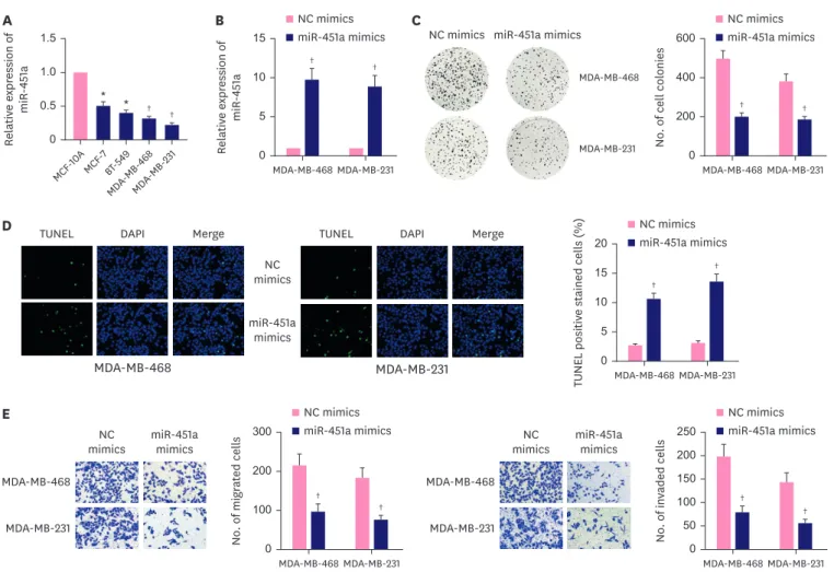
MiR-451a has been reported to be a potential risk marker of BC, but its detailed mechanism remains to be further elucidated [18]. Herein, we used RT-qPCR to detect the expression of miR-451a in BC cells. In contrast to human normal mammary cells (MCF-10A), BC cells presented significantly low level of miR-451a, among which, MDA-MB-468 and MDA-MB-231 presented the lowest level of miR-451a (Figure 1A). Thus MDA-MB-468 and MDA-MB-231 cells were selected for further gain-of function experiments. RT-qPCR verified miR-451a expression was elevated by miR-451a mimics in MCF-10A, MDA-MB-468 and MDA-MB-231 cells (Figure 1B, Supplementary Figure 1A). Colony formation assays revealed that the number of colonies was declined when miR-451a was up-regulated in MCF-10A, MDA- MB-468 and MDA-MB-231 cells (Figure 1C, Supplementary Figure 1B). In addition, the results from TUNEL assays showed that transfection with miR-451a mimics increased apoptosis rate of MDA-MB-468 and MDA-MB-231 cells (Figure 1D). Moreover, it was discovered in transwell assays that the number of migrated and invaded cells was reduced after miR-451a overexpression (Figure 1E). To sum up, miR-451a inhibited cell proliferation, migration and invasion while facilitated cell apoptosis in BC.
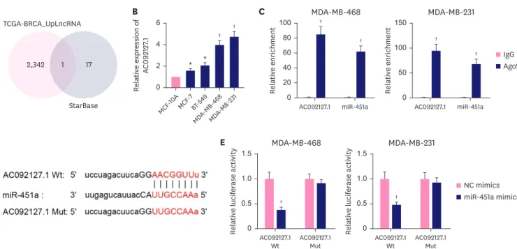
AC092127.1 is a sponge of miR-451a in BC cells
Next, we investigated the upstream mechanism of miR-451a in BC cells. Recent studies have implied that lncRNAs can serve as miRNA sponges. Here, we investigated whether miR-451a could interact with a certain lncRNA in BC cells. According to the data of TCGA (https://
www.cancer.gov/about-cancer) and starBase (http://starbase.sysu.edu.cn) databases, we
found that AC092127.1 was the only lncRNA that could bind with miR-451a and also was
up-regulated in BC tissues (Figure 2A). Next, we identified the high level of AC092127.1 in BC
cells via RT-qPCR (Figure 2B). Moreover, we validated that both AC092127.1 and miR-451a
were enriched in Ago2 groups, indicating that AC092127.1 and miR-451a co-existed in the
RNA-induced silencing complex (RISC) (Figure 2C). Meanwhile, we obtained the binding
sequence of AC092127.1 with miR-451a via starBase database and mutated AC092127.1 for
subsequent experiments (Figure 2D). Through luciferase reporter assays, we found that miR-
451a overexpression reduced the luciferase intensity of AC092127.1-wild type (WT) while had no influence on that of AC092127.1-Mut (Figure 2E). In a word, miR-451a could interact with AC092127.1 in BC cells.
AC092127.1 facilitated BC cell proliferation, migration and invasion via miR-451a To investigate the role of AC092127.1/miR-451a axis in BC cellular processes, we silenced knocked down AC092127.1 and miR-451a with specific short hairpin RNA and inhibitor, respectively (Figure 3A, Supplementary Figure 1C). It was validated in colony formation assays that the proliferation ability was repressed when AC092127.1 was silenced in MCF-10A, MDA- MB-468 and MDA-MB-231 cells, while co-transfection of miR-451a inhibitor reversed this effect (Figure 3B, Supplementary Figure 1D). Furthermore, the results from TUNEL assays indicated that miR-451a inhibitor reduced the elevated apoptosis rate induced by AC092127.1 down-regulation (Figure 3C). In addition, transwell assays revealed that AC092127.1 deletion- mediated suppression on cell migration and invasion were attenuated by co-transfection of miR-451a inhibitor (Figure 3D). In conclusion, AC092127.1 facilitated proliferation, migration and invasion but suppressed apoptosis of BC cells and MCF-10A cells via miR-451a.
Relativ e e xpr ession of miR -451 a 0 5 10 15
MDA-MB-468 MDA-MB-231
B NC mimics
miR-451a mimics
† †
No . of cell col onies
0 200 400 600
MDA-MB-468 MDA-MB-231 NC mimics miR-451a mimics
MDA-MB-468
MDA-MB-231
C NC mimics
miR-451a mimics
† †
Relativ e e xpr ession of miR -451 a 0 0.5 1.0 1.5
MCF-10A MCF-7
BT-549 MDA-MB-
468 MDA-MB-
231
A
† †
* *
TUNEL positiv e stained cells (%)
0 10 5 15 20
MDA-MB-468 MDA-MB-231 NC mimics miR-451a mimics
†
NC
†mimics
TUNEL DAPI Merge TUNEL DAPI Merge
miR-451a mimics
MDA-MB-468 MDA-MB-231
D
No . of migr at ed cells
0 100 200 300
MDA-MB-468 MDA-MB-231 NC mimics miR-451a mimics
†
†
No . of in vaded cells
0 100 50 150 200 250
MDA-MB-468 MDA-MB-231 NC mimics miR-451a mimics
†
†
mimics NC miR-451a mimics MDA-MB-468
MDA-MB-231 mimics NC miR-451a
mimics MDA-MB-468
MDA-MB-231
E
Figure 1. MiR-451a inhibited BC cell proliferation, migration and invasion. (A) MiR-451a was verified to be lowly expressed in BC cells compared with MCF-10A via RT- qPCR analysis. (B) MiR-451a expression was demonstrated to be elevated in MDA-MB-468 and MDA-MB-231 cells transfected with miR-451a mimics via quantitative reverse transcription real-time polymerase chain reaction. (C) Colony formation assays verified the proliferation ability of MDA-MB-468 and MDA-MB-231 cells was repressed after transfection of miR-451a mimics. (D) TUNEL assay revealed the apoptosis rate of miR-451a up-regulated MDA-MB-468 and MDA-MB-231 cells. (E) Transwell assays elucidated migration and invasion abilities of MDA-MB-468 and MDA-MB-231 cells were weakened after miR-451a overexpression.
NC = negative control; TUNEL = terminal-deoxynucleoitidyl transferase mediated nick end labeling; DAPI = 4′,6-diamidino-2-phenylindole; BC = breast cancer.
* p < 0.05,
†p < 0.01.
AEBP2 was targeted by miR-451a in BC cells
Since miRNAs usually exert functions via directly regulating their target genes. Herein, we further explored the downstream gene of miR-451a. On the basis of microT, miRanda, and TargetScan databases, we found 5 messenger RNAs (mRNAs) possibly combined with miR-451a (Figure 4A), among which EMSY, CAB39 and PSMB8 have been reported to be directly targeted by miR-451a in cancers. Thus, we applied RNA pull down assays to detect the correlation of AEBP2 or SAMD48 with miR-451a. Compared with Bio-NC and Bio-miR- 452a-Mut groups, AEBP2 was abundant in Bio-miR-452a-WT groups, while SAMD48 had no obvious abundance (Figure 4B). Subsequently, we validated that AC092127.1, miR-451a and AEBP2 were highly enriched in Ago2 precipitates through RIP assay, suggesting that AC092127.1 might act as a competing endogenous RNA (ceRNA) to regulate miR-451a/AEBP2 axis (Figure 4C). In addition, we discovered the binding sites between AEBP2 3′UTR and miR-451a via starBase database (Figure 4D). Moreover, we found the luciferase intensity was obviously reduced in MDA-MB-468 and MDA-MB-231 cells co-transfected with AEBP2 3'UTR WT and miR-451a mimics, while no significant difference of that in MDA-MB-468 and MDA- MB-231 cells co-transfected with AEBP2 3′UTR Mut and miR-451a mimics (Figure 4E). In brief, AEBP2 was targeted by miR-451a in BC cells.
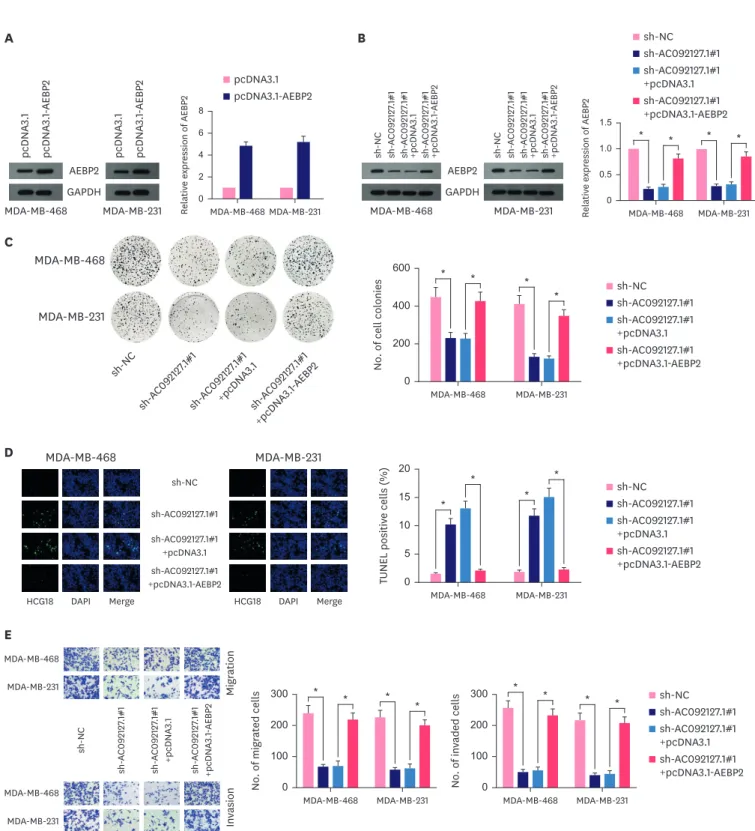
AC092127.1 affected BC cells proliferation, apoptosis, migration and invasion via regulating AEBP2
To further determine the involvement of AEBP2 in AC092127.1-mediated BC cell activities, we designed rescue assays. First, we up-regulated the mRNA and protein levels of AEBP2 in MCF- 10A, MDA-MB-468 and MDA-MB-231 cells and found that overexpression of AEBP2 reversed the inhibited mRNA and protein levels of AEBP2 mediated by AC092127.1 deletion (Figure 5A and B, Supplementary Figure 1E and F). Next, we found that AEBP2 up-regulation rescued the repressed Relativ e e xpr ession of AC 092127 .1
0 2 4 6
MCF-10A MCF-7
BT-549 MDA-MB-
468 MDA-MB-
231
B
†
†
* *
Relativ e enrichment
0 40 80
20 60 100
AC092127.1 miR-451a
C
†
†
MDA-MB-468
IgG Ago2
Relativ e enrichment
0 100 50 150
AC092127.1 miR-451a
†
†
MDA-MB-231 A
TCGA-BRCA_UpLncRNA
StarBase
2,342 1 17
E
Relativ e l ucifer ase activity
0 1.0 0.5 1.5
AC092127.1
Wt AC092127.1 Mut
†
MDA-MB-468
Relativ e l ucifer ase activity
0 1.0 0.5 1.5
AC092127.1
Wt AC092127.1 Mut
†
MDA-MB-231
NC mimics miR-451a mimics
D
Figure 2. AC092127.1 is a sponge of miR-451a in BC cells. (A) AC092127.1 was selected out due to it was up-regulated in BC cells and could bind with miR-452a after intersection of TCGA and starBase databases. (B) Quantitative reverse transcription real-time polymerase chain reaction was performed to examine AC092127.1 expression in BC cells and MCF-10A. (C) AC092127.1 and miR-451a were confirmed to enrich in RNA-induced silencing complex by using RNA immunoprecipitation assays. (D) Binding sites between AC092127.1 and miR-451a were predicted via starBase database. (E) Luciferase reporter assays verified AC092127.1 WT could interact with miR-451a in MDA-MB-468 and MDA-MB-231 cells.
NC = negative control; IgG = immunoglobulin G; WT = wild type; BC = breast cancer.
* p<0.05,
†p<0.01.
proliferation ability of BC cells and MCF-10A cells caused by AC092127.1 depletion (Figure 5C, Supplementary Figure 1G). Meanwhile, TUNEL assays indicated that AEBP2 overexpression reduced the enhanced apoptosis rate of MDA-MB-468 and MDA-MB-231 cells transfected with
NC inhibitor miR-451a inhibitor
Relativ e e xpr ession of miR -451 a 0 1.0 0.5 1.5
MDA-MB-468 MDA-MB-231
* *
Relativ e e xpr ession of AC 092127 .1 0 1.0 0.5 1.5
MDA-MB-468 MDA-MB-231
sh-NC sh-AC092127.1#1 sh-AC092127.1#2
* * * *
A
sh-NC sh-AC092127.1#1 sh-AC092127.1#1 +NC inhibitor sh-AC092127.1#1 +miR-451a inhibitor MDA-MB-468
MDA-MB-231
sh-NC sh- AC 092127
.1#1 sh- AC 092127
.1#1 +NC inhibit or sh- AC 092127
.1#1
+miR -451 a inhibit
or
B
MDA-MB-468 MDA-MB-231
* *
* *
No . of cell col onies
0 400 200 300 100 500
C
sh-NC sh-AC092127.1#1 sh-AC092127.1#1 +NC inhibitor sh-AC092127.1#1 +miR-451a inhibitor MDA-MB-468 MDA-MB-231
*
* *
*
TUNEL positiv e stained cells (%)
0 15
5 10 sh-NC 20
sh-AC092127.1#1 sh-AC092127.1#1 +NC inhibitor sh-AC092127.1#1 +miR-451a inhibitor
TUNEL DAPI Merge TUNEL DAPI Merge
MDA-MB-468 MDA-MB-231
D
sh-NC sh-AC092127.1#1 sh-AC092127.1#1 +NC inhibitor sh-AC092127.1#1 +miR-451a inhibitor MDA-MB-468 MDA-MB-231
* *
* *
No . of migr at ed cells
0 200 100 400 300
MDA-MB-468 MDA-MB-231
* *
* *
No . of in vaded cells
0 200 100 300 MDA-MB-468
MDA-MB-231
MDA-MB-468
MDA-MB-231
sh-NC
Migr ation In vasion
sh-AC092127.1#1 sh-AC092127.1#1 +NC inhibitor sh-AC092127.1#1 +miR-451a inhibitor
Figure 3. AC092127.1 facilitated BC cells proliferation, migration and invasion via miR-451a. (A) Quantitative reverse transcription real-time polymerase chain reaction severally detected the expression of AC092127.1 and miR-451a in MDA-MB-468 and MDA-MB-231 cells transfected with short hairpin RNAs targeting AC092127.1 or miR-451a inhibitor. (B) Colony formation assays discovered the number of colonies of MDA-MB-468 and MDA-MB-231 cells was reduced by sh- AC092127.1#1 and recovered partially by miR-451a inhibitor. (C) TUNEL assays reflected the apoptosis rate of MDA-MB-468 and MDA-MB-231 cells was increased by sh-AC092127.1#1 and largely recovered by miR-451a inhibitor. (D) Transwell assays manifested cell migration and invasion of MDA-MB-468 and MDA-MB-231 cells were restrained by sh-AC092127.1#1 and rescued by miR-451a inhibitor.
NC = negative control; BC = breast cancer; TUNEL = terminal-deoxynucleoitidyl transferase mediated nick end labeling; DAPI = 4′,6-diamidino-2-phenylindole.
* p < 0.01.
sh-AC092127.1#1 (Figure 5D). Finally, through transwell assays, we confirmed that the migration and invasion abilities of MDA-MB-468 and MDA-MB-231 cells inhibited by sh-AC092127.1#1 were restored by co-transfection of pcDNA3.1-AEBP2 (Figure 5E). All above results indicated that AC092127.1 promoted cell proliferation, migration and invasion and suppressed cell apoptosis via regulating AEBP2.
DISCUSSION
BC is the most commonly diagnosed cancer in women and a leading cause of cancer-related mortality [19]. It is a heterogeneous disease in urgent need for developing novel research, classification, and therapy approaches [20]. Therefore, finding novel therapeutic targets are important for improving BC treatment.
Emerging evidences have demonstrated that miRNAs are important regulators in the development of many cancers, including BC [21]. For example, miR-362-3p functions as A
microT miRanda
TargetScan
8 14 136
2 5
5 10
B
Relativ e enrichment
0 40 30 20 10 50
AEBP2 SAMD4B EMSY CAB39 PSMB8
NS NS NS NS
MDA-MB-468
*
Relativ e enrichment
0 40 20 60
AEBP2 SAMD4B EMSY CAB39 PSMB8
NS NS NS
NS
MDA-MB-231
*
Bio-NC Bio-miR-451a
C D
IgG Ago2
Relativ e enrichment
0 100 50 150
AC092127.1 miR-451a AEBP2
MDA-MB-468
Relativ e enrichment
0 100 50 150
AC092127.1 miR-451a AEBP2
MDA-MB-231
E
Relativ e l ucifer ase activity
0 1.0 0.5 1.5
AEBP2 3′UTR
Wt AEBP2 3′UTR Mut
MDA-MB-468
NC mimics miR-451a mimics
*
*
*
Relativ e l ucifer ase activity
0 1.0 0.5 1.5
AEBP2 3′UTR
Wt AEBP2 3′UTR Mut
MDA-MB-231
*
* *
*
*
Figure 4. AEBP2 was targeted by miR-451a in breast cancer cells. (A) Five mRNAs combined with miR-451a were predicted by microT, miRanda, and TargetScan.
(B) RNA pull down assays detected enrichment of 5 mRNAs in bio-miR-451a and bio-NC groups. The results showed only AEBP2 could bind with miR-451a. (C) RNA immunoprecipitation assays demonstrated AC092127.1, miR-451a and AEBP2 co-existed in RNA-induced silencing complex. (D) StarBase database predicted the binding sequences between miR-451a and AEBP2 3′UTR. (E) Luciferase reporter assays indicated AEBP2 3′UTR WT could bind with miR-451a in MDA-MB-468 and MDA-MB-231 cells.
NC = negative control; IgG = immunoglobulin G; mRNA = messenger RNA; WT = wild type; AEBP2 = AE binding protein 2; mRNA = messenger RNA; NS = not significant.
* p < 0.01.
pcDNA3.1 pcDNA3.1-AEBP2
Relative expression of AEBP2
0 6 4 2 8
MDA-MB-468 MDA-MB-231
pcDNA 3.1 pcDNA 3.1 -AEBP 2 pcDNA 3.1 pcDNA 3.1 -AEBP 2
AEBP2 GAPDH
MDA-MB-468 MDA-MB-231
A sh-NC
sh-AC092127.1#1 sh-AC092127.1#1 +pcDNA3.1 sh-AC092127.1#1 +pcDNA3.1-AEBP2
MDA-MB-468
* *
MDA-MB-231
* *
Relative expression of AEBP2
0 1.0 0.5 1.5
AEBP2 GAPDH
MDA-MB-468 MDA-MB-231
sh-NC sh-AC092127.1#1 sh-AC092127.1#1 +pcDNA3.1 sh-AC092127.1#1 +pcDNA3.1-AEBP2 sh-NC sh-AC092127.1#1 sh-AC092127.1#1 +pcDNA3.1 sh-AC092127.1#1 +pcDNA3.1-AEBP2
B
sh-NC sh-AC092127.1#1 sh-AC092127.1#1 +pcDNA3.1 sh-AC092127.1#1 +pcDNA3.1-AEBP2 MDA-MB-468 MDA-MB-231
* *
*
*
No . of cell col onies
0 400 200
MDA-MB-468 600
MDA-MB-231
sh-NC sh- AC 092127
.1#1 sh- AC 092127
.1#1 +pcDNA
3.1 sh- AC 092127
.1#1
+pcDNA 3.1 -AEBP2
C
sh-NC sh-AC092127.1#1 sh-AC092127.1#1 +pcDNA3.1 sh-AC092127.1#1 +pcDNA3.1-AEBP2 MDA-MB-468 MDA-MB-231
*
* *
*
TUNEL positiv e cells (%)
0 15 10 5 20
D
sh-NC
sh-AC092127.1#1 sh-AC092127.1#1 +pcDNA3.1 sh-AC092127.1#1 +pcDNA3.1-AEBP2
HCG18 DAPI Merge HCG18 DAPI Merge
MDA-MB-468 MDA-MB-231
E
sh-NC sh-AC092127.1#1 sh-AC092127.1#1 +pcDNA3.1 sh-AC092127.1#1 +pcDNA3.1-AEBP2 MDA-MB-468 MDA-MB-231
* *
* *
No . of migr at ed cells
0 200 100 300
MDA-MB-468 MDA-MB-231
* * * *
No . of in vaded cells
0 200 100 300 MDA-MB-468
MDA-MB-231
MDA-MB-468
MDA-MB-231
sh-NC
Migr ation In vasion
sh-AC092127.1#1 sh-AC092127.1#1 +pcDNA3.1 sh-AC092127.1#1 +pcDNA3.1-AEBP2
Figure 5. AC092127.1 affected breast cancer cells proliferation, apoptosis, migration and invasion via regulating AEBP2. (A) AEBP2 overexpression in MDA-MB-468 and MDA-MB-231 cells was verified via RT-qPCR and WB. (B) Rescue experiments were carried out in MDA-MB-468 and MDA-MB-231 cells transfected with sh- NC, sh-AC092127.1#1, sh-AC092127.1#1+pcDNA3.1 or sh-AC092127.1#1+pcDNA3.1-AEBP2. RT-qPCR and WB indicated effect of sh-AC092127.1#1 could be offset by pcDNA3.1-AEBP2. (C) Colony formation assays demonstrated the number of colonies of MDA-MB-468 and MDA-MB-231 cells was reduced by sh-AC092127.1#1 and partially recovered by pcDNA3.1-AEBP2. (D) TUNEL assays proved the apoptosis rate of MDA-MB-468 and MDA-MB-231 cells was facilitated by sh-AC092127.1#1 and recovered by pcDNA3.1-AEBP2. (E) Transwell assays detected the properties of migration and invasion of MDA-MB-468 and MDA-MB-231 cells were refrained by sh-AC092127.1#1 and rescued by pcDNA3.1-AEBP2.
AEBP2 = AE binding protein 2; GAPDH = glyceraldehyde-3-phosphate dehydrogenase; RT-qPCR = quantitative reverse transcription real-time polymerase chain reaction; NC = negative control; DAPI = 4′,6-diamidino-2-phenylindole.
* p < 0.01.
a tumor inhibitor in BC [22]. MiRNA-21 and let-7 are potential prognostic biomarkers for patients with BC [23]. Additionally, miR-451a has been reported to be lowly expressed in osteosarcoma and can suppress the growth of osteosarcoma cells by targeting TRIM66 [24]. Moreover, miR-451a is also lowly expressed in colorectal cancer tissues and miR-451a weakens colorectal cancer cell proliferation via interacting with endoplasmic reticulum stress [25]. However, the molecular mechanism involving miR-451a in BC cells has not been clearly elucidated. In the current study, we found that miR-451a demonstrated a low expression level in BC cells. Through gain-of function assays, we validated that miR-451a played a tumor suppressive role in BC.
The recent study has pointed out that lncRNA SNHG12 affects BC progression via interacting with miR-451a [26]. Consistently, our present study also investigated the upstream
mechanism of miR-451a in BC cells. We found that AC092127.1 functioned as a ceRNA to interact with miR-451a in BC cells. As reported previously, the competitive relation between lncRNAs and miRNAs in BC was well documented. SNHG6 targets miR-26a to facilitate BC cell proliferation and invasion [27]. NEAT1 promotes BC cell growth via targeting miR-211 [28]. In this study, we found AC092127.1 was the upstream lncRNA of miR-451a and was highly expressed in BC cells. We also demonstrated that miR-451a inhibitor could restore the inhibiting effects of AC092127.1 deletion on BC cell proliferation, migration and invasion.
Meanwhile, it has been reported that miRNAs function at the post-transcription level usually by base-pairing to the mRNA 3′-untranslated regions [29]. MiR-451a has been reported to repress BC cell proliferation via targeting MIF [11]. Through bioinformatics analysis and RNA pull down assays, we confirmed that AEBP2 could bind with miR-451a in BC cells. AEBP2 has been reported to participate in the cisplatin resistance in ovarian cancer [30]. However, the reports between miRNA and AEBP2 are rare. Our study found for the first time that AEBP2 was the target gene of miR-451a in BC cells. Moreover, we also arranged rescue experiments to validate the interaction between AC092127.1 and AEBP2. We found that AC092127.1 influenced BC cell proliferation, apoptosis, migration and invasion via regulating AEBP2 expression. All above results suggested that AC092127.1, miR-451a and AEBP2 formed a ceRNA network to regulate BC progression.
Conclusively, our study investigated the interaction among miR-451a, AC092127.1 and AEBP2 in BC development. AC092127.1 acted as a ceRNA to upregulate AEBP2 in BC cells through sponging miR-451 (Figure 6). Our findings may provide a novel sight for better understanding of BC.
ACKNOWLEDGMENTS
Thanks for all support.
SUPPLEMENTARY MATERIALS
Supplementary Table 1
The concentration of plasmids used in this study was specified
Click here to view
Supplementary Table 2
The sequences involved in quantitative reverse transcription real-time polymerase chain reaction assay were listed
Click here to view
Supplementary Figure 1
The transfection efficiency of different plasmids and supplementary functional assays. (A) MiR-451a overexpression in MCF-10A cells was verified via RT-qPCR. (B) Colony formation assays demonstrated the number of colonies of MCF-10A cells was decreased after transfecting miR-451a mimics. (C) RT-qPCR detected the expression of AC092127.1 and miR-451a in MCF- 10A cells transfected with sh-AC092127.1 or miR-451a inhibitor. (D) Colony formation assays discovered the number of colonies of MCF-10A cells was reduced by sh-AC092127.1#1 and recovered by miR-451a inhibitor. (E) AEBP2 overexpression in MCF-10A cells was verified via RT-qPCR and WB. (F) Rescue experiments were carried out in MCF-10A cells transfected with sh-NC, sh-AC092127.1#1, sh-AC092127.1#1+pcDNA3.1 or sh-AC092127.1#1+pcDNA3.1-AEBP2.
RT-qPCR and WB indicated effect of sh-AC092127.1#1 could be offset by pcDNA3.1-AEBP2. (G) Colony formation assays demonstrated the number of colonies of MCF-10A cells was reduced by sh-AC092127.1#1 and recovered by pcDNA3.1-AEBP2.
Click here to view
REFERENCES
1. Lu J, Steeg PS, Price JE, Krishnamurthy S, Mani SA, Reuben J, et al. Breast cancer metastasis: challenges and opportunities. Cancer Res 2009;69:4951-3.
PUBMED | CROSSREF
miR-451a
miR-451a
miR-451a AEBP2 Nucleus
Cytoplasm
Proliferation
Migration Invasion
Cell proliferation, invasion and migration
Cell apoptosis
Breast cancer cells AC092127.1
Figure 6. The concept map depicted the role of miR-451a/AC092127.1/AE binding protein 2 axis in breast cancers.
2. Mu Q, Wang H, Zhang M. Nanoparticles for imaging and treatment of metastatic breast cancer. Expert Opin Drug Deliv 2017;14:123-36.
PUBMED | CROSSREF
3. McDonald ES, Clark AS, Tchou J, Zhang P, Freedman GM. Clinical diagnosis and management of breast cancer. J Nucl Med 2016;57 Suppl 1:9S-16S.
PUBMED | CROSSREF
4. Lu TX, Rothenberg ME. MicroRNA. J Allergy Clin Immunol 2018;141:1202-7.
PUBMED | CROSSREF
5. Zealy RW, Wrenn SP, Davila S, Min KW, Yoon JH. microRNA-binding proteins: specificity and function.
Wiley Interdiscip Rev RNA 2017;8:e1414.
PUBMED | CROSSREF
6. Cheng Y, Xiang G, Meng Y, Dong R. MiRNA-183-5p promotes cell proliferation and inhibits apoptosis in human breast cancer by targeting the PDCD4. Reprod Biol 2016;16:225-33.
PUBMED | CROSSREF
7. Kuang WB, Deng QC, Deng CT, Li WS, Zhang YG, Shu SW, et al. MiRNA regulates OCT4 expression in breast cancer cells. Eur Rev Med Pharmacol Sci 2018;22:1351-7.
PUBMED | CROSSREF
8. Minna E, Romeo P, Dugo M, De Cecco L, Todoerti K, Pilotti S, et al. miR-451a is underexpressed and targets AKT/mTOR pathway in papillary thyroid carcinoma. Oncotarget 2016;7:12731-47.
PUBMED | CROSSREF
9. Shen YY, Cui JY, Yuan J, Wang X. MiR-451a suppressed cell migration and invasion in non-small cell lung cancer through targeting ATF2. Eur Rev Med Pharmacol Sci 2018;22:5554-61.
PUBMED | CROSSREF
10. Fan X, Zhao Y. miR-451a inhibits cancer growth, epithelial-mesenchymal transition and induces apoptosis in papillary thyroid cancer by targeting PSMB8. J Cell Mol Med 2019;23:8067-75.
PUBMED | CROSSREF
11. Liu Z, Miao T, Feng T, Jiang Z, Li M, Zhou L, et al. miR-451a inhibited cell proliferation and enhanced tamoxifen sensitive in breast cancer via macrophage migration inhibitory factor. BioMed Res Int 2015;2015:207684.
PUBMED | CROSSREF
12. Renganathan A, Felley-Bosco E. Long noncoding RNAs in cancer and therapeutic potential. Adv Exp Med Biol 2017;1008:199-222.
PUBMED | CROSSREF
13. Ransohoff JD, Wei Y, Khavari PA. The functions and unique features of long intergenic non-coding RNA.
Nat Rev Mol Cell Biol 2018;19:143-57.
PUBMED | CROSSREF
14. Li J, Meng H, Bai Y, Wang K. Regulation of lncRNA and its role in cancer metastasis. Oncol Res 2016;23:205-17.
PUBMED | CROSSREF
15. Zhou W, Ye XL, Xu J, Cao MG, Fang ZY, Li LY, et al. The lncRNA H19 mediates breast cancer cell plasticity during EMT and MET plasticity by differentially sponging miR-200b/c and let-7b. Sci Signal 2017;10:eaak9557.
PUBMED | CROSSREF
16. Kim J, Piao HL, Kim BJ, Yao F, Han Z, Wang Y, et al. Long noncoding RNA MALAT1 suppresses breast cancer metastasis. Nat Genet 2018;50:1705-15.
PUBMED | CROSSREF
17. Zhang M, Wu WB, Wang ZW, Wang XH. lncRNA NEAT1 is closely related with progression of breast cancer via promoting proliferation and EMT. Eur Rev Med Pharmacol Sci 2017;21:1020-6.
PUBMED
18. Chang CW, Wu HC, Terry MB, Santella RM. microRNA expression in prospectively collected blood as a potential biomarker of breast cancer risk in the BCFR. Anticancer Res 2015;35:3969-77.
PUBMED
19. Wei YT, Guo DW, Hou XZ, Jiang DQ. miRNA-223 suppresses FOXO1 and functions as a potential tumor marker in breast cancer. Cell Mol Biol 2017;63:113-8.
PUBMED | CROSSREF
20. Petrovic N, Davidovic R, Bajic V, Obradovic M, Isenovic RE. MicroRNA in breast cancer: the association with BRCA1/2. Cancer Biomark 2017;19:119-28.
PUBMED | CROSSREF
21. Zubor P, Kubatka P, Dankova Z, Gondova A, Kajo K, Hatok J, et al. miRNA in a multiomic context for diagnosis, treatment monitoring and personalized management of metastatic breast cancer. Future Oncol 2018;14:1847-67.
PUBMED | CROSSREF
22. Assiri AA, Mourad N, Shao M, Kiel P, Liu W, Skaar TC, et al. MicroRNA 362-3p reduces hERG-related current and inhibits breast cancer cells proliferation. Cancer Genomics Proteomics 2019;16:433-42.
PUBMED | CROSSREF
23. Elghoroury EA, ElDine HG, Kamel SA, Abdelrahman AH, Mohammed A, Kamel MM, et al. Evaluation of miRNA-21 and miRNA let-7 as prognostic markers in patients with breast cancer. Clin Breast Cancer 2018;18:e721-6.
PUBMED | CROSSREF
24. Ma X, Li D, Gao Y, Liu C. miR-451a inhibits the growth and invasion of osteosarcoma via targeting TRIM66. Technol Cancer Res Treat 2019;18:1533033819870209.
PUBMED | CROSSREF
25. Xu K, Han B, Bai Y, Ma XY, Ji ZN, Xiong Y, et al. MiR-451a suppressing BAP31 can inhibit proliferation and increase apoptosis through inducing ER stress in colorectal cancer. Cell Death Dis 2019;10:152.
PUBMED | CROSSREF
26. Dong Y, Wang G. Knockdown of lncRNA SNHG12 suppresses cell proliferation, migration and invasion in breast cancer by sponging miR-451a. Int J Clin Exp Pathol 2020;13:393-402.
PUBMED
27. Li K, Ma YB, Tian YH, Xu XL, Gao Y, He YQ, et al. Silencing lncRNA SNHG6 suppresses proliferation and invasion of breast cancer cells through miR-26a/VASP axis. Pathol Res Pract 2019;215:152575.
PUBMED | CROSSREF
28. Li X, Wang S, Li Z, Long X, Guo Z, Zhang G, et al. The lncRNA NEAT1 facilitates cell growth and invasion via the miR-211/HMGA2 axis in breast cancer. Int J Biol Macromol 2017;105:346-53.
PUBMED | CROSSREF
29. Fabian MR, Sonenberg N, Filipowicz W. Regulation of mRNA translation and stability by microRNAs.
Annu Rev Biochem 2010;79:351-79.
PUBMED | CROSSREF
30. Zhang Q, Wang W, Gao Q. β-TRCP-mediated AEBP2 ubiquitination and destruction controls cisplatin resistance in ovarian cancer. Biochem Biophys Res Commun 2020;523:274-9.
PUBMED | CROSSREF