213
©The Korean Society of Food Science and Technology
등골나물 추출물이 인간의 유방암세포인 MDA-MB-231 세포의
이동, 침윤 및 부착에 미치는 영향
우은영·박소영·권수진·권규택·김종대
1·임순성·윤정한*
한림대학교 식품영양학과, 1강원대학교 바이오산업공학부 식품생명공학전공
Effect of Eupatorium japonicum Extract on the Metastasis,
Invasion and Adhesion of MDA-MB-231 Human Breast Cancer Cells
Eun Young Woo, So Young Park, Soo Jin Kwon, Gyoo Taik Kwon, Jong Dae Kim1, Soon Sung Lim,and Jung Han Yoon Park*
Department of Food Science and Nutrition, Hallym University
1Department of Food Science and Biotechnology, Kangwon National University
Abstract The metastatic effect of Eupatorium japonicum extract (EJE) on MDA-MB-231 human breast cancer cells was investigated. MDA-MB-231 cells were treated with various concentrations of EJE (0, 5, 10 and 20 µg/mL). EJE inhibited cell migration, invasion and adhesion of MDA-MB-231 cells in dose-dependent manners. Gelatin zymography exhibited that EJE significantly down regulated secretion of matrix metalloproteinase (MMP)-9 and MMP-2. EJE decreased the protein levels of tissue inhibitor of metalloproteinase (TIMP)-1 but increased TIMP-2 levels. Additionally, EJE reduced the protein and mRNA levels of urokinase-type plasminogen activator (uPA), vascular endothelial growth factor (VEGF) and intercellular adhesion molecule (ICAM). In several solvent fractions of EJE, the hexane fraction markedly decreased MDA-MB-231 cell migration. Thus, these finding suggest that EJE may be a potential antimetastatic agent, which can considerably inhibit the metastatic and invasive capacity of breast cancer cells.
Keywords: Eupatorium japonicum, breast cancer, metastasis, matrix metalloproteinase, urokinase-type plasminogen activator
서
론
유방암은 서양에서 가장 흔한 여성암으로 우리나라 역시 유방 암 발생빈도가 1999년에 10만 명 당 24.5%에서 2007년에는 39.9%로 연간 6.6%씩 증가하였고, 최근 9년간 여성암 유병율에 서도 유방암(21%)이 갑상선암(21.9%) 다음으로 2위를 차지하였 다(1). 유방암의 치료는 조기 진단 시 외과적 절제술, 방사선요 법, 화학요법, 호르몬요법 등으로 치료가 가능하나 화학요법이나 방사선요법은 정상세포의 손상 및 면역력 저하 등의 부작용을 일 으키기도 한다(2). 또한 암에 의한 사망의 90%가 암전이에 의한 것으로 유방암의 경우도 겨드랑이 림프절, 뼈, 폐, 뇌, 피부 및 반 대편의 유방 등으로 원격전이 되어 각종 물리화학적 요법을 동 원하여도 완전한 치료가 불가능한 경우가 많다. 그러므로 부작용 이 적고 비교적 안전하다고 알려진 식품소재들을 이용하여 암전 이를 억제하는 것은 효과적인 항암방법이라 할 수 있다. 실제로 미국의 경우, National Cancer Institute에서 울금의 노란 색소 성 분인 curcumin, 대두의 isoflavone인 genistein, 십자화과 식물의 성분인 indole-3-carbinol(I3C)와 phenylethyl isothiocyanate, 토마토의 주요한 carotenoid인 lycopene 등의 식품성분에 대해 임상실험을 진행하고 있다. 등골나물은 국화과에 속하는 여러해살이풀로 한 국·중국·일본 등지에 분포하며 야생(주로 산과 들)에서 서식 한다. 어린 잎은 나물로 먹기도 하고 꽃은 차로 마시기도 하며 한방에서는 황달, 중풍 및 고혈압 등에 사용하는 것으로 알려져 있다(3). 종양이 생성되어 악성 암으로 발전한 후 다른 조직으로 전이 되기 위해서는 세포외 기질의 분해, 신생혈관 형성(angiogenesis), 순환계를 통한 종양세포의 이동 및 새로운 조직에 부착하여 침 윤하는 일련의 과정을 거쳐야 한다(4). 세포외 기질(extracellular matrix, ECM)의 구성성분을 분해하는 유전자로는 matrix metallo-proteinase(MMPs), plasminogen activator(PA), cathepsin 등이 알려 져 있다(5-7). MMP는 zinc와 calcium dependent proteinase로 콜 라겐과 다른 matrix 단백질들을 분해함으로써 암의 전이에 중추 적인 역할을 담당한다(8). 지금까지 알려진 여러 종류의 MMP 중 type IV collagenase인 MMP-2(72 kDa)와 MMP-9(92 kDa)은 기저 막과 ECM의 주요 구조 성분인 type IV collagen을 분해할 뿐 아 니라 신생혈관 형성에도 관여하여 종양의 이동 및 침윤을 유도 한다(9,10). 또한, 종양조직과 악성 종양세포에서 이들 효소의 활 성이 다른 조직이나 정상세포에 비하여 증가되는 것으로 알려져 있다(11). Tissue inhibitor of metalloproteinase(TIMP)는 MMP의 활성을 억제하는 단백질로 암세포에서는 TIMPs와 MMPs의 불균 형으로 인하여 기저막의 분해가 증가된다(12,13). 현재까지 알려 진 TIMPs는 TIMP-1, -2, -3와 -4가 있으며 TIMP-1은 MMP-9과 *Corresponding author: Jung H.Y. Park, Department of Food
Sci-ence and Nutrition, Hallym University, Chuncheon, Gangwon 200-702, Korea
Tel: 82-33-248-2134 Fax: 82-33-256-0199 E-mail: jyoon@hallym.ac.kr
Received October 30, 2010; revised December 9, 2010; accepted December 9, 2010
TIMP-2는 MMP-2와 복합체를 형성하는 것으로 알려져 있다(12, 14). Urokinase-type plasminogen activator(uPA)는 PA의 한 종류로 불활성 zymogen인 plasminogen을 plasmin으로 활성화시키고 활성 화된 plasmin은 extra cellular matrix와 basement matrix를 분해함 으로써 암세포의 이동 및 침윤을 증가시킨다(15,16). 암세포의 부 착에 관여하는 여러 단백질 중 integrin은 세포와 세포, 세포와 기 질 간 결합을 매개하는 단백질로서 세포의 기질에 대한 수용체 로 작용하며(17), intracellular adhesion molecule(ICAM)은 암세포 또는 암세포에 의해 활성화된 내피세포에 의해 생성되어 세포의 이동, 부착 및 혈관형성을 유도하여 암세포의 성장을 증가시킨다 (18). 암세포가 생존 및 성장을 하려면 산소 및 영양분을 공급받 아야 하므로 신생혈관형성은 종양 성장의 필수적인 요소이다. 그 중 vascular endothelial growth factor(VEGF)는 강력한 혈관신생 유도인자로 알려져 있어(19) 암의 치료 시 VEGF 생성을 억제시 키는 것 또한 중요하다. 본 연구에서는 지금까지 생리활성에 대한 연구가 이루어지지 않은 등골나물 추출물이 유방암의 전이에 미치는 영향을 조사하 기 위해 인간의 유방암 세포인 MDA-MB-231 세포의 이동, 침윤 및 부착에 미치는 영향을 조사하고 그 작용기전을 규명하고자 하 였다.
재료 및 방법
세포주 및 실험재료 본 실험에서 사용한 인간의 유방암세포인 MDA-MB-231 세포 는 American Type Culture Collection(ATCC)에서 구입하였다. 세 포 배양에 사용한 Dulbecco’s modified eagle’s medium: Nutrient mixture ham’s F12(DMEM/F12), Fetal bovine serum(FBS), trypsin-EDTA, penicillin-streptomycin은 Cambrex Bio Technology (Walkersville, MD, USA)에서, migration assay에 사용한 8.0 µm pore size의 transwell과 세포배양에 사용한 멸균된 플라스틱 용기 는 Corning Costar(Corning, New York, NY, USA)에서 구입하였 다. Invasion assay에 사용한 Matrigel invasion chamber는 BD Biosciences(San Jose, CA, USA)에서, molecular weight standard, horse radish peroxidase(HRP)-conjugated anti-rabbit, anti-mouse IgG는 Amersham(Arlington Heights, IL, UK)에서 구입하였으며, MMP-9, TIMP-1, -2, ICAM, Integrin-α2, VEGF, β-actin anti-body는 Santa Cruze Biotechnology Inc.(Santa Cruz, CA, USA)에 서 구입하였다. RNase H, Oligo(dT) Primer, 100 bp DNA Lad-der, Superscript II RNase H-Reverse transcriptase는 Invitro-gen(Carlsbad, CA, USA)에서, Amicon® Ultra-15와 ImmobilonTM Western Chemiluminescent HRP Substrate는 Millipore(Billerica, MA, USA)에서, 그리고 단백질 정량을 위한 BCA protein assay kit는 Pierce(Rockford, IL, USA)에서 구입하였다. Real-time PCR 에 필요한 primer는 Bioneer(Daejeon, Korea)에서 주문·제작하였다.등골나물 추출물의 분리
등골나물(Eupatorium japonicum)의 꽃에 70% 에탄올을 가하여 등골나물 꽃을 완전히 침지시킨 후, 70oC에서 환류시키면서 추출
하였다. 추출을 통해 제조된 등골나물 추출액은 감압농축기 (Heidolph, Schwabach, Germany)를 이용하여 감압농축한 뒤, −20oC에서 동결건조하였다. 등골나물 추출물의 분획물은 추출물 30 g에 300 mL의 증류수 와 동일한 양의 메틸렌클로라이드를 첨가하고 분획하여 메틸렌 클로라이드층만을 분리함으로써 메틸렌클로라이드 분획물(MC)을 제조하고, 나머지 용액(물층)에 동일한 양의 헥산을 첨가하고 분 획하여 헥산층만을 분리함으로써 헥산 분획물(HX)을 제조하였 다. 같은 방법으로 에틸아세테이트 분획물(EA)과 부탄올 분획물 (BT)을 제조하고, 나머지 용액은 물 분획물(WT)으로 사용하였 다. 분획을 통해 제조된 용매별 등골나물 분획물은 각각 감압농 축기를 이용하여 감압농축한 뒤, −20oC에서 동결건조하여 제조하 였다. 세포배양 MDA-MB-231 세포는 DMEM/F12 배지에 10% FBS, 100 U/ mL penicillin, 100µg/mL streptomycin, 2 µmoles/L L-glutamate, 2µg/mL insulin을 첨가한 배양액을 사용하여 37oC 습윤한 CO2 incubator(5% CO2/95% air)에서 배양하였다. 세포가 배양접시의 70-80% 정도 차면 phosphate-buffered saline(PBS, pH 7.4)으로 세 포의 단층을 씻어낸 후, 0.25% trypsin-2.65 mM EDTA로 처리하 여 세포를 계대배양하였고 배지는 2-3일 마다 교환하였다. 살아 있는 세포의 수는 MTT assay방법으로 측정하였다(20).
Total cell lysate
MDA-MB-231 세포를 1×10⁶ cells/dish의 밀도로 100 mm dish 에 분주하였다. 24시간 배양 후, 1% charcoal-stripped FBS를 함 유한 DMEM/F12 배지로 serum deprivation하여 혈청에 함유된 여 러 성분들의 효과를 최소화하였다. Serum deprivation 후 등골나 물 추출물을 0, 5, 10 및 20 µg/mL의 농도로 첨가하여 24시간 동 안 세포를 배양하였다. 세포 파쇄액 준비를 위해 배양한 세포는 차가운 PBS(1 mmol/L iodoacetic acid와 1 mmol/L phenylmethyl-sulfonylfluoride(PMSF) 포함)로 헹구고 scraper로 세포를 수집하여 원심분리(2,000 rpm, 2 min, 4oC) 하였다. Pellet에 lysis buffer(20
mmol/L Hepes, pH 7.5, 1% Triton X-100, 150 mmol/L NaCl, 1 mmol/L EDTA, 1 mmol/L EGTA, 100 mmol/L NaF, 10 mmol/L iodoacetic acid, 0.2 mmol/L PMSF, 20 mg/L aprotinin, 10 mg/L antipain, 10 mg/L lupeptin, 80 mg/L benzamidine HCl)를 첨가하 여 4oC에서 40분간 교반하여 lysis시킨 후, 원심분리하여 상층액
을 취하여 실험에 사용할 때까지 −70oC에 보관하였다.
Medium concentration
위와 같은 방법으로 세포를 처리한 다음 무혈청 배지에 등골 나물 추출물을 0, 5, 10 및 20 µg/mL의 농도로 첨가하여 세포를 배양하였다. 24시간 후에 conditioned medium을 모아서 Amicon Ultra-15을 이용하여 centrifugal ultrafiltration(3,500 rpm, 40 min, 4
oC) 방법으로 100배 농축시켰다. 단백질 농도는 BCA protein assay
kit를 이용하여 측정하였다.
Transwell migration, invasion and adhesion assay 암세포의 이동을 조사하기 위하여 transwell cell culture chamber 를 사용하였다. 먼저 transwell membrane의 아래쪽을 0.1% type A gelatin으로 코팅한 후 transwell cell culture chamber의 아랫부 분에 0.1% BSA를 첨가한 1% charcoal-stripped FBS-DMEM/F12 배지를, 윗부분에는 세포와 등골나물 추출물을 첨가하여 37oC에
서 4시간 동안 배양하였다. Polycarbonate filter를 통해 이동한 세 포는 hematoxylin-eosin staining 방법으로 염색하였고 이동된 세 포의 수는 현미경(×100)을 이용하여 정량하였다.
Invasion assay는 matrigel이 미리 코팅된 Matrigel invasion chamber(BD Biosciences)를 사용하여 transwell migration assay와 같은 방법으로 수행하였다.
Adhesion assay를 위해 Type-I collagen coated CytoMatrix Cell Adhesion strip에 세포를 분주하고 등골나물 추출물을 0, 5, 10 및 20µg/mL의 농도로 첨가하여 45분 동안 배양하였다. Strip은 Ca2+/ Mg2+ 을 함유한 PBS로 2회 헹군 다음 crystal violet 용액(0.2% crystal violet in 10% ethanol)으로 5분간 염색하였다. 염색된 세포 는 570 nm에서 흡광도를 찍어 측정하였다.
Western blot analysis
Cell lysate(50µg protein)와 conditioned medium(100배 농축, 50µg protein)을 4-20% 또는 10-20% gradient sodium dodecyl sulfate polyacrylamide gel electrophoresis(SDS-PAGE)로 분리하여 polyvinylidene difluoride(PVDF) membrane에 이동시켰다. Mem-brane은 5% non-fat milk-TBST(20 mmol/L Tris-HCl, pH 7.6, 150 mmol/L NaCl, 0.1% Tween 20)에서 1시간 동안 blocking한 다음 TBST로 10분간 3회 헹구었다. Membrane에 MMPs, TIMPs, uPA 또는 VEGF antibody를 첨가하고 4oC에서 16시간 동안 교반 하여 antibody를 붙인 후, TBST로 10분간 3회 헹구었다. 그 후 membrane을 horse radish peroxidase(HRP)-linked anti-rabbit, mouse, 혹은 goat IgG를 첨가하여 1시간 교반한 후 TBST로 10 분간 3회 헹구었다. Antibody에 결합한 단백질들의 signal은 immo-bilon western chemiluminescent HRP substrate를 이용하여 가시화 하였고 분자량은 molecular weight standard와 비교하여 나타내었다.
Gelatin zymography
MMPs 활성을 조사하기 위하여 농축된 conditioned medium을 0.2% 젤라틴이 포함된 SDS-PAGE에서 전기영동한 후 renaturing buffer(2.5% Triton X-100)로 30분씩 2번 씻은 뒤, 50 mmol/L Tris-HCl(pH 7.5), 0.15 mmol/L NaCl, 10 mmol/L CaCl2, 0.02% Brij35를 포함하는 developing buffer로 37oC에서 48시간 동안 반 응시켰다. 반응이 끝난 젤은 0.25% coomassie brilliant blue solu-tion으로 2시간 동안 염색을 한 후, 탈색하여 젤라틴의 분해 정도 를 관찰하였다.
Real-time PCR
세포를 여러 농도의 등골나물 추출물로 처리하고 total RNA를 Qiagen RNase mini kit(Qiagen, Hilden, Germany)를 사용하여 분 리하였다. RNA(3 µg)는 Superscript II reverse transcriptase(Invitro-gen, Carlsbad, CA, USA)를 사용하여 42oC에서 1시간 45분 동안,
그리고 70oC에서 15분 동안 반응시켜 cDNA를 얻은 후, real-time
PCR을 수행하였다. 사용한 primer는 Table 1에 명시하였다. 통계처리
본 연구의 모든 분석 수치는 mean±SEM으로 나타내었다. 수 집된 결과는 SAS(Statistical Analysis System) Window 8.1 프로 그램(SAS Institute, Cary, NC, USA)을 이용하여 통계 분석하였으 며, 각 실험군의 평균치간의 유의성은 p<0.05 수준에서 Duncan’s multiple range test에 의해 분석하였다.
결과 및 고찰
등골나물 추출물이 MDA-MB-231 세포의 이동 및 침윤에 미 치는 영향
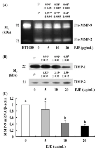
암전이는 암세포의 세포외 기질(extracellular matrix; ECM) 분 해, 이동 및 혈관 내 이동, 암세포의 원격 부위 착상 과정을 통 해 이루어진다(21). 등골나물 추출물이 MDA-MB-231 세포의 전 이에 미치는 영향을 조사하기 위하여 먼저 transwell system을 사 용하여 세포의 이동 및 침윤에 미치는 영향을 조사하였다. MDA-MB-231 세포에 등골나물 추출물을 0, 5, 10 및 20 µg/mL로 처리 하고 4시간 동안 이동한 세포의 수를 확인한 결과 등골나물 추 출물은 MDA-MB-231 세포의 이동을 농도의존적으로 감소시킴을 확인하였다(Fig. 1A). 또한, 세포외 기질 성분으로 알려진 matrigel 로 코팅된 invasion chamber를 사용하여 15시간 동안 세포의 침 윤을 살펴본 결과 세포의 침윤 역시 농도의존적으로 감소함을 확 인하였다(Fig. 1B). 그러나, 같은 시간 동안 등골나물 추출물을 처 리하였을 때 세포의 증식에는 어떠한 영향도 미치지 않는 것으 로 나타났다(Fig. 1C). 이러한 결과를 통해 등골나물 추출물은 세 포독성이 없으면서 유방암세포인 MDA-MB-231의 이동 및 침윤 을 억제하여 암전이 억제제로 개발할 수 있는 좋은 소재가 될 수 있음을 제시한다. 등골나물 추출물이 MDA-MB-231 세포의 MMPs, TIMPs 그 리고 uPA의 분비에 미치는 영향 ECM의 분해는 암전이의 중요작용으로 암세포에서 생성 분비 되는 다양한 proteolytic enzyme에 의해 이루어진다(22). ECM의 분해에 관여하는 중요한 유전자로는 세포외 기질의 구성성분을 분해하는 효소인 MMPs와 plasminogen을 plasmin으로 활성화시 켜 세포외 기질의 분해를 유도하는 uPA가 있다(7). 등골나물 추 출물이 MDA-MB-231 세포의 이동 및 침윤을 억제하였으므로 관 련 유전자의 발현에 미치는 영향을 조사하였다. MMPs의 분비를 조사하기 위하여 gelatin zymography를 수행한 결과 pro MMP-9 과 pro MMP-2는 20 mg/mL의 등골나물 추출물을 처리한 경우에 만 유의적으로 감소하였다(Fig. 2A). MMP-9의 mRNA 수준을 확 인하기 위해 real-time PCR을 수행한 결과 10과 20 µg/mL 등골 나물 추출물은 MMP-9의 transcript 수준을 유의적으로 감소시켰 다(Fig. 2C). 등골나물 추출물이 MMPs의 분비를 억제하였으므로 MMPs의 활성을 저해하는 단백질로 알려진 TIMPs의 발현을 Western blot을 수행하여 확인하였다. Fig. 2B에 따르면 TIMP-1은 20 mg/mL의 등골나물 추출물을 처리한 경우에만 감소하는 것으 로 나타났으며, 그와는 반대로 TIMP-2는 10과 20 mg/mL의 등골 나물 추출물을 처리한 경우에 유의적으로 증가하는 것으로 나타 났다. TIMP-1의 경우는 MMP-9의 저해제로도 알려져 있으나(14), 그 외에도 세포증식 촉진, 세포사멸 억제 등의 다른 여러 가지 기능을 가지고 있어(23) 이러한 결과를 보인 것으로 판단된다. Table 1. Primer sequences used form of PCR amplification
mRNA Primer sequences (5'-3') MMP-9 Sense: GCTCTTCCCTGGAGACCTG
Anti-sense: TTTCGACTCTCCACGCATC ICAM Sense: GCCGGCCAGCTTATACACAA
Anti-sense: CAATCCCTCTCGTCCAGTCG Integrin-α2 Sense: GCACAGCAATGTGGGAATC
Anti-sense: GGCTGAGTTGCAGGTGAGA β-actin Sense: GAGCCGTGTTTCCTTCCATAnti-sense: GTCCCAGTTGGTGACGATG
uPA Sense: GCCATCTACAGGAGGCACCG Anti-sense: GGGTAATCAATGAAGCAGTG VEGF Sense: ATGCCAAGTGGTCCCAGGCT
Anti-sense: CAGGGTCTCGATTGGATGGC GAPDH Sense: CATCAGCAATGCCTCCTGCA
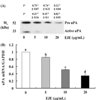
ECM의 분해에 관여하는 또 다른 중요한 유전자인 uPA는 암 세포에서 분비되며 uPA receptor와 결합하여 plasminogen을 plas-min으로 활성화시키며 활성화된 plasmin은 직·간접적으로 ECM 을 분해하여 세포의 이동 및 침윤을 도와준다(24). uPA 단백질의 발현 변화를 확인하기 위하여 Western blot을 수행한 결과 등골
나물 추출물은 uPA 단백질의 발현을 농도의존적으로 감소시켰으 며 특히, 분자량 35 kDa의 active uPA가 현저하게 감소하였다(Fig. 3A). 이러한 결과는 mRNA 수준에서도 확인할 수 있었다(Fig. 3B). 등골나물 추출물이 MDA-MB-231 세포의 VEGF 발현에 미치 는 영향
신생혈관생성은 암전이 과정에 관여할 뿐만 아니라 초기 전암 병소(premalignant lesions)에서부터 암세포의 성장에 필수적인 과 정으로서 인식되고 있으며, vascular endothelial growth fac-tor(VEGF)는 신생혈관생성을 유도하는 중요한 angiogenic factor로 알려져 있다(25). 따라서 등골나물 추출물이 VEGF의 발현에 미 치는 영향을 확인하기 위하여 Western blot 및 real-time PCR을 Fig. 2. EJE decreases MMP and TIMP-1 secretion and increases TIMP-2 secretion in MDA-MB-231 cells. Cells were plated at a density of 1×10⁶ cells/dish in 100 mm dishes. After serum starvation, cells were incubated with 0-20µg/mL EJE for 24 h. 24 h conditioned media were collected and concentrated for gelatin zymography (A) and Western blotting (B). The volumes of media loaded onto the gel were adjusted for equivalent proteins. Photographs of commassie blue stained gel (A) and chemilu-minescent detection of the immunoblots (B), which were representative of three independent experiments, are shown. The relative abundance of each band was quantified and the control levels were set at 1.The adjusted means±SEM (n=3) of each band is shown above each blot. Serum-deprivated MDA-MB-231 cells were incubated with 0-20µg/mL EJE for 12 h. Total RNA was isolated, reverse transcripted, and real time-PCR was conducted. The expression of mRNA was normalized with that of β-actin. Each bar represents the mean±SEM (n=3). Means without a common letter differ, p<0.05 (C).
Fig. 1. EJE decreases migration and invasion of MDA-MB-231 cells. MDA-MB-231 cells were serum-deprived in DMEM/F12 containing 1% charcoal-stripped FBS for 24 h. The lower side of 6.5 mm transwell filter was coated with type IV collagen. Cells were plated into upper compartment at 2.5×10⁴ cells/well and treated with 0-20µg/mL of EJE. The lower compartment was filled with DMEM/F12 containing 1% charcoal-stripped FBS and 0.1% BSA. Cells were incubated for 4 h. The migrated cells were quantified by counting the H&E-stained cells (A). Cells were plated in 6.5 mm matrigel-coated transwell at 2.5×10⁴ cells/well and treated with 0-20 µg/mL EJE. Lower compartment was filled with DMEM/F12 containing 1% charcoal-stripped FBS and 0.1% BSA. Cells were incubated for 15 h (B). MDA-MB-231 cells were plated at 8×10⁴ cells/well in 12-well plates with DMEM/F12 supplemented with 10% FBS and 2 mg/mL insulin. 24 h after plating, the monolayers were serum-deprivated for 24 h in DMEM/F12 containing 1% charcoal-stripped FBS. After serum deprivation, cells were incubated for 4 hours in DMEM/F12 with 1% charcoal-stripped FBS medium containing various concentrations (0-20µg/mL) of EJE. Cell numbers were estimated by the MTT assay (C). Each bar represents the mean±SEM (n=3). Means without a common letter differ, p<0.05.
수행하였다. 그 결과 VEGF의 발현은 5 µg/mL의 등골나물 추출 물을 처리하였을 때 증가하였다가 10과 20 µg/mL 처리시에는 점 차 감소하는 경향을 보였다(Fig. 4A). VEGF의 mRNA수준을 확 인한 결과에서도 Western blot 결과와 마찬가지로 VEGF의
tran-Fig. 5. EJE decreases cell adhesion and decreases integrin-α2 and ICAM expressions in MDA-MB-231 cells. Cells were plated in human collagen type IV-coated CytoMatrixTM Cell Adhesion Strips (Chemicon). Cells were incubated in DMEM/F12 containing 1% charcoal-stripped FBS with 0-20µg/mL EJE for 45 min. Strips were rinsed twice with PBS (containing Ca2+/Mg2+) and stained for 5 min with 0.2% crystal violet in 10% ethanol. Strips were washed 3 times with PBS and solubilized with solubilization buffer (1:1, 50% ethanol:0.1 mol/L NaH2PO4, pH 4.5) until the cell-bound stain is completely solubilized. Cell-bound stains were quantified by determining the absorbance at 570 nm on microplate reader (A). MDA-MB-231 cells were serum-deprived and treated as in Figure 1. Total cell lysates were subjected to immunobloting with antibodies raised against integrin-α2 or ICAM. Photographs of chemilu-minescent detection of the blot, which were representative of three independent experiments, are shown. The relative abundance of each band was quantified and the control levels were set at 1 (B). Real-time PCR was performed as described in Fig. 2 (C). Each bar represents the mean±SEM (n=3). Means without a common letter differ, p<0.05.
Fig. 3. EJE reduces the secretion of uPA in DU145 cells. Cells were plated and treated as described in Fig. 2. 24 h conditioned media were collected and concentrated for Western blotting. The volumes of media loaded onto the gel were adjusted for equivalent proteins. Photographs of chemiluminescent detection of the immunoblots, which were representative of three independent experiments, are shown. The relative abundance of each band was quantified and the control levels were set at 1 (A). Real-time PCR was performed as described in Fig. 2 (B). Each bar represents the mean±SEM (n=3). Means without a common letter differ, p<0.05.
Fig. 4. EJE decreases the secretion of VEGF in MDA-MB-231 cells. MDA-MB-231 cells were plated and treated as described in Fig. 2. 24 h conditioned media were collected and concentrated for Western blotting, which were representative of three independent experiments, are shown. The relative abundance of each band was quantified and the control levels were set at 1 (A). Real-time PCR was performed as described in Fig. 2 (B). Each bar represents the mean±SEM (n=3). Means without a common letter differ, p<0.05.
script 수준이 5 µg/mL의 등골나물 추출물에 의해 증가하는 경향 을 보이다가 10과 20 µg/mL 농도에서는 유의적으로 감소하는 것 을 확인하였다(Fig. 4B). 신생혈관형성을 억제하는 것으로 알려진 소재들이 항암제 또는 암전이 억제제로 개발되고 있는 것으로 보 아(26,27) 신생혈관생성의 중요인자인 VEGF의 발현을 현저히 감 소시킨 등골나물 추출물 역시 종양의 성장 및 전이 억제에 크게 기여할 수 있을 것으로 판단된다. 등골나물 추출물이 MDA-MB-231 세포의 부착능에 미치는 영향 등골나물 추출물이 암세포의 이동 및 침윤을 억제하였으므로 암전이에 또 다른 중요한 요인인 암세포의 부착에 미치는 등골 나물 추출물의 영향을 조사하였다. 등골나물 추출물을 처리하고 human collagen type I이 코팅된 plate에 부착된 MDA-MB-231 세포수를 정량한 결과 등골나물 추출물 처리로 인하여 MDA-MB-231 세포의 부착이 농도의존적으로 감소하였다(Fig. 5A). 종양세 포와 기저막의 matrix와의 유착에는 다양한 adhesion molecule들 이 관여하는 것으로 알려져 있다(28). 그 중 세포와 세포, 세포와 기질간의 결합을 매개하는 단백질인 integrin과 integrin 수용체의 ligand로 알려진 inter-cellular cell adhesion molecule(ICAM)의 발 현을 Western blot을 수행하여 조사하였다. Fig. 5B에서 보는 바 와 같이 등골나물 추출물은 ICAM의 단백질 수준은 현저히 감소 시켰으나 integrin-α2는 변화시키지 않았다. 그러나, ICAM과 inte-grin-α2의 transcript 수준은 모두 농도의존적으로 감소시켰다(Fig. 5C). 이러한 결과를 통해 등골나물 추출물은 integrin-α2와 ICAM 의 발현을 조절함으로써 암세포의 부착을 억제하는 것으로 보인다.
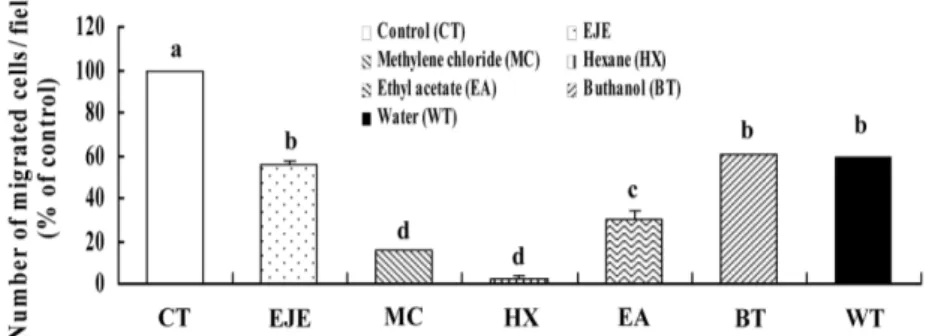
등골나물 추출물의 용매별 분획물이 MDA-MB-231 세포의 이 동에 미치는 영향
MMP-9, -2, uPA 및 TIMP-1의 감소, TIPM-2의 증가 그리고 adhesion molecule들의 감소를 통해 MDA-MB-231 세포의 이동, 침윤 및 부착을 억제시킨 등골나물 추출물에 함유되어 있는 활 성성분을 조사하기 위해 여러 용매를 이용하여 단계적으로 분획 하고 각각의 분획물이 MDA-MB-231 세포의 이동에 미치는 영 향을 조사하였다. 그 결과 비극성 유기용매인 헥산, 에틸아세테 이트, 메틸렌클로라이드를 사용하여 분리한 분획에서 MDA-MB-231 세포의 이동이 각각 97, 69 및 85% 억제되었으며 특히, 헥 산 분획물(HX)에서 세포의 이동이 가장 현저히 억제됨을 확인하 였다(Fig. 6). 보통, 헥산에 의해 분리되어 나오는 물질은 비교적 휘발성이 낮은 완전 비극성 유기화합물로 알려져 있어 등골나물 에 들어있는 비극성 유기화합물이 암세포의 이동을 억제하는데 효과를 나타낼 것으로 판단된다. 이상의 결과를 통해 등골나물 추출물은 MMP-9, MMP-2, uPA 및 TIMP-1의 감소, TIPM-2의 증가 그리고 adhesion molecule들 의 감소를 통해 유방암세포의 전이를 억제하는 것으로 판단된다. 따라서, 등골나물 추출물은 비교적 독성과 부작용이 적은 암전이 억제제로 개발할 수 있는 좋은 소재가 될 수 있음을 제시한다. 이러한 용도로 등골나물 추출물을 사용하기 위해서는 전이 억제 효능을 나타내는 성분의 동정, 동물 모델을 이용한 in vivo 실험 및 좀 더 세밀한 기전 연구 등 추후에도 지속적이고 다양한 연 구의 수행이 필요할 것이다.
요
약
등골나물은 국화과 여러해살이 식물로 한방에서는 고혈압, 폐 렴, 황달, 홍역, 요통 등에 사용한다고 알려져 있다. 본 연구에서 는 등골나물의 꽃 부위를 추출하여 등골나물 추출물이 유방암 세 포인 MDA-MB-231 세포의 이동, 침윤 및 부착에 미치는 영향을 조사하였다. 그 결과 MDA-MB-231 세포의 이동, 침윤 및 부착 은 등골나물 추출물의 농도(0-20 µg/mL)가 증가할수록 현저하게 감소하였다. 등골나물 추출물은 MMP-9, MMP-2의 활성을 억제 하였고, TIMP-1의 발현은 감소시킨 반면 TIMP-2의 발현은 증가 시켰다. 또한, 등골나물 추출물은 uPA, VEGF 그리고 ICAM의 mRNA 및 단백질 수준을 현저히 감소시켰다. 특히, 등골나물 헥 산 분획물이 유방암세포의 이동을 현저하게 억제하였다. 이상의 결과로부터 등골나물 추출물은 MMP-9, MMP-2, uPA, TIMP-1 및 ICAM의 감소, TIPM-2의 증가를 통해 유방암세포의 전이를 억제 하는 것으로 판단된다. 따라서 본 연구는 이러한 효능을 지닌 등 골나물 추출물을 암전이에 효과가 있는 암예방제나 항암제로 개 발할 수 있는 가능성을 제시한다.감사의 글
이 논문은 2010년 정부(교육과학기술부)의 재원으로 한국연구 재단의 지역 거점 연구단 육성사업(의료 바이오 신소재 융복합 연구사업단)과 농촌진흥청 바이오그린21사업(과제번호: PJ007092) 에 의하여 수행되었으며 이에 감사드립니다.문
헌
1. Korea National Cancer Center. Cancer statistics. Incidence of
can-Fig. 6. Hexane fraction of EJE decreases migration of MDA-MB-231 cells. Cells were serum-deprived as described in can-Fig. 1. Cells were plated into upper compartment at 2.5×10⁴ cells/filter and treated with several solvent fraction of EJE (20µg/mL). Lower compartment was filled with DMEM/F12 containing 1% charcoal-stripped FBS and 0.1% BSA. Cells were incubated for 4 h. The migrated cells were quantified by counting the H&E-stained cells. Each bar represents the mean±SEM (n=3). Means without a common letter differ, p<0.05.
cer. Available from: http://www.cancer.go.kr/cms/statics/incidence/ index.html#1. Accessed Oct. 11, 2010.
2. Zhou J, Zhong Y. Breast cancer immunotherapy. Cell. Mol. Immunol. 1: 247-255 (2004)
3. Lee JM. It’s Okay to Become Familiar Slowly: 421 of Wild Edi-ble Greens. Hwan creative company. Seoul, Korea. p. 152 (2009) 4. Choi C. Molecular biological mechanism of tumor invasion and
metastasis. Life Sci. Technol. 28: 16-22 (2003)
5. Yoon SO, Park SJ, Yun CH, Chung AS. Roles of matrix metallo-proteinases in tumor metastasis and angiogenesis. J. Biochem. Mol. Biol. 36: 128-137 (2003)
6. Liao HF, Chen YY, Liu JJ, Hsu ML, Shieh HJ, Liao HJ, Shieh CJ, Shiao MS, Chen YJ. Inhibitory effect of caffeic acid phen-ethyl ester on angiogenesis, tumor invasion, and metastasis. J. Agr. Food Chem. 51: 7907-7912 (2003)
7. Moon HS, Sung SH, Chung HW, Kim SC. Expression of MT1, 2, 3-MMP in invasive cervical cancer. Korean J. Obstet. Gynecol. 49: 1240-1248 (2006)
8. Kimura K, Cheng XW, Nakamura K, Inoue A, Hu L, Song H, Okumura K, Iguchi A, Murohara T, Kuzuya M. Matrix metallo-proteinase-2 regulates the expression of tissue inhibitor of matrix metalloproteinase-2. Clin. Exp. Pharmacol. P. 37: 1096-1101 (2010)
9. Liabakk NB, Talbot I, Smith RA, Wikinson K, Balkwill F. Matrix metalloproteinase 2 (MMP-2) and metalloproteinases 9 (MMP-9) type IV collagenases in colorectal cancer. Cancer Res. 56: 190-196 (1996)
10. Duffy MJ, Maguire TM, Hill A, McDermott E, O’iggins N. Met-alloproteinases role in breast carcinogenesis, invasion, and metastasis. Breast Cancer Res. 2: 252-257 (2000)
11. MacDougall JR, Matrisian LM. Contributions of tumor and stro-mal matrix metalloproteinases to tumor progression, invasion, and metastasis. Cancer Metast. Rev. 14: 351-362 (1995)
12. Xu P, Wang YL, Zhu SJ, Luo SY, Piao YS, Zhuang LZ. Expres-sion of matrix metalloproteinase-2, -9, and -14, tissue inhibitors of metalloproteinase-1, and matrix proteins in human placenta during the first trimester. Biol. Reprod. 62: 988-994 (2000) 13. Yoon HK, Park SM. MMP-2 and MMP-9 expressions in breast
carcinomas and relationship with major prognostic factors. Korean J. Pathol. 38: 79-85 (2004)
14. Lambert E, Dassé E, Haye B, Petitfrère E. TIMPs as multifacial proteins. Crit. Rev. Oncol. Hematol. 49: 187-198 (2003)
15. Ulisse S, Baldini E, Toller M, Marchioni E, Giacomelli L, De Antoni E, Ferretti E, Marzullo A, Graziano FM, Trimboli P,
Biordi L, Curcio F, Gulino A, Ambesi-Impiombato FS, D'Armiento M. Differential expression of the components of the plasminogen activating system in human thyroid tumour derived cell lines and papillary carcinomas. Eur. J. Cancer 42: 2631-2638 (2006)
16. Jogi A, Rono B, Lund IK, Nielsen BS, Ploug M, Hoyer-Hansen G, Romer J, Lund LR. Neutralisation of uPA with a monoclonal antibody reduces plasmin formation and delays skin wound heal-ing in tPA-deficient mice. PLoS One 5:e12746 (2010)
17. Osada K, Seishima M, Kitajima Y, Yaoita H, Mori S. Decreased integrin α2, but normal response to TGF-beta in scleroderma fibroblasts. J. Dermatol. Sci. 9: 169-175 (1995)
18. Ko YS. Function of cell adhesion molecule (CAM) in angiogene-sis. J. Korean Endocr. Soc. 16: 305-312 (2001)
19. Cébe-Suarez S, Zehnder-Fjällman A, Ballmer-Hofer K. The role of VEGF receptors in angiogenesis;complex partnerships. Cell. Mol. Life Sci. 63: 601-615 (2006)
20. Denizot F, Lang R. Rapid colorimetric assay for cell growth and survival: Modifications of the tetrazolium dye procedure giving improved sensitivity and reliability. J. Immunol. Methods 89: 271-277 (1986)
21. Liotta LA, Kleinerman J, Catanawara P, Rynbrandt D. Degrada-tion of basement membrane by murine tumor cells. J. Natl. Can-cer I. 58: 1427-1431 (1977)
22. Woessner JF Jr. Matrix metalloproteinase and their inhibitors in connective tissue remodeling. FASEB J. 5: 2145-2154 (1991) 23. Stetler-Stevenson WG. Tissue inhibitors of metalloproteinases in
cell signaling: Metalloproteinase-independent biological activi-ties. Sci. Signal. 1: re6 (2008)
24. Crippa MP. Urokinase-type plasminogen activator. Int. J. Bio-chem. Cell B. 39: 690-694 (2007)
25. Goh PP, Sze DM, Roufogalis BD. Molecular and cellular regula-tors of cancer angiogenesis. Curr. Cancer Drug Tar. 7: 743-758 (2007)
26. Huang CS, Liao JW, Hu ML. Lycopene inhibits experimental metastasis of human hepatoma SK-Hep-1 cells in athymic nude mice. J. Nutr. 138: 538-543 (2008)
27. Kunnumakkara AB, Anand P, Aggarwal BB. Curcumin inhibits proliferation, invasion, angiogenesis and metastasis of different cancers through interaction with multiple cell signaling proteins. Cancer Lett. 269: 199-225 (2008)
28. Springer TA. Adhesion receptors of the immune system. Nature 346: 425-434 (1990)