ABSTRACT
Purpose: It has been previously reported that breast tumor incidence, growth, and metastasis
are stimulated by high-fat diet but reduced by caloric restriction. However, few studies have elucidated the effects of dietary change from a high-fat diet after breast cancer initiation.
Therefore, in this study, we attempted to provide practical assistance to breast cancer prevention and management by investigating the effects of dietary change from a high-fat diet to normal diet on breast cancer growth and metastasis.
Methods: The experimental animals were divided into 2 groups (high-fat diet control [HFC]
group and diet restriction [DR] group) and consumed a high-fat diet for 8 weeks. 4T1 cells were transplanted into subcutaneous fat or tail vein to measure the growth and metastasis of breast cancer. The HFC and DR groups continuously ingested either high-fat diet or AIG-93G diet for 5 weeks or 3 weeks, respectively. Cell proliferation and apoptosis markers from tumor tissues were analyzed by Western blot analysis. The data were analyzed using the SPSS 25.0 package program.
Results:
The results show that the DR group significantly reduced breast tumor initiation, growth, and tumor tissue weight compared to the HFC group. The DR group suppressed tumor growth by decreasing proliferation and inducing apoptosis through down-regulation of Bcl-xL and up-regulation of caspase-3 activity. Furthermore, the DR group significantly reduced numbers of metastasized tumors in lung tissues.
Conclusion: These results suggest that dietary change from a high-fat diet to normal diet
decreased breast growth by reducing cell proliferation and inducing apoptosis and metastasis.
Taken together, these results indicate that dietary change to a low-fat and balanced diet might suppress breast tumor growth and metastasis even after tumor diagnosis.
Keywords: breast cancer, high-fat diet, apoptosis, metastasis
Research Article
Received: May 18, 2020 Revised: Jun 16, 2020 Accepted: Jun 18, 2020 Correspondence to Jung-Hyun Kim
Department of Physical Education, Chung-Ang University, 84 Heukseok-ro, Dongjak-gu, Seoul 06974, Korea.
Tel: +82-2-820-5378 E-mail: [email protected]
© 2020 The Korean Nutrition Society This is an Open Access article distributed under the terms of the Creative Commons Attribution Non-Commercial License (http://
creativecommons.org/licenses/by-nc/3.0/) which permits unrestricted non-commercial use, distribution, and reproduction in any medium, provided the original work is properly cited.
ORCID iDs Seung hwa Park
https://orcid.org/0000-0003-3318-0784 InKyung Jung
https://orcid.org/0000-0003-3914-4143 Jung-Hyun Kim
https://orcid.org/0000-0001-5882-4094 Funding
This work was supported by the Ministry of Education of Republic of Korea and the National Research Foundation of Korea (NRF- 2017S1A5B8067020).
Conflict of Interest
There are no financial or other issues that might lead to conflict of interest.
Effect of dietary changes from high-fat diet to normal diet on breast cancer growth and metastasis
Seung hwa Park , InKyung Jung , and Jung-Hyun Kim
Department of Physical Education, Chung-Ang University, Seoul 06974, Korea
고지방식이에서 일반식이로의 전환이 유방암의 성장 및 전이에 미치는 영향
박성화 , 정인경 , 김정현
중앙대학교 체육교육과
서론
1990
년 이후 한국인의 사망원인
1위는 악성종양으로 해마다 암으로 인한 사망률이 증가하고
있으며
,발생하는 암의 종류도 생활 패턴과 식생활의 서구화로 인해 변화되고 있다
[1,2].최
근 연구 보고에 따르면
,우리나라의 경우 서구 사회에서 많이 발생하는 암인 유방암
,전립선 암
,대장암의 발병률이 꾸준히 증가하는 추세로 나타났으며
[3],특히 유방암은 우리나라 여 성들에게 가장 빠르게 증가하는 암으로 보고되었다
[3].서구 사회의 경우
,유방암은 연령이 증가할수록 발병률이 증가하는 것으로 나타났으나
,우리나라의 경우
40대 이하의 여성이 전
체 환자의
55.7%를 차지할 만큼 젊은 여성의 발병률이 높아지고 있으며
[2],갑상선암 다음으
로 발병률이 높은 암으로 보고되었다
[3].유방암 발생은 초경
,폐경
,출산 시기
,수유 경험 등 유전적
·환경적인 요인에 의해 영향을 받 으며
,고지방식
,고칼로리로 대변되는 서구화된 식생활과 비활동적인 생활습관에 의해 발병
률이 증가되는 것으로 보고되고 있다
[4].서구화된 식습관과 비활동적인 생활습관은 체지
방의 양을 증가시킴으로써 비만을 유발하며
,이로 인해 체내 염증반응이 증가하여 암의 생성 과 성장을 촉진시키는 것으로 나타났다
[5-8].비만은 유방암의 위험 인자일 뿐 아니라 유방암의 치료 및 재발
,생존에도 영향을 주는 환 경요인으로 알려져 있다
[9].국민건강영양조사 결과에 의하면 전체 유방암 생존자의
2/3가 과체중 이상으로 나타났으며
,체지방률이
30%를 넘는 경우도
89.2%인 것으로 보고되었다
[10].또한
, Rooney와
Wald [11]는
50%–96%의 여성이 유방암 진단 후 평균
2.5–5.2 kg의 체중 이 증가하였으며
,체중 증가에 따른 비만은 유방암 유발에 대한 위험률을 증가시킬 뿐 아니 라 유방암 진단 후 예후를 악화시키고 삶의 질을 저하시킨다고 보고하였다
.비만인 여성의 경우 체지방으로 부터 에스트로겐 분비가 증가되어 유방암의 발생 위험이 높아질 뿐 아니라
유방암 진단 후 체중이 증가하게 되면 암 재발률이
1.5배
,사망률이
1.6배 증가하는 것으로 알 려져 있어 비만이 유방암의 성장과 전이의 위험을 증가시키는 것으로 나타났다
[2].특히
,비 만을 유발하는 주요 요인 중 고지방식이는 유방암의 생성과 성장
,전이를 촉진시키는 것으로 보고되어 유방암 생성 및 전이 억제를 위해 체중 조절과 함께 건강하고 균형 있는 식이를 섭 취하는 것이 매우 중요한 것으로 보고되었다
[12-14].유방암을 조기에 발견하면 수술이나 방사선 및 화학적 요법으로 치료가 가능하나 다양한 부
작용을 겪게 되며
,전이가 된 경우
,치료가 어려울 뿐 아니라 여러 합병증에 의해 사망에 이르
게 될 수 있다
.따라서
,유방암 발견 시 유방암의 성장과 전이를 안전하게 억제할 수 있는 방안
이 강구되고 있으며
,다양한 기능성 식품의 섭취나 식이 조절
,열량 제한 등이 안전하게 유방
암의 성장과 전이를 억제하는 것으로 보고되었다
[15,16].그러나
,대부분의 연구에서는 유방
암 생성 이전의 열량 제한에 의한 암 억제 효과나 식이 섭취 시간 제한에 의한 영향을 검증하
였으며
,유방암의 생성이나 전이 이후 열량이나 식이 제한에 대한 연구는 매우 미흡한 실정
이다
.이에 본 연구에서는 정상식이로의 전환이 고지방식이로 인해 촉진된 유방암의 성장과
전이에 미치는 영향과 그 기전을 알아보고자 한다
.연구방법
연구설계
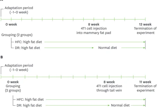
고지방식이에서 일반식이로의 전환이 유방암세포의 성장
(Fig. 1A)과 전이
(Fig. 1B)에 미치는
영향을 알아보기 위해
Fig. 1과같이 실험을 진행하였다
.실험 동물을
1주간 기본 식이
(AIN-93G)
로 실험 환경에 적응시킨 후 임의적으로 고지방식이 유방암 유도군
(high-fat diet control;HFC)
과 고지방식이 후 일반식이로 전환한 유방암 유도군
(diet restriction; DR)으로 나누었 다
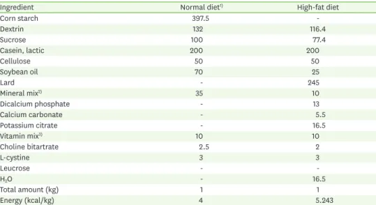
.일반식이와 고지방식이의 조성은
Table 1과 같다
.모든 군은
8주간 고지방식이로 비만을
유발한 후 유방암 세포인
4T1세포를 유방 조직에 이식하거나 꼬리 정맥을 통해 전이시켰으
며
, DR군은 식이를
AIN-93G로 전환하고
,대조군
(HFC)은 고지방식이를 계속 유지시켰다
.유
방암 성장 실험은 유방암 세포 이식
5주 후
,전이 실험은
3주 후에 종료하였으며
,본 동물 실험 은 중앙대학교의 동물윤리위원회에 의해서 승인받아 실시되었다
(승인번호
: 2016-00099).실험동물
실험 동물은
Koatech (Seoul, Korea)에서
8주령의
Balb/c female mouse 20마리씩 나누어 분양
받아 사용하였으며
,온도
22 ± 1°C,습도
55 ± 1°C, 12시간 간격으로 점등과 소등을 반복한 동물 실험실에서 사육되었다
.체중과 유방암의 크기는 매주
2회 측정하였다
.세포배양및유방암과전이유도
Balb/c mouse
에 유방암을 유도하기 위해
4T1 (CRL-2539™)세포를 이승민 교수님
(연세대학
교
)께 받아 사용하였다
. 4T1세포는
37°C, 5% CO2, 90%이상 습도가 유지된 항온기에서
10%A
Adaptation period (−1~0 week)
4T1 cell injection
into mammary fat pad Termination of
experiment
0 week 8 week 13 week
Normal diet Grouping (2 groups)
HFC: high fat diet DR: high fat diet
B
Adaptation period (−1~0 week)
4T1 cell injection
through tail vein Termination of experiment
0 week 8 week 11 week
Normal diet Grouping
(2 groups)
HFC: high fat diet DR: high fat diet
Fig. 1. Experimental study design. After 1 week of adaptation, 8-week-old Balb/c mice were fed a high-fat diet for 8 weeks, and then 4T1 mouse mammary tumor cells were transplanted into their mammary fat pads (1 × 10⁴) (A) or injected into tail vein (5 × 10³). After tumor cell injection (B), DR group was fed a normal diet and HFC group was continuously fed a high-fat diet until the experiments ended (n = 10 per group).
HFC, high-fat diet control; DR, diet restriction.
fetal bovine serum (Gibco; Thermo Fisher Scientific, Waltham, MA, USA), 1% penicillin strepto- mycin (WelGENE, Daegu, Korea), 1% non-essential amino acid (WelGENE)
가 포함된
RoswellPark Memorial Institute 1640 (WelGENE)
배지를 이용하여 계대 배양하였다
.원발성 종양
(pri-mary tumor)
을 유도하기 위해
4T1세포를
trypsin처리하여 단세포의 형태로 만든 후
, mam-mary fat pad
에
1 × 10⁴개를 피하 주사하였다
.또한
,유방암의 전이를 유도하기 위해 적외선 램
프로
Balb/c mouse의 체온을 상승시켜 혈관을 확장시킨 후
, 5 × 10³개의
4T1세포를 꼬리 정맥
을 통해 전이시켰다
. 유방암의개시및성장측정암의 개시는 종양이 처음 촉진된 시점을 말하는 것으로
8 mm3이상 일 때를 기준으로 계산
하였다
.유방암의 부피는 버니어 캘리퍼스
(Mitutoyo, Kawasaki, Japan)를 이용하여 주
2회 종
양의 가장 긴 지름과 가장 짧은 지름을 측정하였으며
,피하 두께를 제외하기 위해 지름에서
1 mm를 빼고 지름을 계산하였다
.종양의 부피는
4/3πr3공식으로 계산하였다
[17].시료채취
유방암 유도
5주
,유방암 전이
3주 후 조직 채취를 위해 실험동물을 전신 마취제
(isoflu-rane)
로 마취시킨 후 종양
,간
,비장
,신장 주변 지방조직을 적출하여 차가운
phosphatebuffer saline
로 혈액을 제거한 후 여과지로 수분을 제거하고 장기의 무게를 측정하였다
.Table 1. Ingredients of the experimental diets (g/kg diet)
Ingredient Normal diet1) High-fat diet
Corn starch 397.5 -
Dextrin 132 116.4
Sucrose 100 77.4
Casein, lactic 200 200
Cellulose 50 50
Soybean oil 70 25
Lard - 245
Mineral mix2) 35 10
Dicalcium phosphate - 13
Calcium carbonate - 5.5
Potassium citrate - 16.5
Vitamin mix3) 10 10
Choline bitartrate 2.5 2
L-cystine 3 3
Leucrose - -
H2O - 16.5
Total amount (kg) 1 1
Energy (kcal/kg) 4 5.243
1)All amounts were based on an AIN-93G diet. 2)Mineral mix: calcium carbonate (35.7%), potassium phosphate monobasic (25.0%), potassium citrate monohydrate (2.8%), sodium chloride (7.4%), potassium sulfate (4.66%), magnesium oxide (2.43%), ferric citrate (0.606%), zinc carbonate (0.165%), manganous carbonate (0.063%), cupric carbonate (0.031%), potassium iodate (0.001%), sodium selenite (0.001%), ammonium paramolybdate (0.001%), sodium metasilicate (0.145%), chromium potassium sulfate (0.028%), lithium chloride (0.002%), boric acid (0.008%), sodium fluoride (0.006%), nickel carbonate hydroxide tetrahydrate (0.003%), ammonium vanadate (0.001%), and sucrose (20.95%). 3)Vitamin mix: nicotinic acid (0.3%), calcium pantothenate (0.16%), pyridoxine-HCl (0.06%), riboflavin (0.06%), folic acid (0.02%), D-biotin (0.002%), vitamin B12 in 0.1% mannitol (0.25%), DL-α-tocopherol acetate, 500 IU/g (1.50%), retinol palmitate, 500,000 IU/g (0.08%), vitamin D3 (50,000 IU/g) (0.02%), vitamin K (0.007%) and sucrose (97.47%).
유방암의폐전이측정
폐에 전이된 유방암의 수와 크기를 측정하기 위해 폐를
India black ink (Talens, Apeldoorn,Netherlands)
로 염색하였다
.이를 위해 희생된 실험동물의 기도 부위 조직과 근육을 제거한
후
,기도 삽관하여 기도를 확보하고
,폐 조직이 염색될 때까지
15%로 희석된
India black ink를
기도를 통해 주입하였다
.염색된 폐 조직을
Fekete's solution으로 고정시킨 후
,전이된 유방암 의 수와 크기를 현미경을 통해 측정하였다
.자료수집방법
본 실험 자료의 통계처리 및 분석을 위해
SPSS 25.0을 이용하였으며
,모든 실험값은 반복 실
험에 대한 평균
(mean) ±표준오차
(standard error)로 나타내었다
.두 군 간의 통계적 유의성
검증은 독립 표본
t-검정을 통해 분석하였으며
,두 군 간의 유의한 차이는
p < 0.05수준에서 검증하였다
.결과
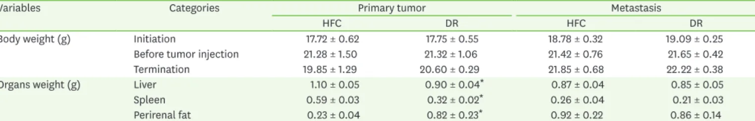
고지방식이에서일반식이로의전환이유방암성장및전이후체중과조직무게에미치는 영향
유방암 생성 전후 체중 및 조직 무게의 변화를 살펴본 결과는
Table 2와 같다
.체중은 실험 전
,암세포 이식 전
,실험 종료일로 구분하여 유방암과 전이군 모두 제시하였다
.유방암 생성군 의 경우 체중은 실험 전
,암 생성 전
,실험 후 모두 유의미한 차이를 보이지 않았으며
,두 군 모
두
8주간의 고지방 식이 이후
20%정도 체중이 증가하였다
.또한
,유방암 생성
5주 후
HFC군
은 체중의
6.72%가 감소한 반면
, DR군은
3.38%만이 감소하여 식이전환 군의 체중감소가 적
게 나타났으나
,두 군 간 유의적인 차이는 보이지 않았다
.조직의 무게를 측정한 결과
,고지 방식이군의 간과 비장 무게가 유의적으로 증가하였으나
,신장 주변의 지방조직은 유의미하 게 감소하였다
.유방암 전이의 경우
, HFC군과
DR군의 실험 전
,유방암 전이 전과 실험 후 체 중 모두 유의미한 차이를 보이지 않았으며
,간과 비장
,신장 주변의 지방 조직의 무게에도 차 이가 없는 것으로 나타났다
.일반식이로의전환이유방암의개시와성장에미치는영향
고지방식이 섭취 후 일반식이로의 전환이 유방암의 개시와 성장에 미치는 영향을 알아본 결
과는
Fig. 2와같다
.유방암 이식 후 종양이 처음 촉진된 시기를 알아본 결과
, HFC군은
9.6일
,DR
군은
11.6일로 나타나 일반식이로 전환한
DR군의 유방암 개시가 고지방식이 대조군에 비
Table 2. Effects of high fat diet on body and organs weights in Balb/c mice injected with 4T1 cells
Variables Categories Primary tumor Metastasis
HFC DR HFC DR
Body weight (g) Initiation 17.72 ± 0.62 17.75 ± 0.55 18.78 ± 0.32 19.09 ± 0.25
Before tumor injection 21.28 ± 1.50 21.32 ± 1.06 21.42 ± 0.76 21.65 ± 0.42
Termination 19.85 ± 1.29 20.60 ± 0.29 21.85 ± 0.68 22.22 ± 0.38
Organs weight (g) Liver 1.10 ± 0.05 0.90 ± 0.04* 0.87 ± 0.04 0.85 ± 0.05
Spleen 0.59 ± 0.03 0.32 ± 0.02* 0.26 ± 0.04 0.21 ± 0.03
Perirenal fat 0.23 ± 0.04 0.82 ± 0.23* 0.92 ± 0.22 0.86 ± 0.14
Data are shown as mean ± SD. HFC and DR represent diet change from 8 weeks of high fat diet to normal diet.
HFC, high-fat diet control; DR, diet restriction.
*p < 0.05.
해 유의미하게 지연된 것으로 나타났다
(Fig. 2A).또한
,고지방식이에서 일반식이로의 전환
이 유방암의 성장에 미치는 영향을 알아본 결과
(Fig. 2B), DR군의 유방암 성장이
HFC군에 비
해 유의미하게 낮아졌으며
(p < 0.05),이러한 차이는 암 이식 이후
17일부터 나타나 실험 종료 시까지 지속되었다
.마지막으로 식이 전환이 유방암 조직의 무게에 미치는 영향을 알아본 결
과
(Fig. 2C),유방암 조직 무게는
HFC군이
1.29 g, DR군이
0.51 g으로 나타나 식이전환으로 인
해 유방암 조직 무게가
61%감소한 것으로 나타났으며
,이 차이는 유의미하였다
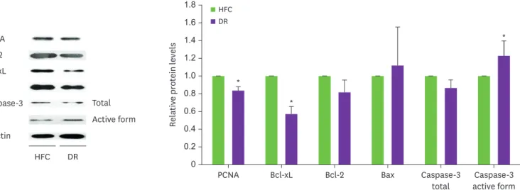
(p < 0.001).일반식이로의전환이유방암의성장조절인자의발현에미치는영향
고지방식이에서 일반식이로의 전환이 유방암의 성장에 미치는 기전을 알아보기 위해 세포
의 성장과 사멸을 조절하는 인자의 발현을 측정한 결과는
Fig. 3과같다
.세포의 분열을 나타
내는
proliferating cell nuclear antigen (PCNA)의 경우
DR군의 발현이
HFC군에 비해 유의미하 게 감소한 것으로 나타났다
.또한
, DR군의 경우 세포사멸을 억제하는 인자인
Bcl-xL의 발현 이 유의미하게 감소하였으며
,세포 사멸을 유도하는
caspase-3의 활성을 유의하게 증가한 것
으로 나타났다
. Bcl-2와
Bax의 발현은
DR군과
HFC군 간에 유의미한 차이는 보이지 않았으
나
, DR군에서
Bcl-2는 감소하는 경향을
, Bax는 증가하는 경향을 보였다
.따라서
,일반식이로
의 전환은 유방암 세포의 분열을 억제하고
,세포 사멸을 촉진함으로써 유방암의 성장을 억 제하는 것으로 판단된다
.일반식이로의전환이유방암의전이에미치는영향
유방암세포가 폐에 전이된 정도를 측정하기 위해 폐를 염색한 후 전이된 폐를 적출하여 전
이된 암의 수와 암의 크기를 분석한 결과는
Fig. 4와같다
.먼저 전이 유무를 살펴본 결과
,모
든 동물에서 폐전이가 야기되었으며
,폐에 전이된 유방암의 수와 부피를 비교한 결과
(Fig. 4)HFC
군은 평균
50.9개의 암이 폐로 전이되었으나
, DR군은 평균
26.1개의 암이 전이된 것으로
C
Tumor weight (g)
1.6 1.4 1.2 1.0 0.8 0.6 0.4 0.2
0 HFC DR
*
A
Days until tumor initiation
14 12 10 8 6 4 2
0 HFC DR
*
*
*
*
*
* *
B
Tumor volume (mm3) 900 800 700 600 500 400
200 300
100
0 5 10 15 20 25 30 35
Time (day) HFC
DR
Fig. 2. Effects of DR on breast tumor latency (A), growth (B), and tumor weight (C) using primary tumor model. Mice were fed the high-fat diet for eight weeks then tumor cells were transplanted into the mice. After transplantation, the mice were fed either high-fat diet or normal diet (n = 10 per group). (A) After inoculation with 1 × 10⁴ 4T1 cells, latency was calculated as the time for the injected tumor cells to reach a size of 8 mm3. (B) Tumor volume was measured twice a week and calculated until they reached 1,000 mm3. (C) Tumor weight was measured after termination of experiments. Values are presented as the mean ± standard error of the mean. HFC and DR represent diet change from 8 weeks of high fat diet to normal diet.
HFC, high-fat diet control; DR, diet restriction.
*p < 0.05.
나타나 두 군 간에 유의적인 차이
(p < 0.05)가 있었다
.폐에 전이된 유방암의 부피를 계산해 본 결과
,고지방식이군이
DR군보다
70%부피가 컸으나
,유의미한 차이는 보이지 않았다
.이 러한 결과는 일반식이로의 전환이 지속적으로 고지방식이를 섭취하는 것보다 유방암의 전
이를 약
48%정도 억제한다는 것을 의미하는 것으로 유방암의 전이를 억제하기 위해서는 지
방과 에너지 밀도가 낮은 식사를 해야 할 것으로 판단된다
. 1.61.8
1.4 1.2 1.0 0.8 0.6 0.4 0.2 0
*
*
*
Relative protein levels
HFC DR
PCNA Bcl-xL Bcl-2 Bax Caspase-3
total Caspase-3
active form PCNA
Bcl-xL Bcl-2
Bax
β-actin
Total Active form Caspase-3
HFC DR
Fig. 3. Effect of DR on tumor cell proliferation and apoptosis using primary tumor tissues. Tumor tissues from the 2 groups were used to measure PCNA, Bcl-2, Bcl-xL, Bax, and caspase-3 expression via Western blot. β-actin was detected as a loading control. HFC and DR represent diet change from 8 weeks of high fat diet to normal diet.
PCNA, proliferating cell nuclear antigen; HFC, high-fat diet control; DR, diet restriction.
*p < 0.05.
B
Tumor number
70 60 50 40 30 20 10
0 HFC DR
*
C
Metastasized tumor volume (mm3) 70 60 50 40 30 20 10
0 HFC DR
A
HFC DR
Fig. 4. Effect of DR on breast tumor metastasis on lung tissue. Mice were fed the high-fat diet for eight weeks then 5 × 10³ 4T1 cells were injected into the tail vein. After metastasis, the mice were fed either high-fat diet or normal diet for 3 weeks and then experiment was terminated (n = 10 per group). (A) Lung tissues were stained with India black ink. (B) Number of metastasized breast tumor was calculated. (C) Total metastasized breast tumor volumes were calculated. HFC and DR represent diet change from 8 weeks of high fat diet to normal diet.
HFC, high-fat diet control; DR, diet restriction.
*p < 0.05.
고찰
식습관 및 생활습관의 변화로 비만율이 증가하면서 비만으로 인한 만성질환의 위험도 함께 증가하고 있으며
,이중 유방암의 발병율도 증가하고 있다
.이에 식생활이나 생활습관을 바 꾸고
,체중을 조절함으로써 유방암의 생성과 전이를 억제하고자 하는 방안이 강구되고 있 다
.비만을 유발하는 다양한 요인 중 고지방식이는 비만뿐 아니라 유방암 생성 및 전이를 촉 진시키는 것으로 보고되었으며
,만성적인 식이 제한이나 주기적인 식이 제한은 유방암의 성 장 및 전이를 억제하는 것으로 나타났다
.그러나
,식이 제한의 시점이나 고지방식이 이후 식 이 제한으로의 전환에 대한 연구는 매우 제한적이다
.따라서
,본 연구에서는 유방암 생성 시 점에 식이를 고지방식이에서 일반식이의 전환이 유방암의 생성과 전이에 미치는 영향과 그 기전을 검증하였다
.선행 연구에 따르면 비만인 유방암 환자의 경우 인슐린 저항성이 높아 혈중 인슐린의 농도
가 높아지게 되며
,높아진 혈중 인슐린이
insulin-like growth factor-1의 합성과 활성을 높임
으로써 유방암의 성장을 촉진시키는 것으로 보고되었다
[18,19].과분비된 인슐린은
insulinreceptor substrate (IRS)-1
를 활성화시키고
,활성화된
IRS-1은 암세포의 생존 기전을 지속적으
로 높여 암세포의 분열을 촉진하고
,사멸을 억제하여 암세포의 성장이 높아지는 것으로 설명
되고 있다
[20,21].또한
,비만인 여성의 경우
,지방세포로부터 에스트로겐이 과다 합성
·분비
되어 유방암 세포의 증식을 높이는 것으로 보고되고 있어 비만이 유방암의 생성과 성장을 촉
진하는 주요 인자로 설명되고 있다
[22].비만뿐 아니라 지속적인 고지방식이도 유방암의 생
성에 영향을 미치는 것으로 보고되었다
.고지방식이 섭취는 암의 개시 시기를 짧게 단축시켰 으며
,암세포의 분열과 혈관 형성 및 전이를 증가시키는 것으로 나타났다
[14].또한
,고지방 식이 섭취는 혈중 지방의 농도를 높여 암세포 내 대식세포를
M2-type으로 분화 시킴으로써 암세포의 성장과 전이를 촉진시키는 것으로 보고되었다
[23].특히
,고지방식이에 의해 체중
증가가 일어나지 않는
obesity-resistant동물 모델의 경우 유방암의 개시가 짧아지고
,암의 성
장이 더욱 증가하는 것으로 나타났다
[24].고지방식이와는 달리 만성적인 식이 제한이나 열량 제한은 발암물질에 의한 유방암 생성 이나 유전자 형질 전환 마우스의 유방암 생성을 억제하는 것으로 보고되었다
. 10%–40%의 열량 제한은 발암물질로 유발한 유방암의 생성을 억제하는 것으로 나타났으며
, 4개월간
의
20%열량 제한은
MMTV-TGF-mice의 유방암 생성을
96%감소시켰다고 보고되었다
[25].또한
,같은 고지방식이 섭취를 섭취하더라도 섭취 시간을
12시간으로 제한하는 경우 랩틴
,monocyte chemoattractant protein-1
과 혈관신생합성 조절 인자의 발현이 낮아져 유방암의 성
장을 유의적으로 억제하였으며
[13],고지방식이 후
30%섭취량을 제한한 식이 제한이 유방 조직의 염증반응을 억제할 뿐 아니라
aromatase의 활성을 감소시켜 유방암의 위험을 낮추는
것으로 나타났다
[12,26].최근에는 열량 섭취 제한뿐 아니라 식이 섭취와 금식을 반복하는 식
이 방법의 효과에 대한 연구도 진행되었는데
,식이와 금식의 반복은 유방암의 개시와 암 발 생을 감소시키고
,수명도 증가시키는 것으로 나타났다
[13,27-30].본 연구에서는
8주간의 고 지방식이 섭취 후 유방암 생성이 일반식이로의 전환에 의해 변화하는지를 측정하였으며
,지 속적인 고지방식이에 비해 일반식이로의 전환이 유방암 개시를 지연시켰을 뿐 아니라 암 성
장 속도와 조직 무게를
60%이상 감소시키는 것으로 나타났다
.이러한 결과는 장기간의 열량
제한뿐 아니라 유방암 생성 이후 열량이나 지방 섭취의 제한 또한 유방암의 성장을 억제할 수
있다는 것을 의미하는 것으로
,비만인 유방암 환자들이 암 진단 이후에라도 체중 조절과 함 께 고지방식이를 지방 함량을 낮춘 균형식으로 바꾸는 것이 암의 성장을 억제하는데 도움이
될 것으로 판단된다
[31].또한
, DR군의 신장 주위 지방의 무게가
HFC군보다 유의미한 것으
로 나타났는데
,이는
HFC군의 경우 빠른 유방암 세포의 성장으로 인해 체중이 감소
(−6.7%)하게 되면서 근육과 체지방 분해가 촉진되었기 때문으로 판단된다
[32].본 연구에서는 유방암 생성 후 고지방식이에서 일반식이로 전환한
DR군의
PCNA발현이
약
20%정도 감소하였으며
, Bcl-xL의 발현 감소로
caspase-3의 활성이 유의하게 증가하였다
.PCNA
는 세포 분열에 필수적인 인자로
DR군의
PCNA발현 감소는 유방암 발생시 식이를 저
지방식이로 전환하면 암세포의 분열을 억제할 수 있음을 의미한다
.또한
, Bcl-xL은
Bcl-2 fam- ily에 속하는 단백질이며
, Bcl-2 family는 미토콘드리아의 외막에서 미토콘드리아의 기능을 조절할 뿐 아니라 세포사멸을 억제하는 인자와 사멸을 촉진하는 인자 간의 균형을 조절하며
세포 사멸을 조절하는 것으로 알려져 있다
[33].따라서
,외부 요인에 의해
Bcl-2 family에 의한
균형이 깨지면 미토콘드리아 내부에서
cytochrome c가 세포질로 방출되어
caspase의 활성을
증가시켜 세포 사멸을 유발하는 것으로 보고되고 있다
[34-36].본 실험에서
DR군의 경우 세
포 사멸을 억제하는
Bcl-2와
Bcl-xL의 발현이
20%–40%정도 감소하였으며
,활성화된 형태의
caspase-3
의 발현이 유의적으로 증가한 것으로 나타나 일반식이로의 전환이
Bcl-2 family유
전자의 발현에 영향을 주어 세포 사멸을 촉진하였을 것으로 판단된다
.다른 선행연구에서도 장기간 섭취하는 지방의 함량이나 종류를 조절하면 암세포의 분열과 사멸에 영향을 주는 것 으로 보고되었다
[14,37]. 10%지방 함유 식이를 섭취한 군의
PCNA발현이 고지방식이 군보 다 유의적으로 감소하였으며
[14], 20%의 옥수수 기름 식이나 올리브 오일 식이를 섭취시킨 후 발암물질로 유방암을 유도한 연구 결과에서는 올리브 오일을 섭취한 군의 암세포 분열이 억제되고 사멸이 증가되었다고 보고되었다
[37].따라서
,유방암을 예방하고 진단 후 암의 성 장을 억제하기 위해서는 지방의 함량을 낮추고
,지방의 종류도 포화지방산이나
n-6가 다량 함유된 지방보다는
n-3나
n-9지방산으로 대체하는 것이 도움이 될 수 있을 것으로 판단된다
.본 연구에서는 일반식이로의 전환이 유방암의 전이를 억제하는 것으로 나타났으며
,전이 된 암세포의 수를
48%가량 감소시켰다
.다른 선행연구에서도 고지방식이가 암세포의 전이 를 촉진하는 것으로 보고하였다
[14,38]. 10주간 고지방식이나 일반식이 섭취 후
Lewis lungcarcinoma
세포를 전이시킨 결과
,고지방식이군의 전이된 암의 수와 크기가 유의하게 증가
한 것으로 나타났으며
,이는 고지방식이 섭취로
pro-inflammatory cytokines의 발현과 신생혈 관합성 관련 인자의 발현이 증가하였기 때문으로 설명하였다
[38].또한
,고지방식이는 암세
포의 전이를 조절하는 기저막 분해 효소인
matrix-metalloproteinase-9와
adhesion molecules의 발현을 증가시켜 유방암의 전이를 촉진시키며
, 10%지방식이는 암의 전이를
40%이상 억
제하는 것으로 나타났다
[14].그러나 대부분의 선행연구에서는
10–16주의 장기간의 식이 조
절에 의한 효과였으며
,유방암 전이 후 고지방식이에서 일반식이로 전환시킨 식이섭취가 암
의 전이에 미치는 영향을 규명한 연구는 매우 미흡하다
.특히
,유방암 환자나 생존자의 경우
다른 암과 달리 과체중 비율이 높고
,유방암 수술 이후에도 체중이 증가하는 경우가 매우 높
다
.이에 유방암 생존자들을 위한 가이드라인에서는 과체중이거나 비만인 경우 식이 조절과
신체활동을 통해 체중을 감소시키고
,정상체중을 유지하라고 권고하고 있으나 이를 실천하
기에 많은 어려움이 있는 것으로 보고되었다
.본 연구 결과 고지방식이 섭취가 유방암의 전
이를 증가시켰으나
,고지방식이를 섭취하였다 하더라도 유방암 생성이나 전이시 지방의 섭
취량을 줄이고 균형 있는 식이를 섭취하는 것이 암의 전이를 감소시킬 수 있을 것으로 판단 된다
.본 연구에서는 고지방식이에서 일반식이로의 전환이 유방암의 성장 및 전이에 미치는 영향을 규명하였으나
,명확한 기전에 대한 연구가 더 필요할 것으로 사료된다
.요약
본 연구에서는 일반식이로의 식이 제한이 고지방식이로 촉진된 유방암의 성장과 전이에 미 치는 영향과 그 기전을 알아보았다
. 8주간 고지방식이를 섭취시킨 후 유방암을 유방 조직에 이식하거나 꼬리 정맥으로 통해 전이시켰으며
,암 생성 및 전이 후
HFC군은 고지방식이를
, DR군은 일반식이로 전환시켜 사육하였다
.본 연구 결과
,원발성 유방암의 경우
, HFC군과
DR군의 체중에는 차이가 없었으나 간과 비장
,신장 주변의 지방세포의 무게에서 유의한 차이 를 보였다
. DR군이
HFC군에 비해 유방암의 개시를 지연시켰으며
,유방암의 성장과 무게를 유의하게 감소시켰다
.일반식이로의 전환에 의한 유방암 성장 억제는 세포 분열 억제와 세포
사멸을 조절하는 인자의 발현 감소에 의한
caspase-3의 활성 촉진에 의한 것으로 나타났다
.또
한
,식이 제한은 폐로 전이된 유방암의 수를 유의미하게 감소시켰다
.본 실험 결과
,일반식이 로의 제한은 고지방식이로 촉진된 유방암의 성장과 전이를 억제하는 효과를 나타내며
,이는 유방암 세포의 성장 억제와 사멸 유도에 의한 것으로 나타났다
.따라서
,유방암의 성장이나 전이를 억제하기 위해서는 총 섭취 열량을 조절하고
,지방의 섭취량을 줄이는 균형 잡힌 식 이를 섭취하도록 해야 할 것으로 사료된다
.REFERENCES
1. Statistics Korea. Korean Statistical Information Service Resource Page [Internet]. Daejeon: Statistics Korea; 2020 [cited 2020 Jan 29]. Available from: http://kosis.kr/statisticsList/statisticsListIndex.
do?menuId=M_01_01 &vwcd=MT_ZTITLE &parmTabId=M_01_01.
2. National Cancer Center. National Cancer Information Center Resource Page [Internet]. Goyang: National Cancer Center; 2020 [cited 2018 Feb 20]. Available from: http://www.cancer.go.kr.
3. National Cancer Center. National Cancer Center & Korea Central Cancer Registry [Internet]. Goyang:
National Cancer Center; 2020 [cited 2019 Dec 23]. Available from: https://ncc.re.kr/cancerStatsView.
ncc?bbsnum=498&searchKey=total&searchValue=&pageNum=1.
4. Nkondjock A, Ghadirian P. Risk factors and risk reduction of breast cancer. Med Sci (Paris) 2005; 21(2):
175-180.
PUBMED | CROSSREF
5. Morris PG, Hudis CA, Giri D, Morrow M, Falcone DJ, Zhou XK, et al. Inflammation and increased aromatase expression occur in the breast tissue of obese women with breast cancer. Cancer Prev Res (Phila) 2011; 4(7): 1021-1029.
PUBMED | CROSSREF
6. Prado CM, Baracos VE, McCargar LJ, Reiman T, Mourtzakis M, Tonkin K, et al. Sarcopenia as a
determinant of chemotherapy toxicity and time to tumor progression in metastatic breast cancer patients receiving capecitabine treatment. Clin Cancer Res 2009; 15(8): 2920-2926.
PUBMED | CROSSREF
7. Schapira DV, Clark RA, Wolff PA, Jarrett AR, Kumar NB, Aziz NM. Visceral obesity and breast cancer risk.
Cancer 1994; 74(2): 632-639.
PUBMED | CROSSREF
8. Thune I, Brenn T, Lund E, Gaard M. Physical activity and the risk of breast cancer. N Engl J Med 1997;
336(18): 1269-1275.
PUBMED | CROSSREF
9. Picon-Ruiz M, Morata-Tarifa C, Valle-Goffin JJ, Friedman ER, Slingerland JM. Obesity and adverse breast cancer risk and outcome: mechanistic insights and strategies for intervention. CA Cancer J Clin 2017;
67(5): 378-397.
PUBMED | CROSSREF
10. Seo JS, Park HA, Kang JH, Kim KW, Cho YG, Hur YI, et al. Obesity and obesity-related lifestyles of Korean breast cancer survivors. Korean J Health Promot 2014; 14(3): 93-102.
CROSSREF
11. Rooney M, Wald A. Interventions for the management of weight and body composition changes in women with breast cancer. Clin J Oncol Nurs 2007; 11(1): 41-52.
PUBMED | CROSSREF
12. Bhardwaj P, Du B, Zhou XK, Sue E, Harbus MD, Falcone DJ, et al. Caloric restriction reverses obesity- induced mammary gland inflammation in mice. Cancer Prev Res (Phila) 2013; 6(4): 282-289.
PUBMED | CROSSREF
13. Sundaram S, Yan L. Time-restricted feeding mitigates high-fat diet-enhanced mammary tumorigenesis in MMTV-PyMT mice. Nutr Res 2018; 59: 72-79.
PUBMED | CROSSREF
14. Kim EJ, Choi MR, Park H, Kim M, Hong JE, Lee JY, et al. Dietary fat increases solid tumor growth and metastasis of 4T1 murine mammary carcinoma cells and mortality in obesity-resistant BALB/c mice.
Breast Cancer Res 2011; 13(4): R78.
PUBMED | CROSSREF
15. Selvakumar P, Badgeley A, Murphy P, Anwar H, Sharma U, Lawrence K, et al. Flavonoids and other polyphenols act as epigenetic modifiers in breast cancer. Nutrients 2020; 12(3): 761.
PUBMED | CROSSREF
16. Demark-Wahnefried W, Rogers LQ, Gibson JT, Harada S, Frugé AD, Oster RA, et al. Randomized trial of weight loss in primary breast cancer: impact on body composition, circulating biomarkers and tumor characteristics. Int J Cancer 2020; 146(10): 2784-2796.
PUBMED | CROSSREF
17. Faustino-Rocha A, Oliveira PA, Pinho-Oliveira J, Teixeira-Guedes C, Soares-Maia R, da Costa RG, et al.
Estimation of rat mammary tumor volume using caliper and ultrasonography measurements. Lab Anim (NY) 2013; 42(6): 217-224.
PUBMED | CROSSREF
18. LeRoith D, Novosyadlyy R, Gallagher EJ, Lann D, Vijayakumar A, Yakar S. Obesity and type 2 diabetes are associated with an increased risk of developing cancer and a worse prognosis; epidemiological and mechanistic evidence. Exp Clin Endocrinol Diabetes 2008; 116 Suppl 1: S4-S6.
PUBMED | CROSSREF
19. Peyrat JP, Bonneterre J, Hecquet B, Vennin P, Louchez MM, Fournier C, et al. Plasma insulin-like growth factor-1 (IGF-1) concentrations in human breast cancer. Eur J Cancer 1993; 29A(4): 492-497.
PUBMED | CROSSREF
20. Wu W, Ma J, Shao N, Shi Y, Liu R, Li W, et al. Co-targeting IGF-1R and autophagy enhances the effects of cell growth suppression and apoptosis induced by the IGF-1R inhibitor NVP-AEW541 in triple-negative breast cancer cells. PLoS One 2017; 12(1): e0169229.
PUBMED | CROSSREF
21. Porter HA, Perry A, Kingsley C, Tran NL, Keegan AD. IRS1 is highly expressed in localized breast tumors and regulates the sensitivity of breast cancer cells to chemotherapy, while IRS2 is highly expressed in invasive breast tumors. Cancer Lett 2013; 338(2): 239-248.
PUBMED | CROSSREF
22. Gérard C, Brown KA. Obesity and breast cancer - role of estrogens and the molecular underpinnings of aromatase regulation in breast adipose tissue. Mol Cell Endocrinol 2018; 466: 15-30.
PUBMED | CROSSREF
23. Kim KJ. Changes of IL-6 and TNF-α production by macrophages and monocytes after restricted diet and exercise training intervention in diet-induced obese mice. Exer Sci 2010; 19(2): 115-130.
CROSSREF
24. Cleary MP, Grande JP, Maihle NJ. Effect of high fat diet on body weight and mammary tumor latency in MMTV-TGF-alpha mice. Int J Obes Relat Metab Disord 2004; 28(8): 956-962.
PUBMED | CROSSREF
25. Zhu Z, Haegele AD, Thompson HJ. Effect of caloric restriction on pre-malignant and malignant stages of mammary carcinogenesis. Carcinogenesis 1997; 18(5): 1007-1012.
PUBMED | CROSSREF
26. Brown KA, McInnes KJ, Hunger NI, Oakhill JS, Steinberg GR, Simpson ER. Subcellular localization of cyclic AMP-responsive element binding protein-regulated transcription coactivator 2 provides a link between obesity and breast cancer in postmenopausal women. Cancer Res 2009; 69(13): 5392-5399.
PUBMED | CROSSREF
27. Cleary MP, Hu X, Grossmann ME, Juneja SC, Dogan S, Grande JP, et al. Prevention of mammary tumorigenesis by intermittent caloric restriction: does caloric intake during refeeding modulate the response? Exp Biol Med (Maywood) 2007; 232(1): 70-80.
PUBMED
28. Rogozina OP, Bonorden MJ, Seppanen CN, Grande JP, Cleary MP. Effect of chronic and intermittent calorie restriction on serum adiponectin and leptin and mammary tumorigenesis. Cancer Prev Res (Phila) 2011; 4(4): 568-581.
PUBMED | CROSSREF
29. Rogozina OP, Nkhata KJ, Nagle EJ, Grande JP, Cleary MP. The protective effect of intermittent calorie restriction on mammary tumorigenesis is not compromised by consumption of a high fat diet during refeeding. Breast Cancer Res Treat 2013; 138(2): 395-406.
PUBMED | CROSSREF
30. Cleary MP, Grossmann ME. The manner in which calories are restricted impacts mammary tumor cancer prevention. J Carcinog 2011; 10(1): 21.
PUBMED | CROSSREF
31. Friedenreich CM, Woolcott CG, McTiernan A, Ballard-Barbash R, Brant RF, Stanczyk FZ, et al. Alberta physical activity and breast cancer prevention trial: sex hormone changes in a year-long exercise intervention among postmenopausal women. J Clin Oncol 2010; 28(9): 1458-1466.
PUBMED | CROSSREF
32. Baracos VE, Martin L, Korc M, Guttridge DC, Fearon KC. Cancer-associated cachexia. Nat Rev Dis Primers 2018; 4(1): 17105.
PUBMED | CROSSREF
33. Du J, Chen GG, Vlantis AC, Chan PK, Tsang RK, van Hasselt CA. Resistance to apoptosis of HPV 16-infected laryngeal cancer cells is associated with decreased Bak and increased Bcl-2 expression. Cancer Lett 2004; 205(1): 81-88.
PUBMED | CROSSREF
34. Barisić K, Petrik J, Rumora L. Biochemistry of apoptotic cell death. Acta Pharm 2003; 53(3): 151-164.
PUBMED
35. Antonsson B, Martinou JC. The Bcl-2 protein family. Exp Cell Res 2000; 256(1): 50-57.
PUBMED | CROSSREF
36. Schreiber V, Dantzer F, Ame JC, de Murcia G. Poly(ADP-ribose): novel functions for an old molecule. Nat Rev Mol Cell Biol 2006; 7(7): 517-528.
PUBMED | CROSSREF
37. Escrich R, Costa I, Moreno M, Cubedo M, Vela E, Escrich E, et al. A high-corn-oil diet strongly stimulates mammary carcinogenesis, while a high-extra-virgin-olive-oil diet has a weak effect, through changes in metabolism, immune system function and proliferation/apoptosis pathways. J Nutr Biochem 2019; 64:
218-227.
PUBMED | CROSSREF
38. Yan L, Sundaram S, Mehus AA, Picklo MJ. Time-restricted feeding attenuates high-fat diet-enhanced spontaneous metastasis of Lewis lung carcinoma in mice. Anticancer Res 2019; 39(4): 1739-1748.
PUBMED | CROSSREF