서 론
우리나라에서 가장 문제가 되고 있는 질병 중 하나인 암은 2010년 원인별 사망자수 결과 1위를 차지하였다.1) 그 중 유방 암은 2008년 발병율이 여성의 경우 갑상선암에 이어 2위를 차 지할 정도로 우리 사회 심각한 건강 문제 중 하나이다.2) 1999 년부터 2008년까지 10만 명 당 유방암 발병율은 연간 6.5%씩 증가되고 있으며, 이후에도 꾸준히 증가되고 있다.3) 수많은 의 학 연구에서 유방암은 다른 종류의 암과 비교하여 식사성 원 인과 매우 밀접한 관련성을 보고하고 있다.4) 여러 역학조사 및 case control study에 따르면 식품 성분들이 암의 발생 및 예방
에 중요하게 작용하고 있으며 특히, 고지방과 고단백질 식사는 유방암의 발병을 증가시키고 채소와 과일의 섭취 증가는 유방 암 발병을 예방한다고 하였다.5) 식품 성분 중 유방암 예방에 효 과가 있는 것은 콩의 제니스테인,6) EPA (eicosapentaenoic acid), DHA (docosahexaenoic acid)7) 등이 보고되고 있다. 암의 치 료 방법 중 화학요법은 종양세포 이외에 정상세포까지 손상시 키며 오심, 구토 등의 여러 부작용이 보고되고 있어 최근에는 부작용이 없는 천연물의 항암 효과에 대해 많은 연구가 활발 히 진행되고 있다.8,9)
색이 진한 과일이나 채소 등에 함유되어 있는 색소 성분들 이 여러 질병 예방과 치료에 효과가 있다고 보고되면서 색소 성분이 포함된 과일이나 채소의 소비가 증가되고 있다.10,11) 그 http://dx.doi.org/10.4163/jnh.2013.46.6.503
Delphinidin이 인체 유방암세포 MDA -MB-231의 세포증식 억제와 세포사멸 유도에 미치는 영향*
서은영
§장안대학교 식품영양과
Delphinidin inhibits cell proliferation and induces apoptosis in MDA-MB-231 human breast cancer cell lines
*Seo, Eun Young
§Department of Food and Nutrition, Jangan University, Gyeonggi-do 445-756, Korea
ABSTRACT
Breast cancer is the most common malignancy in women, both in the developed and developing countries. Anthocya- nins are natural coloring of a multitude of foods, such as berries, grapes or cherries. Glycosides of the aglycons delphin- idin represent the most abundant anthocyanins in fruits. Delphinidin has recently been reported to inhibit the growth of human tumor cell line. Also, delphinidin is a powerful antioxidant that reportedly exerts beneficial effects in patients with advanced cancer by reducing the level of reactive oxygen species and increasing glutathion peroxidase activity. This study investigates the effects of delphinidin on protein ErbB2, ErbB3 and Akt expressions associated with cell proliferation and Bcl-2, Bax protein associated with cell apoptosis in MDA-MB-231 human breast cancer cell line. MDA-MB-231 cells were cultured with various concentrations (0, 5, 10, and 20 μmol/L) of delphinidin. Delphinidin inhibited breast cancer cell growth in a dose dependent manner (p < 0.05). ErbB2 and ErbB3 expressions were markdly lower 5 μmol/L delph- inidin (p < 0.05). In addition, total Akt and phosphorylated Akt levels were decreased dose-dependently in cells treated with delphinidin (p < 0.05). Futher, Bcl-2 levels were dose-dependently decreased and Bax expression was significantly increased in cells treated with delphinidin (p < 0.05). In conclusion, I have shown that delphinidin inhibits cell growth, proliferation and induces apoptosis in MDA-MB-231 human breast cancer cell lines. (J Nutr Health 2013; 46(6): 503 ~ 510) KEY WORDS: delphinidin, breast cancer, inhibit proliferation, induce apoptosis.
Received: Nov 14, 2013 / Revised: Nov 23, 2013 / Accepted: Dec 3, 2013
*This work was supported by grant from Jangan University in 2013.
§To whom correspondence should be addressed.
E-mail: [email protected]
© 2013 The Korean Nutrition Society
This is an Open Access article distributed under the terms of the Creative Commons Attribution Non-Commercial License (http://creative- commons.org/licenses/by-nc/3.0/) which permits unrestricted non-commercial use, distribution, and reproduction in any medium, provided the original work is properly cited.
중 anthocyanin은 flavonoid 중 하나로 색이 진한 청색, 보라 색, 적색을 띠는 색소 성분이며 식품에는 베리류, 적포도, 체리, 적색 감자와 적색 양배추 등에 많이 함유되어 있다.10) Antho- cyanin은 탁월한 항산화력이 보고되고 있으며, 지질과산화를 억제하고 활성산소종 (SOD) 생성을 감소시키는 효과가 있다.12) 또한 anthocyanin은 암의 진행 단계에서도 생리적인 억제 효 과가 있으며, 다양한 선종 세포 (carcinoma cell)의 기능을 둔 화시킨다.13,14)고 하였다. Anthocyanin을 가수분해하면 당 부 분 (단당류, 소당류)과 비당 부분인 anthocyanidin으로 분해 된다.15) 당 부분인 anthocyanidin은 약 20종으로 구분되는데 그 중에서도 delphinidin과 cyanidin은 우수한 항산화력을 보 고하고 있다.16) 이전 연구에서 anthocyanin 중 cyanidin이 인 체 유방암 세포 MDA-MB-231 세포의 암 전이 과정 중 이동성 과 침윤성의 효과가 관찰되었다.17) 대부분의 연구에서는 적색 과일이나 채소에서 색소를 추출하여 사용하였으며,18,19) 유방암 에서의 암세포 증식과 성장, 세포 사멸에 미치는 영향에 대한 연구는 매우 제한적으로 보고되고 있다. 그러므로, 본 연구에 서는 anthocyanin 중에서 항산화력이 뛰어난 delphinidin을 처리하였을 때 전이성인 강한 유방암 세포인 MDA-MB-231 cell의 암세포 성장과 세포사멸에 미치는 영향을 알아보고자 하였다.
연 구 방 법
세포 배양실험에 사용된 인체 유방암 세포 MDA-MB-231 cell은 Ame- rican Type Culture Collection (ATCC, Rockville, MD, USA) 에서 구입하였다. 세포는 습윤한 5% CO2, 37℃ incubator에서 Dulbecco’s modified Eagle’s medium/Nutrient Mixture Ham’s F12 (DMEM/F12, Gibco/BRL. Gaithersburg, MD, USA), 10%
FBS, 100 units/ml penicillin, 100 μg/mL streptomycin (Gib- co/BRL)이 포함되도록 medium을 만들어 배양하였다.17) 세포 가 80~90% 정도 dish를 덮으면 phosphatate buffered saline solution (PBS)으로 2번 씻어내고 trypsin-EDTA (Gibco/BRL) 를 처리하여 세포를 모은 후 계대 배양하고 medium은 2~3일 마다 교환하였다.20) Delphinidin (Roth, Karlsruhe, Germany) 은 dimethyl sulphoxide (DMSO, Fisher scientific)에 100 mM 로 stock을 만들어 냉동 보관하여 사용하였고, 대조군을 포함 하여 모든 well에는 DMSO의 농도가 동일하게 포함되도록 하 였다.
세포 증식
Delphinidin의 첨가 농도 증가에 따른 세포의 성장을 알아
보기 위해 24 well plate에 2.5 × 10⁴ cells/mL의 농도로 plat- ing 하고, 48시간 후에 fetal bovine serum (FBS)을 첨가하지 않은 serum free medium (SFM, 0.1% BSA, 5 μg/mL trans- ferrin, 5 ng/mL selenium, 1,000 units/mL penicillin, 1,000 μg/mL streptomycin, Gibco)으로 medium을 교환하였다. 24 시간 serum starvation 시킨 후, SFM에 delphinidin을 0, 5, 10, 20 μmol/L로 처리하여 medium을 교환하였다. Treatment 후 0, 24, 96시간이 경과한 후 MTT assay 방법으로 세포 증 식 정도를 측정하였다. MTT [3-(4,5-dimethylthiazol-2-yl)-2,5- diphenyl tetrazolium bromide, Sigma]를 1 mg/mL의 농도 로 well 당 1 mL씩 넣고 37℃, 5% CO2 incubator에서 3시간 incubation시킨 후 iso-propanol 0.5 mL에 용해시킨 다음 490 nm에서 microplate reader를 이용하여 흡광도를 측정하 였다.21,22)
단백질 발현
세포 성장, 세포 사멸과 관련된 단백질 발현을 알아보기 위 해 western blot을 실시하였다. MDA-MB-231 cell을 5 × 10⁵ cells/mL의 농도로 100 mm dish에 분주하고 48시간 후에 SFM 으로 교환하여 세포를 starvation하였다. 24시간 후 SFM에 delphinidin을 0, 5, 10, 20 μmol/L로 농도로 첨가하여 treat- ment하였다. 48시간 후에 새로운 treatment 용액으로 교환하 고, 24시간 후에 차가운 rinse buffer (PBS, 1 mM PMSF, 1 mM sodium orthovanadate)를 이용하여 세척하고 cell을 모 아 1,000 rpm에서 5분간 원심분리하였다. 차가운 lysis buffer (137 mM NaCl, 20 mM Tris-Cl, 1% triton X-100, 10% glyc- erol, 1 mM sodium orthovanadate, 1 mM PMSF, 20 μg/mL aprotinin, 10 μg/mL antipain, 10 μg/mL leupeptin, 80 μg/
mL benzamidine HCl)를 넣어 세포를 파괴한 후, 12,000 rpm 에서 10분간 원심분리하여, 상층액을 모아 시료로 사용하였 다. 단백질을 정량하고 4~20% gradient sodium dodecyl sul- fate polyacrylamide gel electrophoresis (SDS-PAGE)에서 단 백질을 분리한 후, immobilonTM-P membrane (Millipore, Bed- ford, MA, USA)에 4℃, overnight으로 transfer하였다. Mem- brane은 5% milk/TBST (20 mM Tris-HCl, 137 mM NaCl, 0.1% Tween 20, pH 7.4)로 실온에서 1시간 incubation 한 후 알아보고자하는 단백질의 antibody (EerB2, EerB3, Akt, pAkt, Bcl-2, Bax; Santacruz, USA)를 사용하여 incubation시켰다.
TBST로 씻어낸 후 다시 anti-mouse 1 g horseradish peroxi- dase/TBST 또는 anti-rabbit 1 g horseradish peroxidase/TBST (Amersham Buckinghamshire, England)으로 incubation시 켰고, SupersignalR West Dura extended Duration Substrate (Pierce, lL, USA) 사용하여 발색시킨 후 X-Omat film (Ko-
dak)으로 현상하여 high molecular weight marker (Amers- ham, England)로 분자량을 비교하여 분석하였다. 각 밴드는 imaging program인 Image J Launcher (Provided by NCBI) 를 이용하여 밀도를 측정하였으며, control군의 발현 정도를 100%로 나타낸 후 delphinidin을 처리한 농도별로 control군 에 대비하여 결과를 정리하였다. 실험은 독립적으로 3번 반복 하여 통계처리하였다.21)
통계 처리
본 연구의 실험으로 얻어진 결과는 SAS 프로그램을 이용 하여 각 실험군의 평균과표준 편차로 계산되었고, 각 군 간의 차이는 ANOVA 분석 후 α = 0.05 수준에서 Duncan’s multi- ple range test를 실시하여 유의성을 검증하였다.17)
결 과
세포 성장 억제 효과 (MTT assay)
인체 유방암 세포인 MDA-MB-231에 delphinidin을 0, 5, 10, 20 μmol/L로 처리하고 0, 24, 96시간 후에 살아있는 세포를 염 색하여 흡광도를 측정하였다. 그 결과 delphinidin을 처리한 후 24시간에는 세포 증식에 delphinidin이 영향을 주지 않았으 나, 96시간 후에는 처리 농도가 증가할수록 세포 증식이 억제 되었다. Delphinidin을 처리한 96시간 후에는 처리 농도 5, 10, 20 μmol/L 농도에서 control에 비해 8.4%, 20%, 28%로 세포 증식이 유의적으로 억제되었다(p < 0.05)(Fig. 1).
세포 증식과 관련된 단백질 발현 (Western blot)
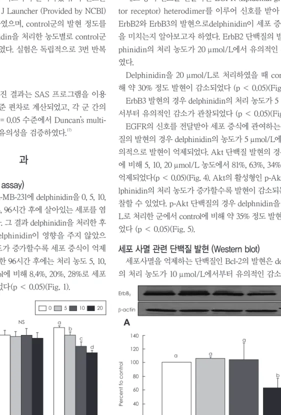
세포 증식 신호를 전달하는 EGFR (Epidemal growth fac- tor receptor) heterodimer를 이루어 신호를 받아 전달하는 ErbB2와 ErbB3의 발현으로delphinidin이 세포 증식에 영향 을 미치는지 알아보고자 하였다. ErbB2 단백질의 발현에 del- phinidin의 처리 농도가 20 μmol/L에서 유의적인 감소를 보 였다.
Delphinidin을 20 μmol/L로 처리하였을 때 control에 비 해 약 30% 정도 발현이 감소되었다 (p < 0.05)(Fig. 2).
ErbB3 발현의 경우 delphinidin의 처리 농도가 5 μmol/L에 서부터 유의적인 감소가 관찰되었다 (p < 0.05)(Fig. 3).
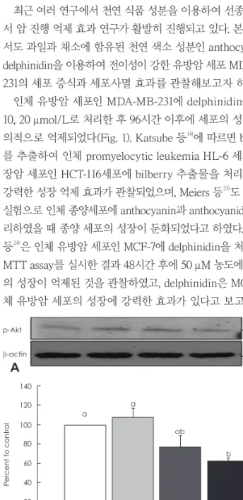
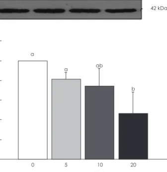
EGFR의 신호를 전달받아 세포 증식에 관여하는 Akt 단백 질의 발현의 경우 delphinidin의 농도가 5 μmol/L에서부터 유 의적으로 발현이 억제되었다. Akt 단백질 발현의 경우 control 에 비해 5, 10, 20 μmol/L 농도에서 81%, 63%, 34%로 발현이 억제되었다(p < 0.05)(Fig. 4). Akt의 활성형인 p-Akt 역시 de- lphinidin의 처리 농도가 증가할수록 발현이 감소되는 것을 관 찰할 수 있었다. p-Akt 단백질의 경우 delphnidin을 20 μmol/
L로 처리한 군에서 control에 비해 약 35% 정도 발현이 억제되 었다 (p < 0.05)(Fig. 5).
세포 사멸 관련 단백질 발현 (Western blot)
세포사멸을 억제하는 단백질인 Bcl-2의 발현은 delphinidin 의 처리 농도가 10 μmol/L에서부터 유의적인 감소를 보였으
Fig. 1. Effect of delphinidin on cell proliferation in MDA-MB-231 cells.
MDA-MB-231 cells were plated at a density of 2.5 × 10⁴ cells/ml in 24 well plate with DMEM/F12 supplemented with 10% FBS for 48 hour, the monolayers were serum-starved with DMEM/F12 supple- mented with 5 μg/mL transferrin, 5 μg/mL selenium, and 1 mg/mL bovine serum albumin for 24 hour. After serum starvation, the mo- nolayer were incubated in serum free medium with 0, 5, 10, 20 μ M delphinidin. Viable cell numbers were estimated by the MTT as- say. Each bar represents the mean ± SE from three independent experiments. Comparison among different concentrations of del- phinidin that yielded significant differences (p < 0.05) are indicat- ed by different letters above each bar.
1.4 1.2 1 0.8 0.6 0.4 0.2 0
Absorbance
(490 nm
)
NS NS a
b c
d
0 24 96 Time (hr)
0 5 10 20
ErbB2
β-actin
185 kDa
42 kDa
A
140 120 100 80 60 40 20 0
Percent fo control
0 5 10 20 Delphinidin (uM)
a a
a
b
B
Fig. 2. Effects of delphinidin on ErbB2 expression in MDA-MB-231 cells. Cell lysates were subjected to immunoblotting with an an- tibody against ErbB2 and β-actin. A: The photographs of chemi- luminescent detection of the blots, which were representative of three independent experiments are shown. B: Quantitative anal- ysis of immunoblots. The relative abundance of each band was estimated by densitometric scanning of the exposed films. Each bar represents the mean ± SE (n = 3). Comparisons between groups that yielded significant differences (p < 0.05) are indicat- ed by different letters above each bar.
나(p < 0.05)(Fig. 6), 세포사멸을 유도하는 단백질인 Bax는 delphinidin의 처리에 유의적인 차이가 관찰되지 않았다 (p < 0.05)(Fig. 7). 그러나, 세포사멸의 지표로 많이 이용되고 있는
Bcl-2/Bax ratio로 살펴보면 20 μmol/L에서부터 유의적으로 감소되었으며, control에 비해 50% 이하로 감소되었다 (p < 0.05)(Fig. 8).
고 찰
최근 여러 연구에서 천연 식품 성분을 이용하여 선종 단계에 서 암 진행 억제 효과 연구가 활발히 진행되고 있다. 본 연구에 서도 과일과 채소에 함유된 천연 색소 성분인 anthocyanin계 delphinidin을 이용하여 전이성이 강한 유방암 세포 MDA-MB- 231의 세포 증식과 세포사멸 효과를 관찰해보고자 하였다.
인체 유방암 세포인 MDA-MB-231에 delphinidin을 0, 5, 10, 20 μmol/L로 처리한 후 96시간 이후에 세포의 성장이 유 의적으로 억제되었다(Fig. 1). Katsube 등14)에 따르면 bilberry 를 추출하여 인체 promyelocytic leukemia HL-6 세포와 대 장암 세포인 HCT-116세포에 bilberry 추출물을 처리한 결과 강력한 성장 억제 효과가 관찰되었으며, Meiers 등23)도 in vitro 실험으로 인체 종양세포에 anthocyanin과 anthocyanidin을 처 리하였을 때 종양 세포의 성장이 둔화되었다고 하였다. Zhang 등24)은 인체 유방암 세포인 MCF-7에 delphinidin을 처리한 후 MTT assay를 실시한 결과 48시간 후에 50 μM 농도에서 세포 의 성장이 억제된 것을 관찰하였고, delphinidin은 MCF-7 인 체 유방암 세포의 성장에 강력한 효과가 있다고 보고하였다.
ErbB3
β-actin
200 kDa
42 kDa
A
140 120 100 80 60 40 20 0
Percent fo control
0 5 10 20 Delphinidin (uM)
a
ab
bc
c
B
Fig. 3. Effects of delphinidin on ErbB3 expression in MDA-MB-231 cells. MDA-MB-231 cells were treated with delphinidin for 48 hr as described in Fig. 1. Cell lysates were subjected to immunoblotting with an antibody against ErbB3 and β-actin. A: The photographs of chemiluminescent detection of the blots, which were represen- tative of three independent experiments are shown. B: Quanti- tative analysis of immunoblots. The relative abundance of each band was estimated by densitometric scanning of the exposed films. Each bar represents the mean ± SE (n = 3). Comparisons between groups that yielded significant differences (p < 0.05) are indicated by different letters above each bar.
Akt β-actin
60 kDa 42 kDa
A
140 120 100 80 60 40 20 0
Percent fo control
0 5 10 20 Delphinidin (uM)
a b
b
c
B
Fig. 4. Effects of delphinidin on Akt expression in MDA-MB-231 cells.
Cells were treated as described in Fig. 1 and protein samples were analyzed by immunoblotting with anti-Akt protein antiboby or β- actin. A: The photographs of chemiluminescent detection of the blots, which are representative of three independent experiments, are shown. B: Quantitative analysis of immunoblots. The relative change in Akt band on Western blots was quantitated by densi- tometric analysis. Each bar represents the mean ± SE (n = 3). Com- parisons between groups that yielded significant differences (p < 0.05) are indicated by different letters above each bar.
p-Akt β-actin
60 kDa 42 kDa
A
140 120 100 80 60 40 20 0
Percent fo control
0 5 10 20 Delphinidin (uM)
a a
ab
b
B
Fig. 5. Effects of delphinidin on p-Akt expression in MDA-MB-231 cells. Cells were treated as described in Fig. 1 and protein sam- ples were analyzed by immunoblotting with antiphospho-Akt pro- tein or β-actin antiboby. A: The photographs of chemiluminescent detection of the blots, which were representative of three inde- pendent experiments, are shown. B: Quantitative analysis of im- munoblots. The relative change in p-Akt band on western blots was quantitated by densitometric analysis. Each bar represents the mean ± SE (n = 3). Comparisons between groups that yielded significant differences (p < 0.05) are indicated by different let- ters above each bar.
또한, Martin 등25)과 Koide 등26)의 연구에서도 delphinidin은 암세포의 성장을 억제하였으며 세포 주기의 진행이 delphini- din의 처리 농도에 따라 억제되었다고 하였다. 인체 embryonic fibroblast cell을 기질로 하여 세포 주기 중 S 단계에서 delph- inidin을 처리하였을 때 다음 단계인 G2 단계로의 진행이 완 전히 차단되었다고 하였다. Marko 등27)은 anthocyanidin 중 delphinidin, malvidin, cyanidin, peonidin을 처리하여 IC50
을 측정한 결과 각 처리 물질의 농도가 35~90 μM에서 종양세 포의 성장이 억제되었다고 보고하였다. 이러한 많은 연구 결과 들과 본 연구 결과로 볼 때 delphinidin은 암세포의 세포 성장 억제에 강력한 효과가 있는 것으로 보여진다.
Epidermal growth factor receptor (EGFR) 과발현은 암의 증후가 악화되었을 때표현되는 지표로 암 예방과 치료에 중요 한 목표 중 하나이다.28,29) EGFR family에는 ErbB1, ErbB2, ErbB3, ErbB4가 있으며, 인산화되면서 성장신호를 전달하여 tyrosine kinase가 활성화된다. 인체 유방암 환자의 약 67%에 서 EGFR family 중 하나 또는 그 이상의 유전자 부호의 과발 현이나 변형이 관찰되었으며,30) 인체 유방암의 20~30%에서 EG- FR1과 EGFR2의 과발현이 관찰되었고, EGFR1의 과발현은
estrogen 작용이 낮아지는 것과 높은 관련성이 있다고 하였
다.31-33) De Potter 등34)과 Gusterson 등35)에 따르면 암 발전에 종
양을 더 악화시키는 데에는 epidermal growth factor (EGF) 가 관여한다고 하였다. 세포 외 성장 관련 리간드 (ligand)와 결 합하는 EGFR family는 세포 내 신호전달 경로와 연결되어 증 식, 분화, 세포의 이동성, 생존 등 여러 생물학적 작용을 조절 한다. 그 중 ErbB2는 ErbB3와 heterodimer를 이루어 인산화 되어 세포 내로 신호를 전달하게 된다.36-38) EGFR family의 과 발현이나 변형도 다양한 인체 종양의 발암 과정에 중요하게
Bcl-2 β-actin
28 kDa 42 kDa
A
120 100 80 60 40 20 0
Percent fo control
0 5 10 20 Delphinidin (uM)
a
a ab
b
B
Fig. 6. Effects of delphinidin on Bcl-2 expression in MDA-MB-231 cells. Cells were treated as described in Fig. 1 and protein sam- ples were analyzed by immunoblotting with an Bcl-2 or β-actin antiboby. A: photographs of chemiluminescent detection of the blots, which were representative of threeindependent experi- ments, are shown. B: quantitative analysis of immunoblots. The rel- ative abundance of each bar was estimated by densitometric scaning of the exposed films. Each bar represents the mean ± SE (n = 3). Comparisons between groups that yielded significant differences (p < 0.05) are indicated by different letters above each bar.
Bax β-actin
28 kDa
42 kDa
A
140 120 100 80 60 40 20 0
Percent fo control
0 5 10 20 Delphinidin (uM)
NS
B
Fig. 7. Effects of delphinidin on Bax expression in MDA-MB-231 cells.
Cells were treated as described in Fig. 1 and protein samples were analyzed by immunoblotting with an Bax or β-actin antiboby. A:
photographs of chemiluminescent detection of the blots, which were representative of three independent experiments, are shown.
B: quantitative analysis of immunoblots. The relative abundance of each bar was estimated by densitometric scaning of the ex- posed films. Each bar represents the mean ± SE (n = 3). Compa- risons between groups that yielded significant differences (p < 0.05) are indicated by different letters above each bar.
1.2 1 0.8 0.6 0.4 0.2 0
Ratio
a
a a
b
0 5 10 20 Delphinidin (uM)
Fig. 8. Effects of delphinidin on Bcl-2/Bax ratio in MDA-MB-231 cells.
Each bar represents the mean ± SE (n = 3). Comparisons between groups that yielded significant differences (p < 0.05) are indicat- ed by different letters above each bar.
작용하며, 암환자의 악화된 증상과 관련이 있다고 하였다.36) Graus-Porta 등39)의 연구에서는 ErbB2와 결합하는 ErbB3 가 결여된 경우 이종 이성화 (heterodimerization)나 인산화 전 이과정이 활성화되지 못해 세포 내로의 신호전달이 원활하지 못하였다고 보고하였다. Meiers 등23)에 따르면 anthocyanidin 인 cyanidin과 delphinidin는 EGFR의 tyrosine kinase 활성 에 강력한 억제자로 EGFR 감소는 연속적으로 신호전달을 받 는 MAPK (Mitogen-activated protein kinases)의 활성을 효 과적으로 둔화시킨다고 하였다. 그 외 다른 연구40)에서도 del- phinidin을 뛰어난 EGFR의 억제제로 연속적 신호전달을 억 제하는 효과가 있다 하였고, 전사인자 Elk1의 인산화로 측정 된 MAPK 활성도 억제되었다고 하였다. 본 연구에서도 delph- inidin의 처리 농도가 증가될수록 ErbB2, ErbB3의 발현이 대 조군에 비해 유의적으로 감소되었다. 또한, 세포성장 신호 경 로 중 EGFR 신호를 전달받는 Akt의 발현에서도 본 연구의 delphinidin 처리 농도 중 가장 저농도인 5 μM에서 유의적으 로 발현이 억제된 것으로 볼 때, delphinidin은 유방암 세포성 장 중 EGFR의 발현을 억제하여 연속적인 신호 전달을 받는 Akt의 발현과 신호 전달에 매우 강력한 억제제로 작용한다고 할 수 있다.
세포사멸 또는 계획된 세포 죽음은 종양 억제 및 치료에 중 요 기전이며, 일반적으로 세포성장과 세포사멸의 장애나 이상 으로 정상세포가 빠르게 preplastic 또는 neoplastic 세포로 성장하여 암으로 발전하게 된다. 세포사멸 또는 세포 주기의 제어는 인체를 선종으로 진행하거나 발전시키고 신생물 또는 신생물로의 진행 전이나 손상으로부터 보호하는데 뛰어난 방 법이라 하였다.41-43) Bax와 Bcl-2유전자는 세포사멸 경로에서 중 요한 작용자로 고려되어지고 있다.44,45) Bcl-2는 Bcl-x family 중 하나로 세포사멸로부터 세포를 억제하는 역할을 하며, 여러 연구에서 세포사멸에서의 Bcl-2는 Bax에 의해 영향을 받는 것 으로 나타났다. Bcl-2는 homodimer로 세포사멸을 억제하는 유전자이며, Bax는 세포사멸을 유도하는 유전자로 알려져 있 다. 이러한 두 유전자의 발현이 세포죽음이나 생존을 결정하 는데 중요하게 작용한다.46) Afaq 등46)은 HaCat 세포에 delph- nidin을 처리하였을 때 세포사멸을 유도하는 Bax는 유의적 으로 감소하고, 세포사멸을 억제하는 Bcl-2는 증가하였으며, Bax/Bcl-2 비율은 세포사멸을 유도하지 않는 방향으로 변화 되었다고도 하였다. 그러나 대부분의 연구에서 anthocyanidin 의 처리는 세포사멸을 효과적으로 유도하는 것으로 나타났다.
Lazze´ 등47)은 자궁암 세포인 HeLa에 delphinidin을 100, 150, 200 μM 농도로 처리하여 A.I. (apoptotic index)를 통해 세포 사멸을 측정한 결과, delphinidin의 농도가 증가함에 따라 각 각 25.4, 35.5, 40.6% 세포사멸이 유도되었다고 하였다. Katsu-
be 등23)도 인체 leukemia 세포 HL-60에 베리에서 추출한 an- thocyanidin계 delphinidin을 처리한지 6시간 후, 세포사멸이 유도되었고 하였고, Shih 등48)은 인체 adenocarcinoma 세포 인 AGS에 anthocyanidin 중 malvidin을 처리한 결과, 처리 농 도에 따라 Bax/Bcl-2 비율이 상승되었다고 하였다. 본 연구결 과에서도 delphnidin은 세포사멸을 억제하는 Bcl-2를 유의 적으로 억제하였고, Bcl-2/Bax 비율에서는 delphinidin을 20 μmol/L로 처리하였을 때부터 유의적으로 감소된 것으로 볼 때, anthocyanin계 delphinidin은 인체 유방암 세포인 MDA- MB-231 세포의 세포사멸을 유도하는 것으로 사료된다.
이상의 결과들을 종합해보면, 인체 유방암 세포인 MDA- MB-231에서 delphinidin은 암세포의 증식을 억제하고, 세포 사멸을 유도하는 것으로 사료되며, 이는 delphinidin 등의 색 소 성분을 이용한 기능성식품 개발 가능성을 제시하는 것으 로 앞으로 동물과 인체를 대상으로 하는 더 많은 연구 진행이 요구되어진다.
요약 및 결론
본 연구는 식물의 대표적인 색소 성분인 anthocyanin계 색 소 중 delphinidin이 인체 유방암 세포 MDA-MB-231에서 암 세포의 성장과 증식, 세포사멸에 미치는 영향을 알아보고자 실 시되었다. 실험 결과 delphinidin의 첨가량이 증가할수록 세포 성장이 억제되었다. Delphinidin은 세포 증식 경로에 관여하 는 ErbB2 와 ErbB3의 발현을 억제하였고, 연속적인 신호 전 달을 받는 Akt의 발현이나 이들 유전자의 m RNA 수준에서 도 발현이 억제되었다. 또한 세포사멸에도 delphinidin은 유의 적으로 뚜렷하게 세포사멸을 유도하는 것이 관찰되었다. 결론 적으로, delphinidin은 인체 유방암 세포인 MDA-MB-231에서 암세포의 증식을 억제하고 세포사멸을 유도하는 효과가 있는 것으로 사료된다.
Literature cited
1) National Health Insurance Service (KR). Analysis of cancer pa- tients in 2008 [Internet]. Seoul: National Health Insurance Ser- vice; 2010 [cited 2010 Jun 5]. Available from: http://www.nhis.
or.kr/cms/board/board/Board.jsp?act=VIEW&communityKey=
B0070&boardId=20108
2) National Cancer Information Center (KR). Types of breast cancer [Internet]. Goyang: National Cancer Information Center; 2013 [cit- ed 2013 February 1]. Available from: http://www.cancer.go.kr/mbs/
cancer/jsp/cancer/cancer.jsp?cancerSeq=4757&menuSeq=4761
&viewType=all&id=cancer_020112000000
3) Korean Breast Cancer Society. Breast cancer facts & figures 2006- 2008. Seoul: Korean Breast Cancer Society; 2008
4) Doll R, Peto R. The causes of cancer: quantitative estimates of avoi-
dable risks of cancer in the United States today. J Natl Cancer Inst 1981; 66(6): 1191-1308
5) Wu C, Ray RM, Lin MG, Gao DL, Horner NK, Nelson ZC, Lampe JW, Hu YW, Shannon J, Stalsberg H, Li W, Fitzgibbons D, Por- ter P, Patterson RE, Satia JA, Thomas DB. A case-control study of risk factors for fibrocystic breast conditions: Shanghai Nutri- tion and Breast Disease Study, China, 1995-2000. Am J Epidemiol 2004; 160(10): 945-960
6) Magee PJ, McGlynn H, Rowland IR. Differential effects of isofla- vones and lignans on invasiveness of MDA-MB-231 breast can- cer cells in vitro. Cancer Lett 2004; 208(1): 35-41
7) Schley PD, Jijon HB, Robinson LE, Field CJ. Mechanisms of ome- ga-3 fatty acid-induced growth inhibition in MDA-MB-231 hu- man breast cancer cells. Breast Cancer Res Treat 2005; 92(2):
187-195
8) Klass CM, Shin DM. Current status and future perspectives of chemoprevention in head and neck cancer. Curr Cancer Drug Tar- gets 2007; 7(7): 623-632
9) Lila MA. From beans to berries and beyond: teamwork between plant chemicals for protection of optimal human health. Ann N Y Acad Sci 2007; 1114: 372-380
10) Harborne JB. Comparative biochemistry of the flavonoids. New York (NY): Academic Press; 1967
11) Bae EA, Moon GS. A study on the antioxidative activities of Ko- rean soybeans. J Korean Soc Food Sci Nutr 1997; 26(2): 203-208 12) Ding M, Feng R, Wang SY, Bowman L, Lu Y, Qian Y, Castrano- va V, Jiang BH, Shi X. Cyanidin-3-glucoside, a natural product derived from blackberry, exhibits chemopreventive and chemo- therapeutic activity. J Biol Chem 2006; 281(25): 17359-17368 13) Hou DX. Potential mechanisms of cancer chemoprevention by
anthocyanins. Curr Mol Med 2003; 3(2): 149-159
14) Katsube N, Iwashita K, Tsushida T, Yamaki K, Kobori M. Induc- tion of apoptosis in cancer cells by Bilberry (Vaccinium myrtil- lus) and the anthocyanins. J Agric Food Chem 2003; 51(1): 68-75 15) Yoon TH, Lee SW. Stability of anthocyanins in foods. Korean J
Food Sci Technol 1979; 11(1): 63-73
16) Kong JM, Chia LS, Goh NK, Chia TF, Brouillard R. Analysis and biological activities of anthocyanins. Phytochemistry 2003; 64 (5): 923-933
17) Chu SK, Seo EY, Kim WK, Kang NE. Effect of cyanidin on cell motility and invasion in MDA-MB-231 human breast cancer cells.
Korean J Nutr 2008; 41(8): 711-717
18) Li L, Adams LS, Chen S, Killian C, Ahmed A, Seeram NP. Euge- nia jambolana Lam. Berry extract inhibits growth and induces apoptosis of human breast cancer but not non-tumorigenic breast cells. J Agric Food Chem 2009; 57(3): 826-831
19) Hui C, Bin Y, Xiaoping Y, Long Y, Chunye C, Mantian M, Wen- hua L. Anticancer activities of an anthocyanin-rich extract from black rice against breast cancer cells in vitro and in vivo. Nutr Can- cer 2010; 62(8): 1128-1136
20) Weng MS, Ho CT, Ho YS, Lin JK. Theanaphthoquinone inhibits fatty acid synthase expression in EGF-stimulated human breast cancer cells via the regulation of EGFR/ErbB-2 signaling. Toxi- col Appl Pharmacol 2007; 218(2): 107-118
21) Seo EY, Lee HS, Kim WK. Effect of [6]-gingerol on inhibition of cell proliferation in MDA-MB-231 human breast cancer cells.
Korean J Nutr 2005; 38(8): 656-662
22) Landesberg R, Cozin M, Cremers S, Woo V, Kousteni S, Sinha S, Garrett-Sinha L, Raghavan S. Inhibition of oral mucosal cell wound healing by bisphosphonates. J Oral Maxillofac Surg 2008;
66(5): 839-847
23) Meiers S, Kemény M, Weyand U, Gastpar R, von Angerer E, Mar- ko D. The anthocyanidins cyanidin and delphinidin are potent in- hibitors of the epidermal growth-factor receptor. J Agric Food Chem
2001; 49(2): 958-962
24) Zhang Y, Vareed SK, Nair MG. Human tumor cell growth inhi- bition by nontoxic anthocyanidins, the pigments in fruits and veg- etables. Life Sci 2005; 76(13): 1465-1472
25) Martin S, Andriambeloson E, Takeda K, Andriantsitohaina R.
Red wine polyphenols increase calcium in bovine aortic endo- thelial cells: a basis to elucidate signalling pathways leading to nitric oxide production. Br J Pharmacol 2002; 135(6): 1579-1587 26) Koide T, Hashimoto Y, Kamei H, Kojima T, Hasegawa M, Terabe
K. Antitumor effect of anthocyanin fractions extracted from red soybeans and red beans in vitro and in vivo. Cancer Biother Ra- diopharm 1997; 12(4): 277-280
27) Marko D, Puppel N, Tjaden Z, Jakobs S, Pahlke G. The substitu- tion pattern of anthocyanidins affects different cellular signaling cascades regulating cell proliferation. Mol Nutr Food Res 2004;
48(4): 318-325
28) Sainsbury JR, Malcolm AJ, Appleton DR, Farndon JR, Harris AL. Presence of epidermal growth factor receptor as an indicator of poor prognosis in patients with breast cancer. J Clin Pathol 1985; 38(11): 1225-1228
29) Sainsbury JR, Farndon JR, Needham GK, Malcolm AJ, Harris AL. Epidermal-growth-factor receptor status as predictor of early recurrence of and death from breast cancer. Lancet 1987;
1(8547): 1398-1402
30) Koenders PG, Beex LV, Geurts-Moespot A, Heuvel JJ, Kienhuis CB, Benraad TJ. Epidermal growth factor receptor-negative tu- mors are predominantly confined to the subgroup of estradiol re- ceptor-positive human primary breast cancers. Cancer Res 1991;
51(17): 4544-4548
31) Slamon DJ, Clark GM, Wong SG, Levin WJ, Ullrich A, McGuire WL. Human breast cancer: correlation of relapse and survival with amplification of the HER-2/neu oncogene. Science 1987;
235(4785): 177-182
32) Slamon DJ, Godolphin W, Jones LA, Holt JA, Wong SG, Keith DE, Levin WJ, Stuart SG, Udove J, Ullrich A, Press MF. Studies of the HER-2/neu proto-oncogene in human breast and ovarian cancer. Science 1989; 244(4905): 707-712
33) Toi M, Osaki A, Yamada H, Toge T. Epidermal growth factor re- ceptor expression as a prognostic indicator in breast cancer. Eur J Cancer 1991; 27(8): 977-980
34) De Potter CR, Beghin C, Makar AP, Vandekerckhove D, Roels HJ. The neu-oncogene protein as a predictive factor for haema- togenous metastases in breast cancer patients. Int J Cancer 1990;
45(1): 55-58
35) Gusterson BA, Machin LG, Gullick WJ, Gibbs NM, Powles TJ, Elliott C, Ashley S, Monaghan P, Harrison S. c-erbB-2 expres- sion in benign and malignant breast disease. Br J Cancer 1988;
58(4): 453-457
36) Sweeney C, Fambrough D, Huard C, Diamonti AJ, Lander ES, Cantley LC, Carraway KL 3rd. Growth factor-specific signaling pathway stimulation and gene expression mediated by ErbB re- ceptors. J Biol Chem 2001; 276(25): 22685-22698
37) Crovello CS, Lai C, Cantley LC, Carraway KL 3rd. Differential signaling by the epidermal growth factor-like growth factors neu- regulin-1 and neuregulin-2. J Biol Chem 1998; 273(41): 26954- 26961
38) Tzahar E, Pinkas-Kramarski R, Moyer JD, Klapper LN, Alroy I, Levkowitz G, Shelly M, Henis S, Eisenstein M, Ratzkin BJ, Sela M, Andrews GC, Yarden Y. Bivalence of EGF-like ligands drives the ErbB signaling network. EMBO J 1997; 16(16): 4938-4950 39) Graus-Porta D, Beerli RR, Daly JM, Hynes NE. ErbB-2, the pre-
ferred heterodimerization partner of all ErbB receptors, is a me- diator of lateral signaling. EMBO J 1997; 16(7): 1647-1655 40) Franke TF, Kaplan DR, Cantley LC. PI3K: downstream AKTion
blocks apoptosis. Cell 1997; 88(4): 435-437
41) Meyskens FL Jr, Szabo E. Diet and cancer: the disconnect be- tween epidemiology and randomized clinical trials. Cancer Ep- idemiol Biomarkers Prev 2005; 14(6): 1366-1369
42) Ramos S, Alía M, Bravo L, Goya L. Comparative effects of food- derived polyphenols on the viability and apoptosis of a human hepatoma cell line (HepG2). J Agric Food Chem 2005; 53(4): 1271- 43) Heo HJ, Lee CY. Strawberry and its anthocyanins reduce oxida-1280 tive stress-induced apoptosis in PC12 cells. J Agric Food Chem 2005; 53(6): 1984-1989
44) Tessitore L, Davit A, Sarotto I, Caderni G. Resveratrol depresses the growth of colorectal aberrant crypt foci by affecting bax and p21(CIP) expression. Carcinogenesis 2000; 21(8): 1619-1622 45) Bunz F, Hwang PM, Torrance C, Waldman T, Zhang Y, Dillehay
L, Williams J, Lengauer C, Kinzler KW, Vogelstein B. Disrup-
tion of p53 in human cancer cells alters the responses to thera- peutic agents. J Clin Invest 1999; 104(3): 263-269
46) Afaq F, Syed DN, Malik A, Hadi N, Sarfaraz S, Kweon MH, Khan N, Zaid MA, Mukhtar H. Delphinidin, an anthocyanidin in pig- mented fruits and vegetables, protects human HaCaT keratino- cytes and mouse skin against UVB-mediated oxidative stress and apoptosis. J Invest Dermatol 2007; 127(1): 222-232
47) Lazzè MC, Savio M, Pizzala R, Cazzalini O, Perucca P, Scovassi AI, Stivala LA, Bianchi L. Anthocyanins induce cell cycle per- turbations and apoptosis in different human cell lines. Carcino- genesis 2004; 25(8): 1427-1433
48) Shih PH, Yeh CT, Yen GC. Effects of anthocyanidin on the inhi- bition of proliferation and induction of apoptosis in human gas- tric adenocarcinoma cells. Food Chem Toxicol 2005; 43(10):
1557-1566