Epigallocatechin Gallate가 인체 유방암 세포인 MDA-MB-231의 세포사멸에 미치는 영향
홍은정․김우경† 단국대학교 식품영양학과
Effect of Epigallocatechin Gallate on Apoptosis in MDA-MB-231 Human Breast Cancer Cells
Eun-Jung Hong and Woo-Kyung Kim†
Dept. of Food Science and Nutrition, Dankook University, Gyeonggi 448-701, Korea
Abstract
Among the numerous polyphenols isolated from green tea, epigallocatechin gallate (EGCG) is a predominate and is considered to be a major therapeutic agent. To elucidate the mechanical insights of anti-tumor effect, EGCG was applied to human breast cancer MDA-MB-231 cells. We investigated the effect of EGCG on protein and mRNA expression of proteins related to cell apoptosis in MDA-MB-231 human breast cancer cell lines.
We also identified caspase-3 activity. We cultured MDA-MB-231 cells in the presence of 0, 5, 10, and 20 μM of EGCG. Protein and mRNA expression of bcl-2 were decreased dose-dependently in cells treated with EGCG. However, protein and mRNA expression of bax were increased (p<0.05). Caspase-3 activities were increased dose-dependently in cells treated with EGCG. These results suggest that EGCG induces cell apoptosis by increase of caspase activity through decreasing of protein and mRNA expression of bcl-2 and increasing of protein and mRNA expression of bax.
Key words: epigallocatechin gallate, breast cancer, apoptosis, bcl-2 family, caspase
†Corresponding author. E-mail: wkkim@dankook.ac.kr
†Phone: 82-31-8005-3172, Fax: 82-31-8005-3172
서 론
Epigallocatechin gallate(EGCG)는 녹차 카테킨 성분 중 에서 65%를 차지하고 있는 주요 성분으로, 암세포의 성장을 억제하거나 전립선암 및 위암 세포의 사멸을 유도하고(1), H891 head와 neck 종양세포와 A431 종양상피세포에서도 세포사멸을 유도한다고 보고되었다(2). EGCG의 항암효과 는 구조내 함유하고 있는 phenolic hydroxy group이 조직과 체액에 있는 free radical을 제거하기 때문이라고 보고되고 있다(3).
암세포의 사멸을 유도하는 기전은 TNF receptor system, 조기발현 유전자의 전사적 활성, 단백 분해 효소 caspase- family cysteine protease, DNA 분절을 일으키는 endonu- clease 등이 관여하는 것으로 보고되고 있다(4). 암세포의 사멸과 관련 있는 단백질인 Bcl-2 family는 세포 밖이나 세 포 내의 상황을 전달하는 중요한 신호 전달체로서 작용하며 미토콘드리아 세포막의 안정성을 조절하는 단백질로 알려 져 있다(5). Bcl-2 family의 하나인 Bcl-2는 원형질막에 붙 어 있을 수 있는 carboxyl기를 가지고 있어 미토콘드리아나
조면소포체와 같은 세포내 소기관의 세포질 쪽에서 발견되 며, NF-κB를 활성화하여 세포 사멸을 억제하는 다양한 단 백질을 발현시킨다(6). 또한 bcl-2는 Raf-1과 결합하여 Raf-1을 미토콘드리아 막으로 옮겨 세포 사멸을 저해한다 (7,8).반면에 bcl-2 family의 또 다른 단백질인 bax는 세포질 에서 미토콘드리아로 이동하면서 미토콘드리아로부터 cy- tochrome C의 분비를 촉진하여 세포 사멸을 촉진하는 기능 을 가지고 있다. 그러므로 bcl-2와 bax의 발현정도가 사멸과 정에서 중요한 역할을 한다.
또한 세포사멸에 관여하는 중요한 효소중 하나인 caspase 는 Ca2+ 의존 단백질 분해 효소로, 활성 부위에 cystein이 있으며, 기질 단백질 내 특정 aspartic acid를 인지하여 바로 다음 펩타이드 결합을 절단하는 효소로서 핵 층을 형성하는 라민을 잘라서 핵 층을 비가역적으로 분해하여 세포 사멸에 서 결정적인 역할을 한다(9).
본 연구에서는 녹차의 주요 성분인 EGCG가 전이성이 강 한 MDA-MB-231 인체 유방암세포에서도 세포 사멸을 억 제하는 단백질인 bcl-2와 세포 사멸을 유도하는 단백질인 bax의 단백질과 mRNA발현과 세포사멸의 마지막 단계인
Table 1. Sequence and RT-PCR program of protein
Protein Primer Sequence Cycle
β-Actin
sense 5'-GTTTGAGACC
TTCAACACCCC-3' 94oC 1 min
60oC 1 min 72oC 1 min
antisense 5'-GTGGCCATCT 35
CCTGCTCGAAGTC-3'
Bcl-2
sense 5'-CGACTTCGCCGA
GATGTCCAGCCAG-3' 95oC 1 min
65oC 1 min 72oC 1 min
antisense 5'-ACTTGTGGCCCAG 35
ATAGGCACCCAG-3'
Bax
sense 5'-CTGACATGTTTT
CTGACGGC-3' 94oC 1 min
55oC 1 min 72oC 1 min
antisense 5'-CTGACATGTTTT 32
TTCTTCCAGA-3' caspase-3의 활성에 영향을 미치는 지를 관찰하였다.
재료 및 방법
세포 배양
본 연구에 사용한 인체 유방암 세포주인 MDA-MB-231 은 American Type Culture Collection(ATCC, Rockville, MD, USA)에서 구입하였다. 배양액은 Dulbecco modified Eagle's medium/ Nutrient Mixture Ham's F12(DMEM/
F12 Gibco/BRL, USA)에 10% fetal bovine serum(FBS, Gibco/BRL)과 1,000 units/mL penicillin, 1,000 μg/mL streptomycin(Gibco/BRL)를 첨가하여 사용하였다. 세포가 80%정도 배양접시를 덮으면 phosphate-buffered saline (PBS, PH 7.4)으로 세포의 표면을 씻어낸 후 0.25% tryp- sin-2.65 mM EDTA(Gibco /BRL)로 처리하여 계대 배양하 였고 배양액은 2~3일마다 교환해 주었다.
단백질 발현
세포 사멸에 관련된 단백질의 발현을 알아보기 위해서 western blot을 실시하였다. MDA-MB-231 세포를 1×10⁷ cells/dish 농도로 분주하여 48시간 배양 후에, FBS를 첨가 하지 않은 serum free media(SFM)로 교체하여 24시간 동안 starvation 시켰다. Starvation 후, SFM에 EGCG를 0, 5, 10, 20 μM 농도로 희석하여 배지를 교환하고 48시간 동안 배양 하였다. 배양 후, 차가운 rinse buffer(PBS, 1 mM PMSF, 1 mM sodium orthovanadate)로 배양접시를 세척하고 세포 를 모아 1,000 rpm에서 5분간 원심분리 하였다. 상층 액을 제거하고 500 mL의 lysis buffer(137 mM NaCl, 20 mM Tris-Cl, 1% Triton X-100, 10% glycerol, 1 mM sodium orthovanadate, 1 mM PMSF, 20 μg/mL aprotinin, 10 μg/
mL antipain, 10 μg/mL leupeptin, 80 μg/mL benzamidine HCl)를 첨가하고 40분간 4oC에서 회전배양기에서 배양시키 고, 12,000 rpm에서 10분간 원심분리 하여 시료를 준비하였 다. 시료는 Bio-rad 방법으로 595 nm에서 흡광도를 측정하
여 단백질을 정량하였으며, 4~20% gradient sodium dodecyl sulfate polyacrylamide gel electrophoresis(SDS-PAGE)에 서 단백질을 분리한 후, immobilonTM-P membrane(Milipore, Bedford, MA, USA)으로 4oC에서 transfer하였다. Membrane 은 5% milk/TBST(20 mM Tris-HCL, 137 mM NaCl, 0.1%
Tween 20, pH 7.4)로 실온에서 1시간 배양한 후, bcl-2와 baxantibody(Santacruz, USA)를 희석한 5% milk/TBST로 3~5시간 동안 실온에서 배양하였다. Anti-mouse Ig horse- radish peroxidase/TBST(Amersham Buckinghamshire, England)로 1시간 배양 후 SupersignalR West Dura ex- tended Duration Substrate(Pierce, IL, USA)를 사용하여 발 색시켜 X-Omat film으로 현상하였다. 밴드는 imaging pro- gram인 Image J Launcher(provided by NCBI)를 이용하여 밀도를 측정하였다.
mRNA 발현
EGCG가 암세포의 사멸에 영향을 주는 단백질의 mRNA 발현에도 영향을 미치는 지를 알아보기 위해서 단백질 발현 과 같은 조건으로 MDA-MB-231 세포를 배양하여 RT- PCR을 실시하였다. 세포에서 RNA를 분리하기 위해서 Tri reagent를 이용하였으며, 분리된 RNA양이 3 μg이 되도록 tube에 준비하고 oligo DT(0.5 μg/μL, invitrogen) 1 μL를 포함하여 총량이 12 μL 되도록 멸균수를 첨가하였다. 이것 을 70oC에서 10분 동안 배양하고 여기에 reaction buffer(5X first standard buffer 4 μL, 0.1 M DTT 2 μL, 10 mM dNTP mix 1 μL) 7 μL를 첨가하여 42oC에서 5분간 배양을 하고, superscript Ⅱ reverse transcriptase(Invitrogen) 0.5 μL를 첨가하여 42oC에서 1시간 45분, 70oC에서 15분간 배양을 하 였다. RNase(Invitrogen) 0.5 μL를 첨가하고 37oC에서 1시간 배양하고 DEPC water를 첨가하여 cDNA를 만들었다.
cDNA에 2.5 λ mix buffer(10X PCR buffer, 50 mM MgCl2
10 mM dNTP mix, autoclaved water)와 sense, antisense primer, tag polymerase를 혼합하여 Table 1처럼 cycle을 실행하는 PCR 반응 후에, ethidium bromide로 염색한 1%
bcl-2 28 kDa
β-actin 42 kDa
(a)(b)
0 5 10 20 (μM)
bcl-2 (364 bp)
β-actin (318 bp) (a)
(b)
0 5 10 20 (μM)
0 20 40 60 80 100 120
(% Control)0 5 10 20 (μM)
a
ab bc
c
0 20 40 60 80 100 120
0 5 10 20 (μM) (% Control)
a ab
bc
c
(A) (B)
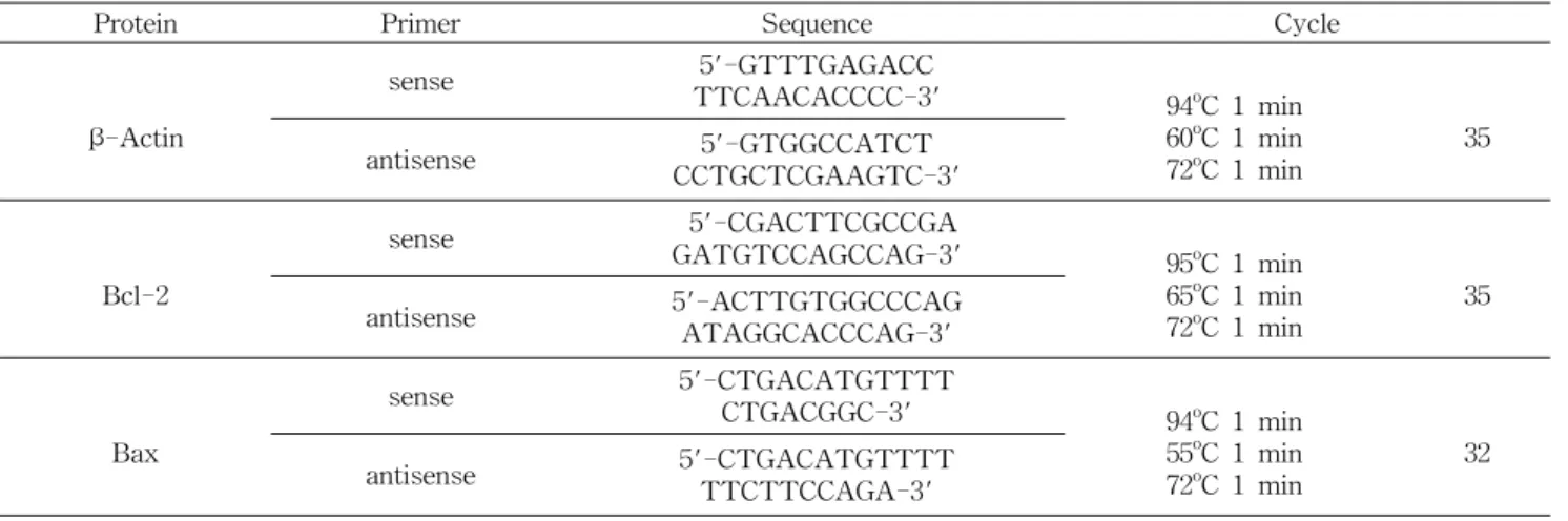
Fig. 1. Effect of EGCG on bcl-2protein and mRNA expression in MDA-MB-231 cells. A: bcl-2protein levels; (a) photograph of bcl-2 protein bands, (b) quantitative analysis of the bands. B: bcl-2mRNA levels; (a) photograph of the bcl-2 mRNA bands, (b) quantitative analysis of the bands. Each bar represents the mean
±
SD (n=3) calculated from three independent experiments.Comparisons among different concentrations of EGCG that yielded significant difference (p<0.05) are indicated by different letters above each bar.
agarose gel로 전기영동을 실시하였다.
Caspase-3 활성
세포 사멸을 유도하는 caspase-3는 cystein proteinase family로써 활성화되어 기질 결합체인 DEVD-pNA를 분해 하는 성질을 이용하여 pNA의 푸른빛으로 활성 정도를 측정 할 수 있다. EGCG를 처리하여 배양한 MDA-MB-231 세포 를 2×10⁶ cells/mL로 맞추고 1,000 rpm으로 원심분리한 후 에 세포만을 얻었다. 세포에 차가운 lysis buffer를 첨가하고 얼음 위에 10분간 두었다가 다시 4oC, 12,000 rpm에서 원심 분리 하여 상층 액을 얻었다. 상층 액을 96 well plate에 옮긴 후, 상층 액에 2X reaction buffer/DTT 용액을 가한 후 37oC, CO2 incubator에서 30분간 배양하였다. 그 후 caspase-3 substrate (DEVD-pNA)를 첨가하여 1시간 동안 incubator 에서 배양한 후에 405 nm로 흡광도를 측정하여 실험 군의 활성을 대조군에 대한 상대적인 활성정도로 표시하였다.
통계처리
본 연구의 실험은 독립적으로 3번 반복하였으며, 얻어진 결과는 SAS 통계 프로그램을 사용하여 평균과 표준편차를 구하였다. 실험군 간의 유의성은 ANOVA 분석 후, p<0.05 수준에서 Ducans's multiple range test를 하여 검증하였다.
결과 및 고찰
Bcl-2, bax의 단백질 및 mRNA 발현
세포 사멸을 억제하는 단백질인 bcl-2의 경우 EGCG를
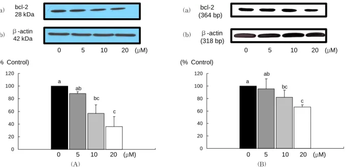
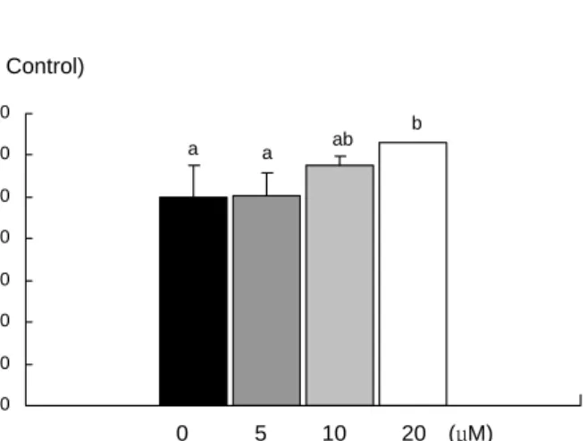
첨가하였을 때 단백질발현이 처리 농도 10 μM 이상부터 유 의적으로 감소하였고, mRNA 발현도 단백질과 같은 경향으 로 10 μM 이상부터 유의적으로 감소하였다(Fig. 1). Bcl-2와 는 반대로 세포사멸을 유도하는 단백질인 bax의 경우에는 단백질의 발현은 10 μM 이상부터, mRNA 발현은 5 μM 이상 부터 유의적으로 증가하였다(Fig. 2). 사멸의 경우 두 단백질 의 비율(bcl-2/bax)이 중요한데 비율을 구하였을 때 EGCG 의 첨가농도가 10 μM 이상이면 비율이 유의적으로 감소하 였다(Fig. 3).
Bcl-2 family는 세포내 중요한 신호 전달체로서 미토콘드 리아의 안정성을 조절하는 단백질로 알려져 있다. Bax는 세 포질에서 미토콘드리아로 이동하면서 미토콘드리아로부터 cytochrome C의 분비를 촉진하여 세포 사멸을 촉진하는 기 능을 가지고 있는 반면에 bcl-2는 미토콘드리아로의 bax의 이동을 억제시킴으로써 세포사멸을 억제시키는 작용을 한 다(5). Zhang 등(10)은 정상 세포에는 bcl-2와 bax가 heter- odimer를 이루어 존재하는데 산화스트레스로 인해 DNA가 손상되어 p53이 증가되면, bax homodimer로 평형이 기울어 세포 사멸이 증가된다고 하였다. Nishikawa 등(11)의 연구 에 의하면 EGCG를 50 μg/mL를 처리하였을 때, 간암세포인 HepG2, HuH-7에서 bcl-2 발현이 감소하고 bax 발현이 증 가하였다고 보고하였다. Baliga 등(12)은 유방암세포인 4T1 에 EGCG를 20~80 μM로 처리하였을 때 bcl-2의 발현이 감소하면서 bax와 cytochrome C, Apaf-3의 발현이 증가되 어 세포 사멸이 유도되었다고 보고하였다. Hsuuw와 Chan (13)의 연구에서는 유방암 세포인 MCF-7에 EGCG를 처리
bax 23 kDa
β-actin 42 kDa
(a)(b)
0 5 10 20 (μM)
bax (289 bp)
β-actin (318 bp) (a)
(b)
0 5 10 20 (μM)
0 50 100 150 200 250
a a
b c
(% Control)0 5 10 20 (μM)
0 50 100 150 200 250
a b
c c
0 5 10 20 (μM) (% Control)
(A) (B)
Fig. 2. Effect of EGCG on baxprotein and mRNA expression in MDA-MB-231 cells. A: baxprotein levels; (a) photograph of bax protein bands, (b) quantitative analysis of the bands. B: baxmRNA levels; (a) photograph of the bax mRNA bands, (b) quantitative analysis of the bands. Each bar represents the mean
±
SD (n=3) calculated from three independent experiments. Comparisons among different concentrations of EGCG that yielded significant difference (p<0.05) are indicated by different letters above each bar.0 0.5 1 1.5 2
a a
b
0 5 10 20 (μM)
c
Fig. 3. Effect of EGCG on bcl-2/bax ratio in MDA-MB-231 cells. Each bar represents the mean
±
SD (n=3) calculated from three independent experiments. Different alphabet represents the significant differences among groups at p<0.05 by Duncan's mul- tiple range test.하였을 때, 저농도(10~50 μM)에서는 Bax 단백질 발현은 증가시키고 bcl-2 단백질 발현을 감소시킴으로써 세포사멸 을 유도하였지만 고농도인(100~400 μM)에서는 bax와 bcl-2의 발현에 영향을 주지 않아서 첨가하는 EGCG 농도에 영향을 받는다고 하였다.
세포사멸의 유도는 NF-κB와 같은 전사 인자의 불활성에 의해 중재되어지기도 한다(14). NF-κB는 전사요인으로서 cytokine의 생성과 세포질 침윤, 세포주기의 활성화, 사멸억 제, 종양생성등과 관련되어 있다(5,14). EGCG는 산소종을 제거하는 기능을 가지고 NF-κB를 불활성화 시킨다고 하며 (15,16), Tamatani 등(17)과 Baeuerie와 Baltomore(18)는 해 마 신경세포인 H891 세포와 A431 인체 상피세포에 EGCG
를 처리하였을 때, NF-κB의 활성이 감소되었다고 보고하였 다. Gupta 등(19)은 A431 인체 상피세포에 EGCG를 처리하 였을 때, Roy 등(20)은 다른 인체 유방암세포인 MDA- MB-486 세포에 EGCG를 처리하면 NF-κB의 불활성화로 bcl-2의 단백질 발현은 감소되고, bax의 단백질 발현이 증가 되어, caspase-3의 활성이 증가되어 세포 사멸이 유도된다 고 보고하였다. 본 연구에서는 단백질 발현뿐만 아니라 EGCG의 처리가 bcl-2의 mRNA 발현을 감소시키고, bax의 mRNA 발현을 유의적으로 증가시키는 것을 확인하였다. 또 한 Kuo와 Lin(21)은 Hep G2의 세포에 EGCG를 처리하였을 때 p53 단백질과 Fas legand의 활성화로 인해 세포주기가 정지되고 세포 사멸이 유도되었다고 보고하였다.
Caspase-3의 활성
세포사멸에서 결정적인 역할을 하는 caspase-3의 활성은 EGCG처리 농도가 증가함에 따라 증가하여 EGCG 첨가농 도가 20 μM 이상일 때 활성이 유의적으로 감소하였다(Fig. 4).
Caspase-3은 caspase family 14종 중 하나이며 apoptosis 의 중요한 역할을 하는 것으로 알려져 있고 32 kDa 효소 전구체의 분리에 의해 유도된 17 kDa 및 12 HkDa hetero- dimer가 활성화된 형태이다. Caspase-3은 caspase-8과 caspase-9의 초기 신호를 증폭시킴으로써 세포사멸에 직접 적으로 관련되어 있다고 알려져 있다(22). Kennedy 등(23) 은 녹차 추출물의 세포 사멸 유도는 caspase의 활성화와 cy- tochrome C 방출이 원인이라고 보고하였고, Zhoa 등(24)은 인체 유방암세포인 T-47D에 EGCG를 처리하였을 때, cas- pase-3이 활성화되어 시간과 농도에 상관없이 세포사멸이
(% Control)
0 20 40 60 80 100 120 140
0 5 10 20 (μM)
a a ab b
Fig. 4. Effect of EGCG on caspase-3 activities in MDA- MB-231 cells. Each bar represent the mean
±
SD (n=3) from three independent experiments. Different alphabet represents the significant difference among groups at p<0.05 by Duncan's mul- tiple range test.유도되었다고 보고하였다. 그리고 저산소의 환경에서 HepG2 cell line에 EGCG를 25 μM, 100 μM을 처리하였을 때, cas- pase-3의 activity가 증가함을 보여주면서 저산소증에 의한 세포사멸 또한 억제시킨다고 보고하였다(25). Roy 등(20)은 MDA-MB-231 cell line과 동일하게 estrogen receptor negative인 유방암 세포 MDA-MB-468 cell line에 EGCG를 처리하였을 때, DNA가 분절되어 세포사멸이 유도되는 것을 확인하였으며, caspase-3 의 활성 또한 증가하였다고 보고 하였다.
요 약
ECCG는 녹차 카테킨의 주요 성분으로 항산화작용으로 인한 항암작용이 보고되고 있다. 본 연구는 EGCG가 전이성 이 강한 인체 유방암 세포인 MDA-MB-231의 세포사멸에 도 영향을 주는지를 알아보고자 하였다. 인체 유방암 세포 배양액에 EGCG를 0, 5, 10, 20 μM로 첨가시켜, 세포사멸과 관련된 단백질들의 단백질과 mRNA 발현, caspase-3 활성 을 관찰하였다. EGCG 첨가 농도가 5 μM 이상부터 세포사멸 을 억제하는 단백질인 bcl-2의 단백질과 mRNA 발현이 감 소하였으며, 세포사멸을 유도하는 단백질인 bax의 단백질과 mRNA 발현은 유의적으로 증가하여 결과적으로 EGCG 첨 가에 따라 bcl-2/bax의 비율이 유의적으로 감소하였다. 또 한 세포사멸의 마지막 단계인 caspase-3의 활성은 EGCG 농도가 증가할수록 유의적으로 증가하였다. 본 연구 결과를 종합해 보면 전이성이 강한 인체 유방암 세포 MDA-MB- 231에서 EGCG는 암세포에서 bcl-2의 발현은 억제시키고 bax의 발현은 증가시키며, caspase-3의 활성을 증가시켜 세 포사멸을 유도하는 것으로 확인하였다.
감사의 글
이 연구는 2006학년도 단국대학교 대학연구비의 지원으
로 연구되었음.
문 헌
1. Fujiki H. 2005. Green tea: health benefits as cancer pre- ventive for humans. Chem Rec 5: 119-132.
2. Darmon AJ, Nicholson DW, Bleackley RC. 1995. Activation of the apoptotic protease CPP32 by cytototic T-cell-derived granzyme B. Nature 377: 466-448.
3. Salah N, Miller NJ, Paganga G, Tijburg L, Bolwell GP, Rice-Evans C. 1995. Polyphenolic flavanols as scavengers of aqueous. Arch Biochem Biophys 322: 339-334.
4. Raff MC, Bares BA, Burne JF, Coles HS, Ishizaki Y, Jacobson MD. 1993. Programmed cell death and the control of cell survival. Science 262: 695-700.
5. Adams JM, Cory S. 1998. The bcl-2 protein family: arbiters of cell survival. Science 281: 1322-1326.
6. Montell DJ. 2006. A kinase gets caspases into shape. Cell 126: 583-596.
7. Marshman E, Ottewell PD, Potten CS. 2001. Caspase acti- vation during spontaneous and radiation-induced apoptosis in the murine intestine. J Patho 195: 285-292.
8. Nagata S. 1997. Apoptosis by death factor. Cell 88: 355-365.
9. Shi YA. 2001. Structural mithocondrial-mediated apoptosis.
Nat Struct Biol 8: 394-401.
10. Zhang HG, Wang J, Yang X, Hsu HC, Mountz JD. 2004.
Regulation of apoptosis proteins in cancer cells by ubiquitin.
Oncogene Rev 23: 2009-2015.
11. Nishikawa T, Nakajima T, Moriguchi M, Jo M, Sekoguchi S, Ishii M, Takashima H, Katagishi T, Kimura H, Minami M, Itoh Y, Kagawa K, Okanoue T. 2006. A green tea poly- phenol, epigalocatechin-3-gallate, induces apoptosis of hu- man hepatocellular carcinoma, possibly through inhibition of Bcl-2 family proteins. J Hepatol 44: 1074-1082.
12. Baliga MS, Meleth S, Katiyar SK. 2005. Growth inhibitory and antimetastatic effect of green tea polyphenols on meta- stasis-specific mouse mammary carcinoma 4T1 cells in vi- tro and in vivo systems. Clin Cancer Res 11: 1918-1927.
13. Hsuuw YD, Chan WH. 2007. Epigallocatechin gallate dose- dependently induces apoptosis or necrosis in human MCF-7 cells. Ann N Y Acad Sci 1095: 428-440.
14. Nomuara M, Shimizu S, Ito T, Narita M, Matsuda H, Tsujimoto Y. 1999. Apoptotic cytosol facilities Bax trans- location to mitochondria that involves cytosolic factors regulated by Bcl-2. Cancer Res 59: 5542-5548.
15. Rice-Evance C. 1999. Implications of the mechanisms of action of tea polyphenols as antioxidants in vitro for che- moprevention in humans. Proc Soc Exp Biol Med 220:
262-266.
16. Chen PC, Wheeler DS, Malhotra V, Odoms K, Denenberg AG, Wong HRA. 2002. Green tea-derived polyphenol, epi- gallocatechin-3-gallate, inhibits IkappaB kinase activation and IL-8 gene expression in respiratory epithelium.
Inflammation 26: 233-241.
17. Tamatani M, Che YH, Che H. 1999. Tumor necrosis factor induces Bcl-2 and Bcl-x expression through NF-κB acti- vation in primary hoppocampal neurons. J Biol Chem 274:
8531-8538.
18. Baeuerie PA, Baltomore D. 1996. NF-κB: ten years. Cell 87: 13-20.
19. Gupta S, Hastak K, Afaq F, Ahmad N, Mukhtar H. 2004.
Essential role of caspases in epigallocatechin-3-gallate- mediated inhibition of nuclear factor kappa B and induction of apoptosis. Oncogene 23: 2507-2522.
20. Roy AM, Baliga MS, Katiyar SK. 2005. Epigallocatechin- 3-gallate induces apoptosis in estrogen receptor-negative human breast carcinoma cells via modulation in protein ex- pression of p53 and Bax and caspase-3 activation. Mol
Cancer Ther 1: 81-90.
21. Kuo PL, Lin CC. 2003. Green tea constituent (-)-epigallo- catechin-3-gallate inhibits Hep G2 cell proliferation and in- duces apoptosis through p53-dependent and Fas-mediated pathways. J Biomed Sci 10: 219-227.
22. Chang HY, Yang X. 2000. Protease for cell suicide: func- tions and regulations of caspases. Microbiol Rev 88: 333- 347.
23. Kennedy DO, Kojima A, Yano Y, Hasuma T, Otani S, Matsui-Yuasa I. 2001. Growth inhibitory effect of green tea extract in Ehrlich ascites tumor cells involves cytochrome c release and caspase activation. Cancer Lett 166: 9-15.
24. Zhao X, Tian H, Ma X, Li L. 2006. Epigallocatechin gallate, the main ingredient of green tea induces apoptosis in breast cancer cells. Front Biol Sci 1: 2428-2433.
25. Park HJ, Shin DH, Chung WJ, Leem K, Yoon SH, Hong MS, Chung JH, Bas JH. 2006. Epigallocathechin galllate re- duces hypoxia-induced apoptosis in human hepatoma cells.
Life Sci 78: 2826-2832.
(2008년 8월 4일 접수; 2008년 9월 2일 채택)