Retinoic Acid-induced Differentiation of Rat Mesenchymal Stem Cells into -Cell Lineage
12
0
0
전체 글
(2) Jae Hyung Kim, et al: Retinoic Acid-induced Stem Cell Differentiation. cells, and up-regulated in -cells(4). Also, Huang et al.(5). cells into diabetic rats. Here, we examined the potential of. found that a pro-endocrine transcription factor, neurogenin. RA to induce differentiation of rat BM-derived MSCs into. 3 (Ngn3), induces expression of essential -cell transcription. the -cell lineage in vitro without using growth factors.. factors, such as paired box gene 4 (Pax4), during pancreatic. Furthermore, we investigated the potential in vivo ther-. development.. apeutic use of differentiated, insulin-secreting MSCs for. Adult stem cells are widely used in medical research due. T1DM.. to their unique properties of self-renewal and ability to differentiate into multiple lineages(6). Two popular types of. MATERIALS AND METHODS. stem cell populations are contained in bone marrow (BM): hematopoietic stem cells and mesenchymal stem cells (MSCs).. 1. Cell isolation and expansion. MSCs are favored for cell-based therapies because of their. Rat BM-derived MSCs (rBM-MSCs) were isolated from. multipotency and immune-modulatory properties, with sev-. male 10-week-old Sprague Dawley (SD) rats (Orient,. eral groups reporting the ability of MSCs to minimize the. Seongnam, Korea). After sacrifice, the femora and tibiae. risk of graft-versus-host disease after allogeneic trans-. were removed. Both ends of the bones were cut off, and. plantation in patients(7-11). MSCs can potentially be in-. BM aspirate was obtained by irrigation of phosphate buf-. duced to differentiate into -cell-like, insulin-producing cells. fered saline (PBS) through the bone using a syringe.. via exposure to specific differentiation factors or genetic. Aspirate was suspended in PBS, filtered through a cell. modification(12). Oh et al.(13) found that introducing. strainer with 100-m pore size, and centrifuged at 800 ×g. MSCs into diabetic mice via subcapsular renal transplan-. for 10 minutes and centrifuged at 800 ×g for 10 minutes.. tation lowered blood glucose levels, demonstrating that BM-. Supernatant was removed, and the remaining pellet was. derived cells are capable of differentiating into insulin-pro-. washed by re-suspending in PBS and centrifuging at 800. ducing cells and suggesting their potential application for. ×g for 5 minutes. After removal of supernatant, the pellet. T1DM treatment.. was re-suspended in low glucose Dulbecco’s Modified. The expansion of -cells from a progenitor/stem cell source. Eagle’s Medium (DMEM-lg, Life Technologies, Carlsbad,. is a practical solution to the shortage of donor islets, as sev-. CA, USA) supplemented with 10% (v/v) heat-inactivated. eral research groups have shown that multipotent MSCs are. fetal bovine serum (FBS; Life Technologies), 100 U/mL. capable of giving rise to functional endoderm(13-16), and. penicillin, 100 g/mL streptomycin, and 25 g/mL am-. numerous studies show that stem cells of various origins can. phortericin B (Antibiotic-Antimycotic, Life Technologies).. differentiate into islet-like cells(17,18). Furthermore, sev-. Re-suspended cells were plated onto a 60-mm dish and cul-. eral studies have demonstrated that progenitor/stem cells,. tivated at 37 C in air containing 5% CO2. The next day, the. including BM-derived cells, can differentiate into insulin-. medium was removed, and cells were washed with PBS six. secreting cells in vitro, and transplantation of these differ-. times to completely remove debris. Attached cells were ex-. entiated cells into diabetic mice reduces hyperglycemia. panded, and the medium was changed every 3∼4 days.. (13,19-21).. Cells at passage 3 were used in the experiments.. o. All-trans retinoic acid (RA) is a signaling molecule involved in the development of neuroectoderm and mesoderm. 2. Induction of pancreatic differentiation. in vertebrates. RA also regulates early pancreatic bud for-. rBM-MSCs were initially plated at a concentration of. mation during embryogenesis and enhances insulin ex-. 2 2,000∼2,500 cells/cm . The next day, serum-free medium. pression in pancreatic -cells(22), and RA signaling is in-. was applied for 48 hours prior to induction of differen-. volved in the emergence of Pdx1-expressing progenitor. tiation. The “base” differentiation medium consisted of. cells(23). Cavallari et al.(24) demonstrated that RA, cou-. DMEM-lg supplemented with FBS, antibiotics, 10 mM nic-. pled with hyaluronic and butyric acids, can improve islet re-. otinamide (Sigma-Aldrich, St. Louis, MO, USA), and 2 mM. vascularization when islets are co-transplanted with stem. L-glutamine (Sigma-Aldrich). Cells were cultivated with. 119.
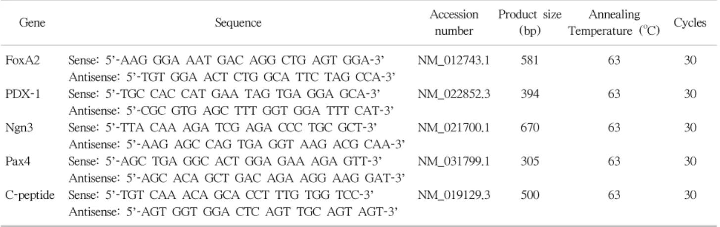
(3) J Korean Soc TransplantㆍSeptember 2015ㆍVolume 29ㆍIssue 3. o. this base medium for 4 days, and then 10 M RA (Sigma-. 30 seconds, and 72 C for 30 seconds or 1 minute for primers. Aldrich) dissolved in dimethyl sulfoxide (DMSO; Sigma-. with product sizes of less or greater than 500 bp, respecti-. Aldrich) was added of the medium (1% of total volume). o vely (Table 1). A final extension step of 72 C for 5 minutes. for 3 days. This 4/3 day cycle was repeated weekly. Cell. was completed after the last cycle. PCR products were load-. morphology was monitored using an Olympus IX71inverted. ed into wells in 1.8% agarose gel for electrophoresis. . microscope (Olympus Optical Co., Tokyo, Japan). 4. Fluorescence-activated cell sorting analysis MSCs were harvested after detachment with 0.25% tryp-. 3. Total RNA isolation and reverse-transcriptase. sin-ethylenediaminetetraacetic acid (Life Technologies). Cells. polymerase chain reaction The RNeasy Kit (Qiagen, Venlo, Netherlands) was used. were washed with 0.2% (w/v) bovine serum albumin (BSA;. to extract total RNA following the manufacturer’s. Sigma-Aldrich) in PBS and collected by centrifugation at. instructions. One microgram isolated total RNA was treated. 2,000 rpm for 5 minutes. Fluorescein isothiocyanate-con-. with 0.5 g OligodT primer 12∼18 mer (Life Technolo-. jugated CD29, CD45, CD90 (BD Biosciences, Franklin Lakes,. o. o. gies) and incubated at 70 C for 5 minutes and then 4 C for. NJ, USA), and CD34 (Santa Cruz Biotechnology, Dallas,. 5 minutes. Four microliters 5×reaction buffer, 2.4 L. TX, USA) antibodies were used for rBM-MSCs. Analysis. MgCl2, 1 L deoxyribonucleotide triphosphates, and 1 L. was performed for at least 10,000 cells/sample using fluo-. ImProm-II reverse transcriptase (Promega, Fitchburg, WI,. rescence-activated cell sorting Calibur and Cell Quest soft-. USA) were added to the mixture. The mixture was then in-. ware (BD Biosicences).. o o o cubated at 25 C for 5 minutes, 42 C for 1 hour, and 70 C. for 5 minutes to synthesize cDNA. The product was main-. 5. Immunocytochemistry. o. tained at 4 C.. rBM-MSCs at week 3 were fixed in 10% formaldehyde. Synthesized cDNA product was quantified and trans-. solution for 30 minutes at room temperature after washing. ferred into AccuPower polymerase chain reaction (PCR). with PBS. Fixed cells were washed three times with PBS,. premix (Bioneer, Daejeon, Korea) with primers. PCR was. permeabilized by incubation in 100% ice-cold methanol for. performed in a DNA thermal cycler 2720 (Life Technolo-. 5 minutes, and washed again washed three times with PBS.. o. gies). An initial denaturation condition of 94 C for 5 mi-. Non-specific binding was blocked with 5% (w/v) BSA sol-. nutes was applied to all primer sets, followed by 35 cycles. ution in PBS by incubation at room temperature for 45. o. of 94 C for 30 seconds, specific annealing temperatures for. minutes. Cells were then incubated in insulin (H-86) pri-. Table 1. Primers for rat bone marrow-derived mesenchymal stem cells Gene FoxA2 PDX-1 Ngn3 Pax4 C-peptide. Sequence Sense: 5’-AAG GGA AAT GAC AGG CTG AGT GGA-3’ Antisense: 5’-TGT GGA ACT CTG GCA TTC TAG CCA-3’ Sense: 5’-TGC CAC CAT GAA TAG TGA GGA GCA-3’ Antisense: 5’-CGC GTG AGC TTT GGT GGA TTT CAT-3’ Sense: 5’-TTA CAA AGA TCG AGA CCC TGC GCT-3’ Antisense: 5’-AAG AGC CAG TGA GGT AAG ACG CAA-3’ Sense: 5’-AGC TGA GGC ACT GGA GAA AGA GTT-3’ Antisense: 5’-AGC ACA GCT GAC AGA AGG AAG GAT-3’ Sense: 5’-TGT CAA ACA GCA CCT TTG TGG TCC-3’ Antisense: 5’-AGT GGT GGA CTC AGT TGC AGT AGT-3’. Accession number. Annealing Product size (bp) Temperature (oC). Cycles. NM_012743.1. 581. 63. 30. NM_022852.3. 394. 63. 30. NM_021700.1. 670. 63. 30. NM_031799.1. 305. 63. 30. NM_019129.3. 500. 63. 30. Abbreviations: FoxA2, forkhead box A2; PDX-1, pancreatic-duodenal homeobox 1; Ngn3, neurogenin 3; Pax4, paired box gene 4; GAPDH, glyceraldehyde 3-phosphate dehydrogenase.. 120.
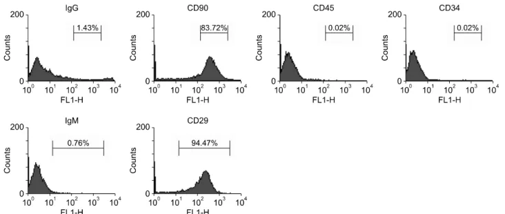
(4) Jae Hyung Kim, et al: Retinoic Acid-induced Stem Cell Differentiation. mary antibody (Santa Cruz Biotechnology) diluted 1:200 in. using references supplied by the manufacturers.. 1% BSA solution overnight on a shaking plate in a cold room. Cells were washed three times with PBS for 5 minutes. 7. Syngeneic graft of rBM-MSCs. at room temperature and then incubated with CruzFluo. Animal study was approved by Department of Laboratory. (CFL)-conjugated (maximum excitation wavelength of 488. Animal Resources, which is an Institutional Animal Care. nm) goat anti-rabbit immunoglobulin G (IgG; Santa Cruz. and Use Committee and a designated Association for Asse-. Biotechnology) secondary antibody diluted 1:200 in 1% BSA. ssment and Accreditation of Laboratory Animal Care Inter-. solution for 1 hour at room temperature in a container cov-. national facility, Yonsei Biomedical Research Institute,. ered with foil. Cells were washed three times with PBS for. Yonsei University College of Medicine, and the entire pro-. 10 minutes. Prior to observation, cell nuclei were stained. cedure was performed under control. Meloxicam (1 mg/kg. with 4’,6-diamidino-2-phenyindole (Sigma-Aldrich). Immuno-. body weight) was daily applied to ameliorate suffering, and. stained cells were observed using an Olympus IX71fluore-. gentamicin was used as antibiotics. Animals were sacrificed. scence light microscope, and images were obtained and ana-. by CO2 gas.. lyzed using a CCD DP71 digital camera and DP Controller software (Olympus Optical Co.).. Male SD rats (250∼300 g, 8∼9 weeks old) were housed at the Department of Laboratory Animal Medicine at Yonsei University College of Medicine, under a 12-hour-. 6. Enzyme-linked immunosorbent assay. day/night cycle with access to water and standard rat chow.. Culture media from differentiated hBM- and rBM-MSCs. Experimental diabetes was induced by intraperitoneal in-. o at week 3 was collected and stored at −20 C. Enzyme-. jection of 80 mg/kg streptozotocin (STZ; Sigma-Aldrich). linked immunosorbent assay (ELISA) analysis was per-. dissolved in 100 mmol/L citrate buffer (pH 4.5) at a volume. formed using the Rat Insulin ELISA Kit (Shibayagi, Shibu-. of 1 L/kg body weight. Glucose levels were checked every. kawa, Japan) following the manufacturers’ instructions.. three days with an Optium blood glucose monitoring device. Reactivity at 450 nm was measured using the Versa Max. (Abbott Laboratories, Abbott Park, IL, USA) using blood. ELISA reader (Molecular Devices, Sunnyvale, CA, USA).. obtained by cutting the tip of the tail. Blood glucose levels. Measured values were converted to insulin concentrations. above 500 mg/dL were recorded as 500.. Fig. 1. Fluorescence-activated cell sorting analysis for characterization of rat mesenchymal stem cells (MSCs) at passage 3. Abbreviation: IgG, immunoglobulin G.. 121.
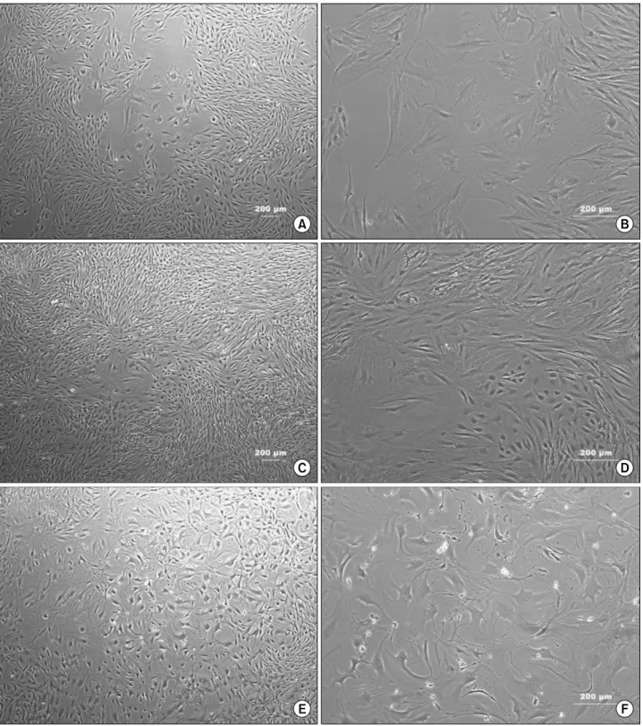
(5) J Korean Soc TransplantㆍSeptember 2015ㆍVolume 29ㆍIssue 3. Diabetic rats (glucose level >450 mg/dL) received syn-. pended in 50 L medium were injected under the surface. geneic grafts of rBM-MSCs that had been cultured for 3. of the left kidney capsule using a 30-gauge needle. Rats in-. weeks. Cells were labeled with the red fluorescent cytoplas-. jected with medium (sham; n=4) or rBM-MSCs (n=9) were. 6. mic stain PKH26 (Sigma-Aldrich), and 1×10 cells sus-. observed over a 3-week period, and rats injected with dif-. Fig. 2. Morphological changes of differentiated rat bone marrow-derived mesenchymal stem cells. (A, B) Week 1. (C, D) Week 2. (E, F) Week 3 (A, C, E: ×40; B, D, F: ×100).. 122.
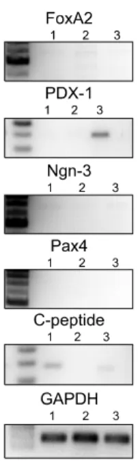
(6) Jae Hyung Kim, et al: Retinoic Acid-induced Stem Cell Differentiation. ferentiated MSCs (n=8) were observed over an 8-week-. RESULTS. period. Non-diabetic rats were observed as positive controls, and untreated STZ-induced rats were observed as negative. 1. Characterization of rBM-MSCs. controls. Glucose levels were checked every 3 days. Neph-. rBM-derived cells were predominately positive for CD29. rectomy of the graft-bearing kidney was performed at the. and CD90 (94.47% and 83.72%, respectively) (Fig. 1) but. end of the observation period, and rats were monitored for. negative for CD34 and CD45 (both 0.02%). Prior to in-. an additional 6 days. . duction of differentiation, cells were spindle-shaped with typical MSC-like morphology (Fig. 2).. 8. Immunohistochemistry Removed graft-bearing kidneys were frozen in plastic. 2. Morphology of differentiated rBM-MSCs. o molds filled with Tissue-Tek OC compound at −70 C and. After RA treatment, morphological changes of rBM-. cryosected at a thickness of 6 m. Slides were incubated in. MSCs were monitored on a weekly basis. rBM-MSCs. acetone for 10 minutes and then washed twice with distilled. showed considerable alterations in their appearance across. water on a shaking plate. ImmEdg Pen (Vector Laborato-. weeks (Fig. 2). Cells typically exhibited polygonal or round. ries, Burlingame, CA, USA) was applied to slides around the. shapes with radial nuclei.. tissue prior to antibody binding. Tissue was incubated with o insulin antibody (H86) diluted 1:200 at 4 C overnight.. 3. Reverse-transcriptase PCR of differentiated rBM-. Slides were washed three times in PBS on a shaking plate. MSCs. for 5 minutes. Tissue was then incubated with horseradish. Primers were selected from representative markers from. peroxidase-conjugated goat anti-rabbit IgG (Life Technolo-. each step of pancreatic differentiation of rBM-MSCs: FoxA2,. gies) diluted 1:250 for 1 hour at room temperature. Slides. PDX-1, Ngn3, Pax4, and C-peptide. Primers for rBM-MSCs. were washed three times in PBS on a shaking plate for 5. corresponded to those for hBM-MSCs: FoxA2, PDX-1,. minutes. Streptavidin (Dako, Glostrup, Denmark) was diluted 1:200 and reacted with the tissue for 1 hour. Slides were washed twice in PBS on a shaking plate for 5 minutes. The 3,3’-diaminobenzidine substrate kit (Vector Laboratories) was used to stain tissue sections following the manufacturer’s instructions. Slides were then washed with distilled water twice for 5 minutes on a shaker, and Accustain Harris Hematoxylin Solution was applied for 1 minute as a counterstain. Slides were washed with flowing water followed by absolute alcohol and xylene and then cover-slipped with Permount (Fisher Scientific, Hampton, NH, USA). Tissue was observed using a fluorescence light microscope. 9. Statistical analysis Quantitative results are expressed as mean±standard deviation. Paired t-tests were used to compare groups using GraphPad Prism 6 for Windows (GraphPad Software, Inc., La Jolla, CA, USA). Statistical significance was set at P< 0.05.. Fig. 3. Reverse-transcriptase polymerase chain reaction of rat bone marrow-derived mesenchymal stem cells (rBM-MSCs). Lanes 1 to 3: differentiated rBM-MSCs from week 1 to 3. Abbreviations: FoxA2, forkhead box A2; PDX-1, pancreatic-duodenal homeobox 1; Ngn3, neurogenin 3; Pax4, paired box gene 4; GAPDH, glyceraldehyde 3-phosphate dehydrogenase.. 123.
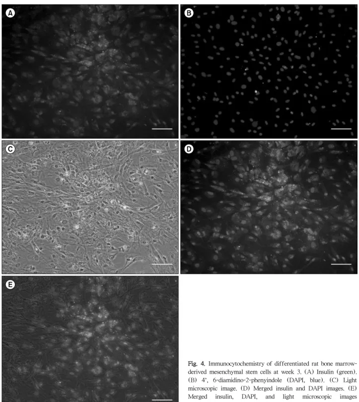
(7) J Korean Soc TransplantㆍSeptember 2015ㆍVolume 29ㆍIssue 3. Fig. 4. Immunocytochemistry of differentiated rat bone marrowderived mesenchymal stem cells at week 3. (A) Insulin (green). (B) 4’, 6-diamidino-2-phenyindole (DAPI, blue). (C) Light microscopic image. (D) Merged insulin and DAPI images. (E) Merged insulin, DAPI, and light microscopic images (magnification, ×100; scale bar=200 m).. Ngn3, Pax4, and C-peptide for rodents. The expression patterns of differentiated rBM-MSCs had no expression of Pax4, minimal expression of FoxA2 and Ngn3, and up-regulation of PDX-1 and C-peptide at week 3 (Fig. 3).. 4. Qualitative and quantitative analysis of insulin expression in hBM- and rBM-MSCs Differentiated rBM-MSCs were incubated with insulin primary antibody and labeled with green fluorescent CFL-488-conjugated secondary antibody. Insulin expression. 124.
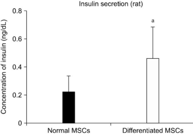
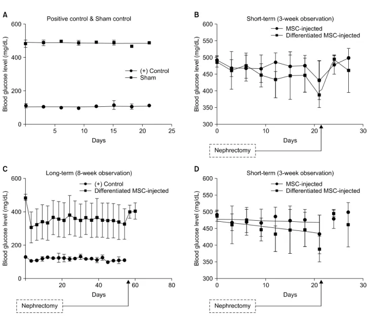
(8) Jae Hyung Kim, et al: Retinoic Acid-induced Stem Cell Differentiation. was clearly observed in differentiated cells (Fig. 4).. for differentiation and plasticity. Furthermore, they may be. ELISA revealed that differentiated rBM-MSCs showed a. an ideal cell type for clinical use because they are more ac-. 2.063-fold increase in insulin secretion, which was signifi-. cessible for harvesting and adaptable to new environments. cantly higher than that observed for MSCs cultivated in. compared with pancreatic islets(17). Over the past several. normal medium (P<0.05; Fig. 5).. years, we have genetically modified cells for T1DM treat-. 5. Syngeneic graft of rBM-MSCs in vivo. However, two major obstacles we encountered were low. ment and established insulin-producing cells in vitro(25,26). Non-diabetic (+control) and STZ-injected rats (sham). transfection efficiency and insufficient viability of cells to. exhibited obvious differences in blood glucose levels, with. establish populations for in vivo trials. Therefore, in the. non-diabetic rats maintaining normoglycemia but diabetic. present study, we focused on enhancing differentiation of. rats exhibiting hyperglycemia (Fig. 6A). Diabetic rats that. cells in vitro while endeavoring to reduce the use of cyto-. received normal MSCs showed no reduction of hyper-. toxic materials and/or growth factors.. glycemia across the 3-week observation period, whereas dia-. Three non-toxic reagents were used to induce -cell dif-. betic rats that received differentiated MSCs showed a mod-. ferentiation of cultivated MSCs: nicotinamide, L-glutamine,. est decrease in blood glucose level (Fig. 7B). Linear re-. and RA. Nicotinamide, also known as vitamin B3, has been. gression analysis revealed that differentiated MSCs reduced. reported to induce endocrine differentiation in pancreatic. blood glucose levels more rapidly than normal MSCs (slope. cells(27). Also, several groups report that L-glutamine in-. for the differentiated MSC-injected group=−1.728±0.7725. duces stem cells to become functional endoderm in hu-. vs. slope for the normal MSC-injected=−0.4935±0.5103). mans(28). Moreover, several groups show that RA receptor. (Fig. 6D).. signaling is required for the development of pancreatic pro-. When we continued to observe differentiated MSC-in-. genitor cells(29,30). As growth factors may induce uncon-. jected rats for an additional 5 weeks, we found that blood. trollable cellular activities in vivo such as oncogenesis(31),. glucose levels remained below 400 mg/dL throughout the. we eliminated growth factors from the cultivation medium. entire observation period (Fig. 6C). Furthermore, rats ex-. and attempted to use a limited number of reagents to ach-. hibited a rise in blood glucose levels after nephrectomy of. ieve sufficient -cell differentiation.. the grafted kidney. . RA, in particular, is an essential substance for -cell differentiation and is closely associated with expression of. 6. Histological observation of grafted rBM-MSCs. PDX-1(29,32). PDX-1, a marker of early pancreatic pro-. rBM-MSCs were labeled with red fluorescent PKH26 prior to their injection. Three weeks after injection, we observed labeled cells mostly along the membrane near the injection site, with some cells partially migrating into the kidney (Fig. 8). Insulin- and hematoxylin-stained images show that grafted cells successfully survived along the inner surface of the tissue membrane (Fig. 7).. DISCUSSION The generation of insulin-producing -cell surrogates from progenitor/stem cells in vitro has remarkable potential for a cell-based therapy for T1DM(13,17-21). However, the. in vivo therapeutic success of mature -cell alternatives has yet to be demonstrated. BM-MSCs exhibit great capacities. Fig. 5. Insulin secretion from differentiated rat bone marrowderived mesenchymal stem cells (MSCs) measured by enzymelinked Immunosorbent Assay at week 3 (n=9). aP<0.05.. 125.
(9) J Korean Soc TransplantㆍSeptember 2015ㆍVolume 29ㆍIssue 3. Fig. 6. Blood glucose levels of rats observed for 3 (A, B) or 8 (C) weeks. (A) Positive control (n=4) and sham control (n=4). (B) Blood glucose level of normal mesenchymal stem cell (MSC)-injected (n=9) and differentiated MSC-injected (n=8) rats. (C) Positive control (n=4) and differentiated MSC-injected (n=8) rats. (D) Linear regression of blood glucose levels from MSC-injected (n=9) and differentiated MSC-injected (n=8) rats. Slope for MSC-injected group=−0.4935±0.5103; slope for differentiated MSC-injected group=−1.728±0.7725.. genitor cells and mature -cells, is a well-established sign of. C-peptide (Fig. 3). Nevertheless, we conclude that these cells. -cell differentiation(32-34), and overexpression of PDX-1. cannot be considered as -cells but only as candidates with. alone may induce differentiation of MSCs into in-. potential. Limitations clearly remain, which we are seriously. sulin-producing cells that are responsive to glucose stim-. taking into account to resolve in the next study.. ulation(17). Consistent with these findings, our reverse-. T1DM is primarily an autoimmune disease, and, as a con-. transcriptase PCR and ELISA results show that PDX-1 ex-. sequence, graft function progressively declines due to the. pressed with insulin secretion (Fig. 5) in rat (Fig. 3) MSCs. loss of functional islets during the early post-transplantation. in vitro. As Ostrom et al.(30) reports, RA is at least partly. period(35). Therefore, the present study focused on MSCs,. required for the development of dorsal pancreatic mesen-. which could play a major role in tissue regeneration through. chyme during early pancreas development. Our results show. localized immune-suppressive effects. To increase the func-. that MSCs from rats secreted insulin (Fig. 5) and express. tional activity of MSCs in vivo, we induced their differ-. 126.
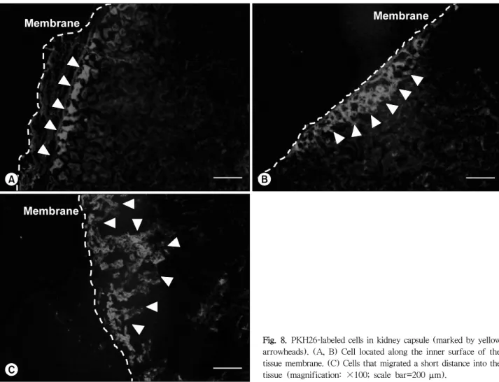
(10) Jae Hyung Kim, et al: Retinoic Acid-induced Stem Cell Differentiation. entiation by culturing MSCs in a customized differentiation. effective than undifferentiated cells in reducing blood glu-. medium for 3 weeks before their graft into the left kidney. cose levels (Fig. 6), and differentiated MSCs successfully. capsule of rats. Differentiated MSCs were modestly more. survived in the kidney for at least 3 weeks (Fig. 7, 8).. Fig. 7. 3,3’-Diaminobenzidine-stained tissue. (A) Cells located along the inner surface of the tissue membrane. (B) Cell that migrated a short distance into the tissue (marked by yellow arrowhead) (magnification: ×100; scale bar=200 m).. Fig. 8. PKH26-labeled cells in kidney capsule (marked by yellow arrowheads). (A, B) Cell located along the inner surface of the tissue membrane. (C) Cells that migrated a short distance into the tissue (magnification: ×100; scale bar=200 m).. 127.
(11) J Korean Soc TransplantㆍSeptember 2015ㆍVolume 29ㆍIssue 3. Furthermore, after nephrectomy of the grafted kidney, rats. REFERENCES. exhibited a return of high blood glucose levels. Although T1DM is an acute disease, we assume that grafted MSCs may enable chronic recovery, aiding in secretion of insulin. Several research groups report that RA can regulate the commitment of embryonic stem cells to establish pancreatic endoderm(29,30,36). However, detailed in vivo evidence of recovered function does not exist, and there has been no. 1) Calne R. Cell transplantation for diabetes. Philos Trans R Soc Lond B Biol Sci 2005;360:1769-74. 2) Ackermann AM, Gannon M. Molecular regulation of pancreatic beta-cell mass development, maintenance, and expansion. J Mol Endocrinol 2007;38:193-206. 3) Melloul D, Marshak S, Cerasi E. Regulation of pdx-1 gene expression. Diabetes 2002;51 Suppl 3:S320-5.. attempt to use MSCs, which entail fewer ethical concerns. 4) Guz Y, Montminy MR, Stein R, Leonard J, Gamer LW, Wright. and are more clinically applicable. Thus, the results of the. CV, et al. Expression of murine STF-1, a putative insulin. present study are compelling, as they clearly show a reduc-. gene transcription factor, in beta cells of pancreas, duodenal. tion in blood glucose levels and the survival of MSCs 3 weeks after injection. We conclude that RA-induced -cell differentiation of BM-MSCs may have great therapeutic potential for T1DM.. epithelium and pancreatic exocrine and endocrine progenitors during ontogeny. Development 1995;121:11-8. 5) Huang HP, Liu M, El-Hodiri HM, Chu K, Jamrich M, Tsai MJ. Regulation of the pancreatic islet-specific gene BETA2 (neuroD) by neurogenin 3. Mol Cell Biol 2000;20:3292-307.. One may argue that the sustained reduction of blood glucose levels in the present study was still too high to be considered normoglycemia. However, we find this data to be meaningful because only a very low number of cells (1× 10⁶ cells per rat) were injected into rats. Increasing the number of injected cells could be explored in future studies as a way to improve the effectiveness of this potential ther-. 6) Fuchs E, Segre JA. Stem cells: a new lease on life. Cell 2000;100:143-55. 7) Anzalone R, Lo Iacono M, Loria T, Di Stefano A, Giannuzzi P, Farina F, et al. Wharton's jelly mesenchymal stem cells as candidates for beta cells regeneration: extending the differentiative and immunomodulatory benefits of adult mesenchymal stem cells for the treatment of type 1 diabetes. Stem Cell Rev 2011;7:342-63.. apeutic tool. Furthermore, diabetic rats initially exhibited. 8) Bacigalupo A, Valle M, Podesta M, Pitto A, Zocchi E, De. excessively high levels of blood glucose, (i.e., typically above. Flora A, et al. T-cell suppression mediated by mesenchymal. 500 mg/dL), meaning that the observed reduction in blood glucose levels could be considered evidence that the differentiated cells aided in the recovery of insulin secretion. It is also noteworthy that blood glucose level appeared to be continuously regulated, without a single instance of a rapid return to hyperglycemia throughout both the short- and long-term observation periods.. CONCLUSION. stem cells is deficient in patients with severe aplastic anemia. Exp Hematol 2005;33:819-27. 9) Jung EJ, Kim SC, Wee YM, Kim YH, Choi MY, Jeong SH, et al. Bone marrow-derived mesenchymal stromal cells support rat pancreatic islet survival and insulin secretory function in vitro. Cytotherapy 2011;13:19-29. 10) Popp FC, Renner P, Eggenhofer E, Slowik P, Geissler EK, Piso P, et al. Mesenchymal stem cells as immunomodulators after liver transplantation. Liver Transpl 2009;15:1192-8. 11) Rasmusson I, Ringden O, Sundberg B, Le Blanc K. Mesenchymal stem cells inhibit lymphocyte proliferation by mitogens and alloantigens by different mechanisms. Exp. In summary, we showed that RA can induce the differ-. Cell Res 2005;305:33-41.. entiation of rBM-MSCs into the -cell lineage, resulting in. 12) Efrat S. Regulation of insulin secretion: insights from en-. cells that have the ability to secrete insulin. Furthermore,. gineered beta-cell lines. Ann N Y Acad Sci 2004;1014:. the success of grafted MSCs in a rat model of diabetes suggests that MSCs can potentially serve as -cell surrogates within therapies for T1DM.. 88-96. 13) Oh SH, Muzzonigro TM, Bae SH, LaPlante JM, Hatch HM, Petersen BE. Adult bone marrow-derived cells trans-differentiating into insulin-producing cells for the treatment of type I diabetes. Lab Invest 2004;84:607-17. 14) Lee KD, Kuo TK, Whang-Peng J, Chung YF, Lin CT, Chou SH, et al. In vitro hepatic differentiation of human mesen-. 128.
(12) Jae Hyung Kim, et al: Retinoic Acid-induced Stem Cell Differentiation. chymal stem cells. Hepatology 2004;40:1275-84. 15) Seeberger KL, Eshpeter A, Rajotte RV, Korbutt GS. Epithelial. human mesenchymal stem cells using recombinant adeno-associated virus. Yonsei Med J 2007;48:109-19.. cells within the human pancreas do not coexpress mesen-. 26) Kim JH, Shin KH, Li TZ, Suh H. Potential of nucleofected. chymal antigens: epithelial-mesenchymal transition is an. human MSCs for insulin secretion. J Tissue Eng Regen Med. artifact of cell culture. Lab Invest 2009;89:110-21.. 2011;5:761-9.. 16) Seo MJ, Suh SY, Bae YC, Jung JS. Differentiation of human. 27) Otonkoski T, Beattie GM, Mally MI, Ricordi C, Hayek A.. adipose stromal cells into hepatic lineage in vitro and in. Nicotinamide is a potent inducer of endocrine differ-. vivo. Biochem Biophys Res Commun 2005;328:258-64.. entiation in cultured human fetal pancreatic cells. J Clin. 17) Zanini C, Bruno S, Mandili G, Baci D, Cerutti F, Cenacchi. Invest 1993;92:1459-66.. G, et al. Differentiation of mesenchymal stem cells derived. 28) Sullivan GJ, Hay DC, Park IH, Fletcher J, Hannoun Z, Payne. from pancreatic islets and bone marrow into islet-like cell. CM, et al. Generation of functional human hepatic endo-. phenotype. PLoS One 2011;6:e28175.. derm from human induced pluripotent stem cells.. 18) Zhou H, Ding S. Evolution of induced pluripotent stem cell technology. Curr Opin Hematol 2010;17:276-80.. Hepatology 2010;51:329-35. 29) Mfopou JK, Chen B, Sui L, Sermon K, Bouwens L. Recent. 19) Hori Y, Rulifson IC, Tsai BC, Heit JJ, Cahoy JD, Kim SK.. advances and prospects in the differentiation of pancreatic. Growth inhibitors promote differentiation of insulin-pro-. cells from human embryonic stem cells. Diabetes 2010;. ducing tissue from embryonic stem cells. Proc Natl Acad Sci U S A 2002;99:16105-10.. 59:2094-101. 30) Ostrom M, Loffler KA, Edfalk S, Selander L, Dahl U, Ricordi. 20) Kang HM, Kim J, Park S, Kim J, Kim H, Kim KS, et al.. C, et al. Retinoic acid promotes the generation of pancreatic. Insulin-secreting cells from human eyelid-derived stem. endocrine progenitor cells and their further differentiation. cells alleviate type I diabetes in immunocompetent mice. Stem Cells 2009;27:1999-2008.. into beta-cells. PLoS One 2008;3:e2841. 31) Li TZ, Kim JH, Cho HH, Lee HS, Kim KS, Lee SW, et al.. 21) Tang DQ, Cao LZ, Burkhardt BR, Xia CQ, Litherland SA,. Therapeutic potential of bone-marrow-derived mesen-. Atkinson MA, et al. In vivo and in vitro characterization. chymal stem cells differentiated with growth-factor-free. of insulin-producing cells obtained from murine bone. coculture method in liver-injured rats. Tissue Eng Part A. marrow. Diabetes 2004;53:1721-32.. 2010;16:2649-59.. 22) Shi Y, Hou L, Tang F, Jiang W, Wang P, Ding M, et al.. 32) Shen CN, Marguerie A, Chien CY, Dickson C, Slack JM,. Inducing embryonic stem cells to differentiate into pancre-. Tosh D. All-trans retinoic acid suppresses exocrine differ-. atic beta cells by a novel three-step approach with activin. entiation and branching morphogenesis in the embryonic. A and all-trans retinoic acid. Stem Cells 2005;23:656-62.. pancreas. Differentiation 2007;75:62-74.. 23) Delaspre F, Massumi M, Salido M, Soria B, Ravassard P,. 33) Peck AB, Ramiya V. In vitro-generation of surrogate islets. Savatier P, et al. Directed pancreatic acinar differentiation. from adult stem cells. Transpl Immunol 2004;12:259-72.. of mouse embryonic stem cells via embryonic signalling. 34) Sumi S, Gu Y, Hiura A, Inoue K. Stem cells and regenerative. molecules and exocrine transcription factors. PLoS One 2013;8:e54243.. medicine for diabetes mellitus. Pancreas 2004;29:e85-9. 35) Rackham CL, Chagastelles PC, Nardi NB, Hauge-Evans AC,. 24) Cavallari G, Olivi E, Bianchi F, Neri F, Foroni L, Valente. Jones PM, King AJ. Co-transplantation of mesenchymal. S, et al. Mesenchymal stem cells and islet cotransplantation. stem cells maintains islet organisation and morphology in. in diabetic rats: improved islet graft revascularization and. mice. Diabetologia 2011;54:1127-35.. function by human adipose tissue-derived stem cells pre-. 36) Micallef SJ, Janes ME, Knezevic K, Davis RP, Elefanty AG,. conditioned with natural molecules. Cell Transplant. Stanley EG. Retinoic acid induces Pdx1-positive endoderm. 2012;21:2771-81.. in differentiating mouse embryonic stem cells. Diabetes. 25) Kim JH, Park SN, Suh H. Generation of insulin-producing. 2005;54:301-5.. 129.
(13)
수치
+5
관련 문서
Carnosol promotes osteoblast differentiation of mouse bone marrow-derived mesenchymal stem cells (mBMSCs) through the activa- tion of bone morphogenetic protein (BMP)
In this study of using a low-intensity ultrasound (LIUS), investigated the effects of LIUS on chondrogenic differentiation of bone marrow-derived mesenchymal stem cells (BM-MSCs)..
Erices et al. reported that hUCB harvests generate mes- enchymal progenitor cells with large ex vivo expansion capac- ity as well as osteogenic and adipogenic differentiation
JVS Original Article J Vet Sci 2015, 16(4), 397 404ㆍhttp //dx doi org/10 4142/jvs 2015 16 4 397 Effect of serum derived albumin scaffold and canine adipose tissue derived mesenchymal
Although a quantita- tive analysis indicated that the expression of neurogenic regulatory genes during RA-induced differentiation was not significantly influenced by
Effect of Fibroblast Growth Factor 23 on Osteoblastic Differentiation and Mineralization of D1 Mesenchymal Stem Cells..
Sustained release of Ascorbate-2- phosphate and Dexamethasone from porous PLGA scaffolds for osteogenic differentiation of mesenchymal stem cells..
Shear Stress Induced by an Interstitial Level of Slow Flow Increases the Osteogenic Differentiation of Mesenchymal Stem Cells through TAZ Activation.. Kyung Min Kim 1 , Yoon Jung Choi
