Role of NFAT5 in Osteogenic Differentiation of Human Adipose Tissue-Derived Mesenchymal Stem Cells
Sun Young Lee, Ji won Yang and Jin Sup Jung*
Department of Physiology, School of Medicine, Pusan National University, Yangsan 626-870, Korea
Received January 23, 2013 /Revised March 19, 2013 /Accepted April 17, 2013Human adipose tissue-derived mesenchymal stem cells (hADSCs) have therapeutic potential, includ- ing the ability to self-renew and differentiate into multiple lineages. Understanding of molecular mechanisms of stem cell differentiation is important for improving the therapeutic efficacies of stem cell transplantation. In this study, we determined the role of nuclear factor of activated T cells (NFAT5) in the osteogenic differentiation of hADSCs. The down-regulation of NFAT5 expression by the trans- fection of a specific siRNA significantly inhibited osteogenic differentiation of hADSCs and decreased the activity of the nuclear factor kappa-light-chain-enhancer of activated B cells (NF-κB) promoter without affecting their proliferation and adipogenic differentiation. The inhibition of NFAT5 expression in- hibited the basal and Tumor Necrosis Factor α (TNF-α) induced activation of NF-κB, but it did not affect TNF- α-induced degradation of the IκB protein. These findings indicate that NFAT5 plays an important role in the osteogenic differentiation of hADSCs through the modulation of the NF-κB pathway.
Key words : Human adipose tissue-derived mesenchymal stem cells (hADSCs), osteogenic differentiation, nuclear factor of activated T cells (NFAT5)
*Corresponding author
*Tel:+82-51-510-8071, Fax:+82-51-510-8076
*E-mail : [email protected]
This is an Open-Access article distributed under the terms of the Creative Commons Attribution Non-Commercial License (http://creativecommons.org/licenses/by-nc/3.0) which permits unrestricted non-commercial use, distribution, and reproduction in any medium, provided the original work is properly cited.
ISSN (Online) 2287-3406 Journal of Life Science 2013 Vol. 23. No. 4. 471~478 DOI : http://dx.doi.org/10.5352/JLS.2013.23.4.471
서 론
줄기세포(Stem cell)는 우리 몸을 구성하는 모든 세포나 조 직으로 분화 할 수 있는 능력을 가지고 있어 이를 이용한 질병 치료에 많은 관심을 받아 오고 있다. 줄기세포는 발생 초기 배반포(blastocyst)에서 얻어지는 배아줄기세포(embryonic stem cell)와 발생과정이 끝난 성체 또는 태반에서 얻어지는 성체줄기세포(adult stem cell)가 있다[14]. 배아줄기세포의 이 용은 생명체 이용이라는 점에서 많은 윤리적인 문제를 안고 있어 실질적인 사용에 제한이 따른다. 반면, 성체줄기세포는 생체 내에 이식된 후 장기 특성에 맞게 분화하는 특이성 및 본래의 세포 특성과는 다른 종류의 세포로 교차 분화할 수 있는 유연성을 가지고 있고, 다양한 세포로 분화될 수 있는 잠재성이 있음이 밝혀지면서 성체줄기 세포를 통한 세포 치료 의 가능성이 높아지고 있다[29].
성체 줄기세포 중 중간엽 줄기세포(adult mesenchymal stem cells, MSC)를 얻기 위한 연구들은 주로 골수에서 이루어 져 왔으며, 골수 줄기세포를 이용하여 다양한 조직으로의 분 화 등 많은 연구가 이루어져 왔다[6, 16, 24, 25, 28]. 그러나
골수에서의 세포 획득은 환자의 고통을 수반하며 임상에 적용 하기 위한 충분한 양의 세포를 얻기 위해서는 여러 번 채취해 야 하는 부담이 존재한다[5, 13]. 따라서 골수와 같은 간엽에서 유래하며 다양한 기질 세포들을 포함 하고 있는 지방조직은 또 다른 줄기세포의 원천으로 각광 받고 있으며[31, 32], 지방 추출물 안에는 줄기세포로 추정되는 세포들이 존재하며, 이를 지방 유래 중간엽 줄기세포(adipose tissue-derived mesen- chymal stem cells, ADSCs)라고 명명 되어지고 있다. 지방조 직은 많은 양의 조직 채취가 용이하여 줄기세포를 수확하는데 좋은 조건을 가지고 있으며, 인체 지방 유래 중간엽 줄기세포 는 배양시 안정적인 성장과 증식을 보여주고 있다[27].
여러 조직으로 분화가 가능한 지방 유래 중간엽 줄기세포에 서 골분화 기전은 이미 잘 알려져 있으며[7, 8, 22], 염증성 골질 환에서의 골조직의 손실과 밀접한 관련이 있는 Tumor ne- crosis factor-α (TNF-α)가 NF-κB 경로를 통해서 인체 골수 유 래 중간엽 줄기세포와 지방 유래 중간엽 줄기세포의 골분화에 관여 한다고 증명되었다[1, 4]. 하지만 이러한 신호전달체계에 서 어떠한 전사 인자를 통해서 지방 유래 중간엽 줄기세포가 골세포로 분화하는지에 대한 메커니즘은 잘 알려져 있지 않다.
NFAT5 (nuclear factor of activated T cells)는 5종류의
NFAT family 중 하나로써 TonEBP (tonicity-responsive en-
hancer binding protein)라고도 불리우며 Rel family로 고장
성에 의해 촉진되는 전사를 매개하는 전사 조절 인자로 발견
되었다[19]. 주로 신장 등의 높은 삼투압 환경에서 세포를 보호
하며 여러 기관에서 세포의 생존, 발달 등에 관련된 기능을
한다[3, 18]. 특히 NFAT5는 세포질의 삼투질 농도에 의해 발현
이 조절되며 활막세포와 혈관내피세포의 증식과 분화에 중요 한 역할을 하는 조절인자라고 보고 되어져 있고[9, 30], 근원세 포의 이주와 분화에도 관여하는 것으로 알려져 있다[15, 21, 26]. 그러나 중간엽 줄기세포에서 NFAT5의 작용은 알려진 바 가 없다.
이에 본 연구에서는 인체 지방 유래 중간엽 줄기세포의 체외배양에서, 골분화 과정에서 NFAT5 의 역할을 규명하고 자 일련의 실험들을 수행 하였으며, TNF-α에 의한 NFAT5의 조절 기작에 대한 연구 결과를 얻었기에 이에 보고하고자 한 다.
재료 및 방법
시약 및 재료
세포 배양액인 α-minimal essential medium (α-MEM)은 Sigma사( St Louis, MO, USA)에서 fetal bovine serum (FBS), penicillin-streptomycin 등의 세포배양용 시약들은 Gibco- BRL (Gaithersburg, MD, USA)에서 구입하여 사용하였다. 실 험에 사용 된 사이토카인 Tumor necrosis factor-α (Recombinant Human TNF-α, 10 ng/ml)는 R&D system (Minneapolis, MN, USA)에서 구입하였다. Dexamethasone, β-glycerophosphate, ascorbate-2-phosphate, IBMX, indomethacin은 Sigma사에서 구입하였다.
세포의 분리 및 배양
중간엽 줄기세포의 분리를 위해 공여자로부터 지방을 공여 받아 실험에 사용하였다. 지방조직을 잘게 쪼갠 후 0.075%
type Ⅰcollagenase로 30분간 처리한 후 세포를 분리하였다.
분리한 세포는 배양액(10% FBS, 100 units/ml penicillin과 100 μg/ml streptomycin을 포함하는 α-MEM에서 배양하였 고, 다음날 새로운 배양액으로 교체하여 배양용기에 달라붙지 않은 세포를 제거하였다. 세포의 배양은 2일마다 새로운 배양 액으로 바꾸어 주면서 배양하였다. 세포가 배양용기에 80~90% 차게 되면 0.25% trypsin (Sigma)를 이용하여 배양용 기에서 떼어낸 후, 새로운 배양용기에 1:10 비율로 희석하여 계대배양 한 후 37℃, 5% CO
2에서 배양 하였다. 이 때를 중간 엽 줄기세포의 1차 계대배양으로 정의 하였다. 나머지 세포는 10% DMSO (Sigma)가 포함된 배양액에서 서서히 냉동시킨 후, 최종적으로 액체질소에 보관하였다.
Small interfering RNA (siRNA) transfection
중간엽 줄기세포에서 siRNA transfection은 비특이적 siRNA (On-Target plus non targeting siRNA, Dharmacon, Inc, CO, USA)와 NFAT5에 특이적인 siRNA (NFAT5 on- TARGET plus SMART pool, Dharmacon, Inc, CO, USA)를 이용하여 DharmaFECT Transfection Reagent로 시행하였 다.
Reverse Transcription-Polymerase Chain Reaction (RT-PCR)
중간엽 줄기세포에서 Trizol (Invitrogen, Gaithersburg, MD, USA)을 이용, total RNA를 분리하여 2 μg의 RNA와 2.5 mM dNTP, oligo dT primer, 5X first strand buffer, RNase inhibitor, M-MLV reverstanscriptase (Promega corporation, Madison, WI, USA)를 사용해서 역전사해서 cDNA를 얻었다.
PCR 반응은 2 μl의 cDNA와 NFAT5 또는 GAPDH에 특이적 인 primer를 이용하여 PCR를 수행하였다. PCR조건은 94℃에 서 30초 denaturation 한 후, 30 cycle로 60℃에서 30초 anneal- ing, 72℃에서 30초 extension 한다. 사용한 primer의 염기서열 은 다음과 같다. 인체 NFAT5 5’-ATGCCCTGATGACTCC ACTC-3’, 5’-CTGCAATAGTGCATCGCTGT-3’, 인체 GAPDH 5’-TCCATGACAACTTTGGTATCG-3’, 5’-TGTAGCCAAAT TCGTTGTCA-3’로써 각각의 PCR 산물을 1.5% agarose gel 상에서 확인하였다.
Real-Time Polymerase Chain Reaction
PCR 산물은 2 X SYBR Green PCR Master Mix (Applied Biosystems, Foster City, CA, USA)를 이용하여 ABI PRISM 7500 (Applied Biosystems, Foster City, CA, USA)을 사용하여 증폭하였다.
사용한 primer의 염기서열은 다음과 같다. Human NFAT5 의 primer는 5’-ATGCCCTGATGACTCCACTC-3’, 5’-CTG CAATAGTGCATCGCTGT-3’이며, Human β-actin의 primer 는 5’-AACACCCCAGCCATGTACG-3’, 5’-GGGAAATCGT GCGTGACAT-3’이며 정량 결과의 분석은 7500 software ver- sion 3.3 (Applied Biosystems Diagnostics)로 하였다.
세포 증식 측정
중간엽 줄기세포 1×10
4개를 6 well plate에 분주하고 2 ml 의 배양액(10% FBS, 100 units/ml penicillin과 100 μg/ml streptomycin을 포함 하는 α-MEM으로 2-3일에 한 번씩 배양 액을 교체하여 3, 4, 5일 동안 배양하였다. 배양시기별 세포를 모아 Hank’s balanced salt solution (Sigma)로 2회 세척 한 후 0.25% trypsin을 이용하여 배양용기에서 떼어낸 후 여러 번 pipetting 하여 단일 세포로 만들었다. 단일 세포와 0.4%
trypan blue dye (Sigma)을 섞은 후 배양 2, 3, 4일 째 되는 날에 혈구계산판을 이용하여 세포 수를 측정하였다.
지방 세포 분화
중간엽 줄기세포 1×10
5개를 12 well plate에 분주하여 하루 동안 배양 한 후 1 ml의 지방분화유도배지(10% FBS, 1 μM dexamethasone, 0.5 mM IBMX, 200 μl indomethacin 을 포함 하는 α-MEM에서 7일간 37℃, 5% CO
2조건에서 배양하였다.
고정 및 세척 후 Oil Red O (Sigma) 용액으로 세포를 염색하였
으며 광학 현미경하에서 지질적화 형성을 관찰하였다.
골세포 분화
중간엽 줄기세포 1×10
5개를 12 well plate에 분주하여 하루 동안 배양 한 후 1 ml의 골분화유도배지(10% FBS, 0.1 uM dexamethasone, 10 mM β-glycerophosphate, 0.5 mM ascor- bate-2-phosphate 를 포함 하는 α-MEM에서 2주간 37℃, 5%
CO
2조건에서 배양하였다. 고정 및 세척 후 Alizarin Red S (Sigma) 용액으로 세포를 염색하였으며 광학 현미경하에서 석 회화 결절 형성을 관찰하였다.
Calcium 농도 측정
골분화유도배지에서 2주간 배양시킨 12 well plate를 PBS 용액으로 3회 세척한 후 hydrichloric acid 0.5 N (Sigma) 용액 을 300 μl을 넣어 상온에서 16시간 반응시켰다. 그리고 96 well plate에 calcium이 추출된 용액 5 μl와 측정시약(BioAssay Systems, Hayward, CA) 200 μl를 상온에서 3분간 반응시킨 후, 612 nm에서 흡광도를 측정하여 분석하였다.
Reporter gene 실험
pNF-κB-Luc, pCMV-β-Gal plasmid (Clontech Laboratories, Inc, Palo Alto, CA, USA)를 중간엽 줄기세포에 Lipofectamine Plus Reagent (Invitrogen, Gaithersburg, MD, USA)를 이용하 여 transfection하였다. Transfection 1-2일 후에 세포를 re- porter lysis buffer (Promega, WI, USA)를 사용하여 lysate를 만든 후, lysate를 원심분리하여 상등액을 취하고 luciferin 기 질(Luciferase Assay System, Promega corporation, Madison, WI, USA)을 이용하여 luciferase activity를 측정하였으며, β- galactosidase 활성은 β-Galactosidase Enzyme Assay System (Promega corporation, Madison, WI, USA)을 이용하여 측정 하였고 이 값을 luciferase 활성 수치를 보정하는데 사용하였다.
Western blot analysis
배양한 세포를 회수 한 후, 2% sodium dodecylsulfate, 10% glycerol, 50 mM DTT를 포함하는 62.5 mM Tris-HCl, pH 6.8 용액에 넣고 5분간 끓인 후 불용성 침전물을 제거 한 후 동량의 단백질을 10% SDS polyacrylamide gel에서 전기 영동 하였다. Gel 내의 단백질을 nitrocellulose membranes (Hybond-ECL; Amersham Pharmacia Biotech, Piscataway, NJ, USA)으로 옮겨 준 후, 5% 탈지우유와 0.1% Tween20이 포함된 tris로 완충된 식염 용액(TBST)으로 상온에서 1시간 반응시킨 후 다중항체(GAPDH, IκB-α; Cell Signaling Tech- nology, Danvers, MA, USA, IκB; Santa Cruz Biotechnology, CA, USA)를 1:1,000으로 희석하여 4℃에서 16시간 동안 반응 시켰다. TBST용액으로 10분씩 3회 세척한 후, peroxidase가 결합된 이차항체(Amersham Pharmacia Biotech)를 넣고 1시 간 반응 시켰다. TBST 용액으로 다시 3회 세척한 후 chem- iluminescence (ELC detection kit, Amersham Pharmacia
Biotech)로 반응시킨 후 LAS-3000으로 감광시켰다.
통계 처리
결과들은 평균 ± 표준오차로 표시하고, 변수의 분석들은 Student’s t-test와 분산분석후 사후검증으로 Duncan test를 사 용하였다. P 값이 0.05 미만인 경우에 통계적으로 유의 하다는 판정을 하였으며, 모든 실험은 독립적으로 3회 이상 실시하여 통계 처리를 하였다.
결 과
인체 지방 유래 줄기세포에서 NFAT5 발현 억제에 의한 세포증식의 효과
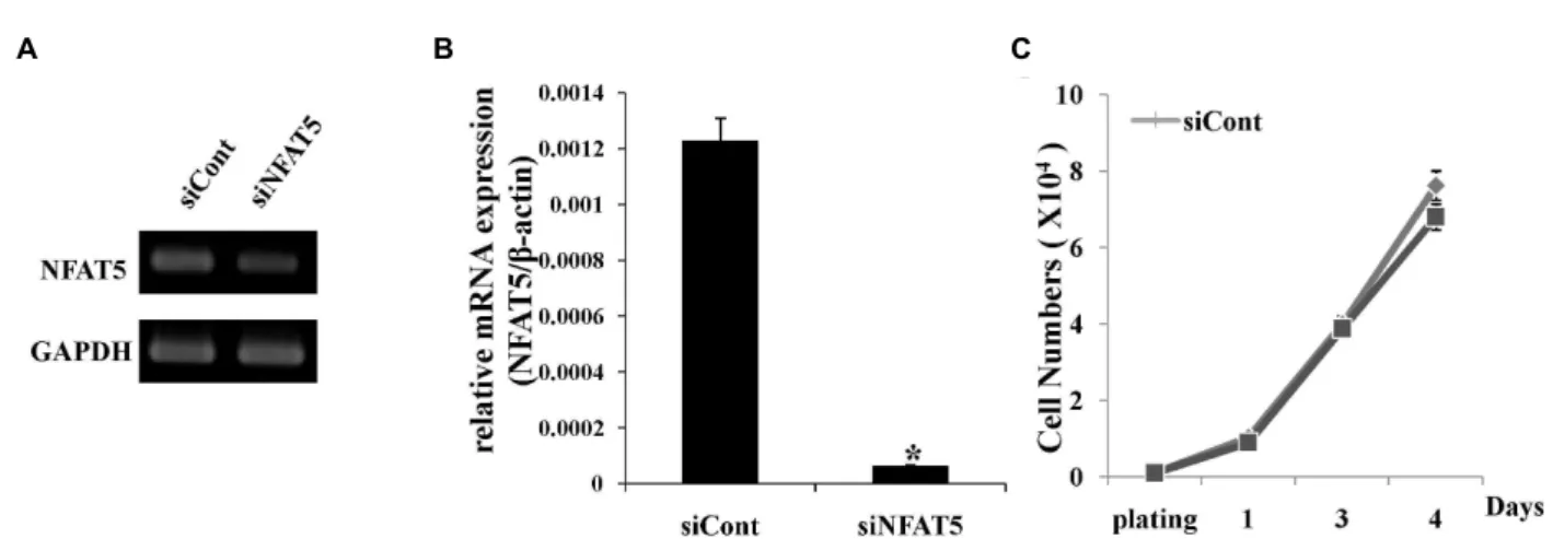
인체 지방 유래 중간엽 줄기세포의 기능에 NFAT5가 미치 는 영향을 확인하기 위해 siRNA를 이용하여 transfection 시킨 뒤, 3일 후 세포를 회수하고 trizol을 이용하여 RNA를 분리한 후 RT-PCR과 real time PCR을 통하여 NFAT5의 mRNA 발현 을 확인 하였다. 그 결과 siNFAT5가 transfection 된 인체 지방 유래 중간엽 줄기세포에서 NFAT5의 mRNA 발현이 siCont이 transfection된 세포와 비교 하였을 때 현저히 감소됨을 확인 하였다(Fig. 1A, B).
줄기세포는 자가증식과 같은 증식력을 가질 뿐만 아니라 다양한 세포로 분화할 수 있는 분화능 등의 특징을 가지고 있으며 이는 다양한 세포 조절 기전에 의해서 조절 되는 것으 로 알려져 있다[6, 16, 24, 25, 28, 29]. 따라서 이러한 줄기세포 의 특징 중 하나인 증식에 NFAT5가 어떠한 영향을 미치는지 알아보기 위하여 NFAT5가 억제 된 인체 지방 유래 중간엽 줄기세포를 1x10
4개로 seeding한 후 2, 3, 4 일 째 각 day 별로 세포를 회수 하여 인체 지방 유래 중간엽 줄기세포의 수를 hematocytometer를 이용하여 측정하였다. 그 결과 NFAT5가 억제 된 인체 지방 유래 중간엽 줄기세포와 siCont이 trans- fection된 세포의 증식에는 큰 차이가 없는 것을 확인 할 수 있었다(Fig. 1C).
NFAT5 발현 억제가 인체 지방 유래 줄기세포의 지방 세 포 분화에 미치는 효과
최근 고농도의 retinoid나 interferon, IL-1, IL-2, TNF-α,
TNF-β와 같은 cytokine이 지방분화 유도를 강력히 억제 한다
고 보고 되어져 있다[2]. 이를 바탕으로 인체 지방 유래 중간엽
줄기세포에서 TNF-α에 의한 지방 분화 억제과정에 NFAT5가
어떠한 영향을 미치는지 알아보기 위하여 NFAT5가 억제 된
인체 지방 유래 중간엽 줄기세포에 TNF-α (10 ng/ml)를 처리
하여 지방세포로의 분화를 유도하여 보았다. 그 결과 siCont
세포와 NFAT5를 억제 시킨 인체 지방 유래 중간엽 줄기세포
에서 지방세포로의 분화에는 큰 차이가 없는 것을 확인 하였
다. 뿐만 아니라 siCont이 transfection된 세포와 NFAT5를 억
A B C
Fig. 1. Effect of NFAT5 down-regulation on the proliferation of hADSCs. NFAT5 expression in NFAT5 siRNA or non-target siRNA-transfected cells were determined by RT-PCR (A) and real time PCR (B). *
p
<0.05 compared with the data of control cells. Prolifeation of siNFAT5 transfected hADSC was determined by direct cell counting at 2, 3, and 4 days after plating.Data represent the mean SEM of three independent experiments (C). Data represent mean±SEM of three different experiments.
*
p
<0.05, compared with control siRNA-transfected hADSCs.Fig. 2. Effect of NFAT5 down-regulation on adipogenic differentiation of hADSCs. On day 7 of adipogenic induction, Lipid drops were observed within the hADSCs. Post-confluent hADSCs were cultured in adipogenic medium. The cells were treated without or with TNF-α (10 ng/ml) for 3 days. Adipogenic differentiation was determined by Oil Red O staining to visualize lipid drops within the cell monolayer (AM, adipogenic differentiation induction). One representative experiment from three independent experiments was shown.
제 시킨 세포에 TNF-α를 처리했을 때에는 TNF-α에 의한 지방 분화 억제효과가 영향을 받지 않았다(Fig. 2).
NFAT5 발현 억제가 인체 지방 유래 줄기세포의 골세포 분화에 미치는 효과
TNF-α에 의해 촉진 되는 인체 지방 유래 중간엽 줄기세포
의 골세포 분화에 NFAT5가 어떠한 영향을 미치는지 알아보
기 위하여 NFAT5가 억제된 인체 지방 유래 중간엽 줄기세포
의 골세포 분화를 유도하였으며, 이때 TNF-α를 농도별로 처리
하였다. 우선 siCont이 transfection세포에서 TNF-α를 농도별
로 처리 하였을 때 골세포로의 분화가 TNF-α의 농도에 비례하
여 증가 됨을 확인 하였다. 그러나 NFAT5를 억제 시킨 세포에
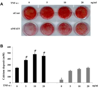
서 골세포로의 분화가 siCont이 transfection된 세포 보다 현저 히 저해 되는 것을 확인하였다. 뿐만 아니라 NFAT5를 억제 시킨 세포에 TNF-α를 처리했을 때 TNF-α에 의한 골분화 증가 가 억제 됨을 확인하였다(Fig. 3A). 그리고 이러한 효과는 인체 지방 유래 중간엽 줄기세포의 골세포 분화 과정 중 생성되는 calcium의 측정을 통해서도 유사한 결과를 나타내는 것을 확 인하였다(Fig. 3B).
NFAT5발현 억제가 인체 지방 유래 줄기세포에서 TNF-α 에 의해 유도 되는 NF-κB활성화에 미치는 효과
TNF-α 신호전달체계는 NF-κB를 통하여 다양한 세포 기능 을 조절 하는 것으로 알려져 있다[18]. 따라서 TNF-α에 의해 유도 되는 NF-κB promoter 활성화에 NFAT5가 어떠한 영향 을 미치는지 알아보기 위하여 인체 지방 유래 중간엽 줄기세 포에 NF-κB promoter를 포함한 luciferase벡터를 transfection 한 후 luciferase 활성을 측정하였다. 그 결과 TNF-α를 처리 하지 않아도 NFAT5가 감소된 인체 지방 유래 중간엽 줄기세
A
B
Fig. 3. Effect of NFAT5 down regulation on osteogenic differ- entiation of hADSCs. (A) On day 14 of osteogenic in- duction, calcification deposits were observed within the hADSCs. Post-confluent hADSCs were cultured in os- teogenic medium. The cells were treated without or with TNF-α for 3 days. Osteogenic differentiation was de- termined by Alizarin Red S staining to visualize calcifi- cation deposits within the cell monolayer (OM, osteo- genic differentiation induction). One representative ex- periment from three independent experiments was shown. (B) Determination of calcium deposition at day 14 after induction of differentiation. Data represent the mean SEM of three independent experiments.#
p
<0.05, compared to nontarget siRNA-transfected cells (siCont).*
p
<0.05, compared to the data of TNF-α-treated control siRNA-transfected cells (siCont-TNF-α).포에서 NF-κB promoter활성이 siCont이 transfection된 세포 보다 감소 되는 것을 확인 하였다. 뿐만 아니라 TNF-α에 의해 서 증가 되었던 NF-κB promoter활성화가 NFAT5를 감소 시 킨 인체 지방 유래 중간엽 줄기세포에서 감소 됨을 확인하였 다(Fig. 4).
NFAT5발현 억제가 인체 지방 유래 줄기세포에서 TNF-α 에 의해 유도되는 IκB 분해 에 미치는 효과
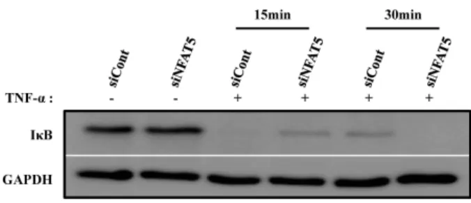
TNF-α에 의한 NF-κB 활성화 과정 중 NFAT5의 역할을 확 인하기 위하여 TNF-α에 의한 IκB 분해에 대한 NFAT5 siRNA 의 작용을 확인 하였다. 이를 위하여 TNF-α를 시간 의존적으 로 처리한 후, 세포를 회수하여 protein lysis buffer로 용해시 킨 후 IκB 발현을 western blot을 통하여 확인하였다. 그 결과 TNF-α에 의해서 유도 되는 IκB 분해분해가 siCont이 transfection된 세포와 비교하여 NFAT5의 발현 억제에 의하 여 유의하게 방지되지 못하였다(Fig. 5).
고 찰
줄기세포는 자가증식이 가능하며 특정 환경이 주어지면 신 경세포나 상피세포, 심근세포, 췌장세포 등 다양한 세포로 분 화할 수 있는 세포로써, 신체의 어떤 조직이나 장기에 질병이 생겼을 때 이러한 줄기세포로부터 분화된 같은 종류의 세포로 병든 세포를 대체하여 다양한 난치성 질환을 치료하는 것이 새로운 의학기술로 제시되고 있다. 줄기세포 중에서도 성체줄
Fig. 4. Effect of NFAT5 down regulation on TNF-α-induced NF- κB activation in hADSCs. The siRNA-transfected cells were co-transfected with NF-κB reporter constructs and β-gal vectors, and then treated with or without TNF-α (10 ng/ml) for 72 hr. Luciferase activity was normalized by β-galactosidase activity. Data represent the mean SEM of three independent experiments.#
p
<0.05, com- pared to nontarget siRNA-transfected cells (siCont). *p
<0.05, compared to the data of TNF-α-treated control siRNA-transfected cells (siCont-TNF-α).Fig. 5. Effect of NFAT5 down regulation on TNF-α-induced IκB degradationin hADSCs. Western blot analysis of total, cytosolic and nuclear proteins extracts obtained from NFAT5 siRNA or non-target siRNA-transfected cells.
The cells were treated with TNF-α (10 ng/ml) for 15 or 30 min. Degradation of IκB was determined by Western blot analysis on cell lysates using anti-IκB antibody.
GAPDH expression was used as a loading control. One representative experiment from three independent ex- periments was shown.
기세포는 골수에서 처음 분리 되었고 생체 외에서 비교적 쉽 게 증식이 가능하며 다양한 세포로 분화 가능한 줄기세포의 하나이다[24, 25]. 성체 줄기세포의 분화능은 지방세포, 골모세 포, 연골세포 등 중배엽성 조직뿐만 아니라 간세포 등 내배엽 성세포, 신경세포 등 외배엽성 세포로도 분화가 가능하다[29].
이러한 특성은 성체 줄기세포가 조직재생 및 세포치료를 위한 유망한 세포 공급원으로 사용될 수 있음을 의미한다. 성체줄 기세포는 골수에서 처음 보고된 이래 지방조직을 포함한 뇌, 비장, 간, 신장, 폐, 골수, 근육, 흉선, 췌장 등 다양한 조직에 존재하고 있음이 보고되었다.
특히 인체 지방 조직은 다른 조직에 비해 쉽게 분리가 가능 하고 대량으로 존재하여 골수에 비해 중간엽 줄기세포의 밀도 가 상대적으로 높아 대량의 세포가 필요한 세포 치료제의 임 상적용의 측면에서 보다 짧은 시간에 세포를 확보 할 수 있는 이상적인 중간엽 줄기세포의 공급원 중 하나이다[7, 8, 22, 27, 31, 32].
앞선 연구를 통해서 인체 지방 유래 중간엽 줄기세포의 골 세포 분화에 TNF-α signal이 NF-κB 경로와 Ras/MAPK 경로 를 통해서 중요하게 관여하는 것을 알 수 있었다[4, 23].
NFAT5는 NFAT1-4와 달리 DNA 결합부위의 결정구조가 NF-κB와 매우 유사하며 Rel family에 속하고 삼투적 자극에 의한 TNF-α에 의해서 발현이 조절되며 세포의 이주와 분화, 증식에 관여한다고 보고되었다[3, 9, 18, 19, 21, 30]. 최근 중간 엽 줄기세포의 분화조절에 중요한 역할을 하는 Transcriptional coactivator with PDZ-binding motif (Taz) 가 NFAT5의 발현 을 활성을 억제함이 보고되었으나, 중간엽 줄기세포에서 NFAT5의 역할은 명확히 밝혀져 있지 않다[11, 12].
본 연구에서는 NFAT5가 인체 지방 유래 중간엽 줄기세포 에서 골세포로의 분화에 관여하는 것으로 알려져 있는 전사인 자인 NF-κB의 활성을 조절 한다는 것을 확인 하였다. 또한
NFAT5를 억제 시킨 인체 지방 유래 중간엽 줄기세포의 골세 포로의 분화가 siCont 세포의 골세포 분화보다 현저히 감소 되는 것을 확인 하였다. 그리고 TNF-α를 처리 하였을 때 NFAT5가 억제된 인체 지방 유래 중간엽 줄기세포의 골세포 로의 분화가 다시 회복 되었으나, TNF-α를 처리한 siCont 세 포의골세포 분화 증가 보다는 억제됨을 확인 하였다. 이러한 결과는 인체 지방 유래 중간엽 줄기세포에서 NFAT5가 NF-κB 의 활성을 조절함으로써 TNF-α에 의한 골분화 작용을 조절 하는 것으로 생각된다.
TNF-α에 의한 IκB 분해에 대하여 siRNA에 의한 NFAT5 억제가 아무런 영향을 미치지 못하였다. 이러한 결과는 TNF-α 에 의한 NF-κB의 활성 조절에서 NFAT5가 IκB 분해의 하부경 로를 조절함을 나타낸다. 실제로 NF-κB의 전사조절작용이 다 양한 경로에 의해 조절됨이 알려져 있으므로[10, 17, 20], NFAT5가 NF-κB에 의한 전사조절작용을 조절할 가능성이 있 으며, 이를 확인하기 위해서는 추가실험이 필요하다.
본 실험에서 인체 지방 유래 중간엽 줄기세포에 NFAT5를 억제시켰을 때 TNF-α에 의한 지방 분화억제효과에는 영향을 미치지 않았다. 이는 TNF-α에 의한 지방 분화의 억제가 NF-κ B 활성 경로와 다른 경로를 통해서 작용함을 시사한다.
이상의 결과를 종합하여 볼 때 인체 지방 유래 중간엽 줄기 세포가 골세포로 분화 하는데 있어서 NFAT5가 NF-κB의 활성 화를 조절하고 이를 통해서 인체 지방 유래 중간엽 줄기세포 의 골분화에 중요한 작용을 미치는 것으로 사료된다.
감사의 글
“이 논문은 부산대학교 자유과제 학술연구비(2년)에 의하 여 연구되었음”으로 이에 감사드립니다.
References
1. Bocker, W., Docheva, D., Prall, W. C., Egea, V., Pappou, E., Rossmann, O., Popov, C., Mutschler, W., Ries, C. and Schieker, M. 2008. IKK-2 is required for TNF-alpha-induced invasion and proliferation of human mesenchymal stem cells.
J Mol Med (Berl)
86, 1183-1192.2. Boone, C., Mourot, J., Grégoire, F. and Remacle, C. 2000.
The adipose conversion process: regulation by extracellular and intracellular factors.
Reprod Nutr Dev
40, 325-358.3. Burg, M., Ferraris, J. and Dmitrieva, M. 2007. Cellular re- sponse to hypertonic stress.
Physiol Rev
87, 1441-1474.4. Cho, H. H., Shin, K. K., Kim. Y. J., Song, J. S., Kim, J. M., Bae, Y. C., Kim, C. D. and Jung, J. S. 2010. NF-kappaB activa- tion stimulates osteogenic differentiation of mesenchymal stem cells derived from human adipose tissue by increasing TAZ expression.
J Cell Physiol
223, 168-177.5. De Ugarte, D. A., Morizono, K., Elbarbary, A., Alfonso, Z., Zuk, P. A., Zhu, M., Dragoo, J. L., Ashjian, P., Thomas, B.,
Benhaim, P., Chen, I., Fraser, J. and Hedrick, M. H. 2003.
Comparison of multi-lineage cells from human adipose tis- sue and bone marrow.
Cells Tissues Organs
174, 101-109.6. Ferrari, G., Cusella-De A. G., Coletta, M., Paolucci, E., Stornaiuolo, A., Cossu, G. and Mavilio, F. 1998. Muscle re- generation by bone marrow-derived myogenic progenitors.
Science
279, 1528-1530.7. Halvorsen, Y. C., Wilkison, W. O. and Gimble, J. M. 2000.
Adipose-derived stromal cells-their utility and potential in bone formation.
Int J Obes Relat Metab Disord
24(Suppl 4), S41-44.8. Halvorsen, Y. D., Franklin, D., Bond, A. L., Hitt, D. C., Auchter, C., Boskey, A. L., Paschalis, E. P., Wilkison, W.
O. and Gimble, J. M. 2001. Extracellular matrix mineraliza- tion and osteoblast gene expression by human adipose tis- sue-derived stromal cells.
Tissue Eng
7, 729-741.9. Halterman, J. A., Kwon, H. M., Zargham, R., Bortz, P. D.
and Wamhoff, B. R. 2011. Nuclear factor of activated T cells 5 regulates vascular smooth muscle cell phenotypic modulation.
Arterioscler Thromb Vasc Biol
31, 2287-2296.10. Hayden, M. S. and Ghosh, S. 2008. Shared Principles in NF-κ B signaling.
Cell
132, 344-362.11. Hong, J.H., Hwang, E. S., McManus, M. T., Amsterdam, A., Tian, Y., Kalmukova, R., Mueller, E., Benjamin, T., Spiegelman, B. M., Sharp, P. A., Hopkins, N. and Yaffe, M.
B. TAZ, a transcriptional modulator of mesenchymal stem cell differentiation.
Science
309, 1074-1078.12. Jang, E. J., Jeong, H., Han, K. H., Kwon, H. M., Hong, J.
H. and Hwang, E. S. TAZ suppresses NFAT5 activity through tyrosine phosphorylation.
Mol Biol Cell
32, 4925-4932.13. Jiang, Y., Jahagirdar, B. N., Reinhardt, R. L., Schwartz, R.
E., Keene, C. D., Ortizgonzalez, X. R., Reyes, M., Lenvik, T., Lund, T., Blackstad, M., Du, J., Aldrich, S., Lisberg, A., Low, W. C., Largaespada, D. A. and Verfaillie, C. M. 2002.
Pluripotency of mesenchymal stem cells derived from adult marrow.
Nature
418, 41-49.14. Jun, Y. J. 2008. Recent development trend and prospects of adipose-derived stem cells on nerve regeneration.
Tissue Engineering and Regenerative Medicine
5, 51-56.15. Kim, J. A., Jeon, U. S., Kwon M. S., Lim, S. W. and Kwon, H. M. 2007. Transcriptional activator TonE-binding protein in cellular protection and differentiation.
Methods Enzymo
l 428, 253-267.16. Kuznetsov, S. A., Friedenstein, A. J. and Robey, P. G. 1997.
Factors required for bone marrow stromal fibroblast colony formation
in vivo. Br J Haematol
97, 561-570.17. Leeman, J. R. and Gilmore, T. D. 2008. Alternative splicing in the NF-kappaB signaling pathway.
Gene
423, 97-107.18. Kwon, M. S., Lim, S. W. and Kwon, H. M. 2009. Hypertonic stress in the kidney: a necessary evil.
Physiology
24, 186-191.19. Miyakawa, H., Woo, S. K., Dahl, S. C., Handler, J. S. and Kwon, H. M. 1999. Tonicity-responsive enhancer binding
protein, a Rel-like protein that stimulates transcription in response to hypertonicity.
PNAS
96, 2538-2542.20. Natoli, G. and Chiocca, S. 2008. Nuclear ubiquitin ligases, NF-kappaB degradation, and the control of inflammation.
Sci Signal
1, pe1.21. O'Connor, R. S., Mills, S. T., Jones, K. A., Ho, S. N. and Pavlath, G. K. 2006. A combinatorial role for NFAT5 in both myoblast migration and differentiation during skeletal mus- cle myogenesis.
J Cell Sci
120, 149-159.22. Ogawa, R., Mizuno, H., Watanabe, A., Migita, M., Shimada, T. and Hyakusoku, H. 2004. Osteogenic and chondrogenic differentiation by adipose-derived stem cells harvested from GFP transgenic mice.
Biochem Biophys Res Commun
313, 871-877.23. Peng, S., Zhou, G., Lu, K. D., Cheung, K. M., Li, Z., Lam, W. M,, Zhou, Z. and Lu, W. W. 2009. Strontium promotes osteogenic differentiation of mesenchymal stem cells through the Ras/MAPK signaling pathway.
Cell Physiol Biochem
23, 165-174.24. Pittenger, M. F., Mackay, A. M., Beck, S. C., Jaiswal, R. K., Douglas, R., Mosca, J. D., Moorman, M. A, Simonetti, D.
W., Craig, S. and Marshak, D. R. 1999. Multilineage poten- tial of adult human mesenchymal stem cells.
Science
284, 143-147.25. Prockop, D. J. 1997. Marrow stromal cells as stem cells for nonhematopoietic tissues.
Science
276, 71-74.26. Roth, I., Leroy, V., Kwon, H. M., Martin, P. Y., Fe´raille, E. and Hasler, U. 2010. Osmoprotective transcription factor NFAT5/TonEBP modulates nuclear factor-κB activity.
Mol Biol Cell
21, 3459-3474.27. Strem, B. A., Hicok, K. C., Zhu, M., Wulur, I., Alfonso, Z., Schreiber, R. E., Fraser, J. K. and Hedrick, M. H. 2005.
Multipotential differentiation of adipose tissue-derived stem cells.
Keio J Med
54, 132-141.28. Woodbury, D., Schwarz, E. J., Prockop, D. J. and Black, I.
B. 2000. Adult rat and human bone marrow stromal cells differentiate in to neurons.
J Neurosci Res
61, 364-370.29. Yang, Y. I., Kim, H. I., Seo, J. Y. and Choi, M. Y. 2007. Adult stem cells as cell therapeutics of angiogenesis.
Tissue Eng Regen Med
4, 484-489.30. Yoon, H. J., You, S., Yoo, S. A., Kim, N. H., Kwon, H. M., Yoon, C. H., Cho, C. S., Hwang, D. and Kim, W. U. 2011.
NFAT5 is a critical regulator of inflammatory arthritis.
Arthritis Rheum
63, 1843-1852.31. Zuk, P. A., Zhu, M., Ashjian, P., De Ugarte, D. A., Huang, J. I., Mizuno, H., Alfonso, Z. C., Fraser, J. K., Benhaim, P.
and Hedric, M. H. 2002. Human adipose tissue is a source of multipotent stem cells.
Mol Biol Cell
13, 4279-4295.32. Zuk, P. A., Zhu, M., Mizuno, H., Huang, J., Futrell, J. W., Katz, A. J., Benhaim, P., Lorenz, H. P. and Hedrick, M. H.
2001. Multilineage cells from human adipose tissue: im- plications for cell-based therapies.