Since Broxmeyer et al.3)showed experimental evidence that human umbilical cord blood (hUCB) is a rich source of hematopoietic stem/progenitor cells (HSPCs), hUCB has been known as an excellent source of HSPCs for transplan- tation. To date, more than 3,000 cases of hUCB hematopoi- etic stem cells (HSCs) transplantation have been performed
worldwide for patients with various hematological and gene- tic disorders8,21).
MSCs are the best candidates for the tissue engineering and cellular therapy of orthopedic musculoskeletal tissues17). Bone marrow (BM) is well known for a good source of mes- enchymal stem cells (MSCs) at the present time. Despite the fact that bone marrow represents the main available source of MSCs, the use of bone marrow-derived cells is not always acceptable due to the high degree of viral infection and the significant drop in cell number and proliferative/differenti- ation capacity with age. Thus, the search for possible alter- native MSC sources remains to be validated. Umbilical cord blood is a rich source of hematopoietic stem/progenitor cells.
hUCB has the advantage of easy availability and relative
537 537
Isolation, Characterization and Tri-lineage Differentiation of Mesenchymal Stem Cells from Human Umbilical Cord Blood
Sung-Eun Yang, M.D.*, Yoon-Sun Yang, M.D.*, and Chul-Won Ha, M.D.
Department of Orthopedic Surgery, Samsung Medical Center, Sungkyunkwan University, Samsung Biomedical Research Institute, Seoul; Department of Research & Development for Cellular Therapy, Medipost Biomedical Research Institute*, Yongin, Korea
537 537 Address reprint requests to
Chul-Won Ha, M.D.
Department of Orthopedic Surgery, Samsung Medical Center, Samsung Biomedical Research Institute, Sungkyunkwan University School of Medicine, 50 Ilwon-dong, Gangnam-gu, Seoul 135-710, Korea
Tel: +82.2-3410-0275, Fax: +82.2-3410-0084 E-mail: [email protected]
*This research was supported partly by Grant for Development of Biological Product and Materials funded by the Ministry of Commerce, Industry, and Energy, Republic of Korea.
Purpose: Human umbilical cord blood (hUCB) is well known for a good source of hematopoietic stem cells (HSCs). However, the presence of mesenchymal stem cells (MSCs) in hUCB is still not well approved by many authors. We hereby report the isolation and characterization of MSCs from hUCB, as well as their differentiation into osteogenic, chondrogenic and adipogenic lineages.
Materials and Methods: Mononuclear cells were isolated from each hUCB harvest (n=411) by density gradient centrifugation, and suspended in -minimum essential medium supplemented with 10% fetal bovine serum (FBS). The cell population was expanded by successive sub-cultivation under the same condition. The cell population that showed more than 1,000-fold expansion at fifth to eighth passage was inspected with known surface antigens of MSCs and other cell lineage. The isolated MSCs were cultured in osteogenic, chondrogenic, and adipogenic condition to identify their potential to differentiate into differ- ent mesenchymal cell lineage.
Results: Ninety five out of 411 hUCB units (23.1%) generated the MSC-like cell population during initial cultivation. Nine cell populations (2.2%) showed more than 1,000-fold expansion capacity at fifth to eighth passage. These cells positively expressed all known MSC-related antigens. They did not express any of myeloid, endothelial, or histo-compatibility antigens. All of the MSCs isolated showed the potential to differ- entiate into osteogenic, chondrogenic, and adipogenic lineages.
Conclusion: Our study supports that hUCB does contain MSCs, which can be differentiated into differ- ent cell lineages. We believe hUCB will be a good source of MSCs with the advantage of availability and relative abundance. We think hUCB should not be considered as a medical waste, and it will serve as a good source of cells for tissue engineering and cellular therapy in the future.
Key Words: Umbilical cord blood, Mesenchymal stem cells, Osteogenic differentiation, Chondrogenic differentiation, Adipogenic differentiation
abundance, however, it is still controversial whether hUCB contains MSCs6,16,30).
In the present study, we report the isolation, expansion, and characterization of MSCs from hUCB, as well as their potential to differentiate into three different mesodermal lin- eages: osteogenic, chondrogenic, and adipogenic lineage.
MATERIALS AND METHODS
1. Isolation and expansion of MSCs
hUCB was collected on delivery from 411 different indi- viduals with informed consent. Mononuclear cells were iso- lated by centrifugation in a Ficoll-Hypaque (Sigma, St. Louis, MO, USA) density gradient centrifugation (1.077 g/cm3).
The separated mononuclear cells were washed, suspended in -minimum essential medium ( -MEM, Gibco BRL, Carlsbad, CA, USA) supplemented with 10% fetal bovine serum (FBS, HyClone, Logan, UT, USA). The cells were then seeded in tissue culture plate at a concentration of 5× 10⁶cells/cm2. Cultures were maintained at 37℃in a humidi- fied atmosphere containing 5% CO2. The culture medium was changed at every 3 days. hUCB-derived mononuclear cells showed fibroblast-like adherent layers during the cul- ture, and the adherent cell reached 80% confluence between 1 to 3 weeks. The cells were trypsinized (0.25% trypsin, HyClone), washed, re-suspended in the same culture medi- um, and sub-cultured at a concentration of 5×10⁴cells/cm2. The cell population from each hUCB harvest was expanded by successive sub-cultivation under the same condition. Fifth to eighth passage cells that showed more than 1,000-fold expansion were studied for immuno-phenotyping of surface antigens and the potential to differentiate into multiple cell lineages.
2. Immuno-phenotyping of hUCB-derived MSC For surface antigen phenotyping, aliquots of the hUCB- derived MSCs after treatment with 0.25% trypsin were washed three times with phosphate-buffered saline (PBS, pH 7.4). Cells were stained with fluorescein isothiocyanate (FITC)- or phycoerythrin (PE)-coupled antibodies. Labeled cells were analyzed by FACSCalibur flow cytometer (Becton Dickinson, San Jose, CA, USA) using the CELLQUEST software (Becton Dickinson, San Jose, CA, USA).
The anti-human antibodies used were CD45-FITC, CD34- FITC, CD14-FITC, HLA-DR-FITC, CD13-PE (Becton
Dickinson, San Jose, CA, USA); CD29-PE, CD44-PE, CD31-FITC, CD106-PE, CD90-FITC, CD51/61-FITC, CD64-FITC (Pharmingen, Los Angeles, CA, USA). As isotypic control, mouse IgG1-FITC, IgG1-PE, IgG2a-FITC (Becton Dickinson), mouse IgG2b-PE, IgG1-FITC, IgG1-PE (Pharmingen) were used. Cells were also immunolabeled with the following anti-human antibodies; SH2, SH3, and SH4. SH2, SH3, and SH4 antibodies made from each hyb- ridoma cell line (American Type Culture Collection, Rock- ville, MD, USA) for indirect assays. As a secondary antibody, anti-mouse IgG-FITC (Jackson ImmunoResearch, West Grove, PA, USA) was used.
3. Osteogenic, chondrogenic, and adipogenic differen- tiation
To induce osteogenic differentiation, cells were cultured in the osteogenic medium. The medium was composed of -MEM supplemented with 10% FBS, 0.1 M dexametha- sone (Sigma), 10 mM -glycerol phosphate (Sigma), and 50 M L-ascorbic acid 2-phosphate (Sigma)10). Medium was changed twice a week.
To induce chondrogenic differentiation, approximately 2
×105cells were placed in 15 mL polypropylene tube and centrifuged at 500×g, 5 minutes, at room temperature to form a pellet. Then, the pallet was treated with chondrogenic medium. Medium was changed twice a week. Chondrogenic medium consisted of serum free, high-glucose Dulbecco’s modified Eagle’s medium (DMEM, Gibco BRL) supple- mented with 100 nM dexamethasone (Sigma), 50 g/mL L- ascorbic acid (Sigma), 100 g/mL sodium pyruvate (Sigma), 40 g/mL L-proline (Sigma), 10 ng/mL transforming growth factor 3 (TGF- 3, Sigma), 500 ng/mL bone morphogenic protein 6 (BMP-6, R&D Systems, Minneapolis, MN, USA), and 50 mg/mL ITS+ premix (Becton Dickinson, 6.25 g/ mL insulin, 6.25 g/mL transferrin, 6.25 ng/mL selenious acid, 1.25 mg/mL bovine serum albumin (BSA), and 5.35 mg/mL linoleic acid22).
To induce adipogenic differentiation, cells were treated with adipogenic medium. Medium changes were carried out twice a week. Adipogenic medium consisted of DMDM (Gibco BRL) supplemented with 10% FBS (Hyclone), 0.5 mM 3-isobutyl-1-methylxanthine (IBMX, Sigma), 1 M dexamethasone (Sigma), 0.2 mM indomethacin (Sigma), and 10 M h-insulin (Sigma)19).
4. Evaluation of Trilineage Differentiation
The onset of osteogenesis was evaluated by the expression of alkaline phosphatase (ALP), using ALP staining kit (85L- 3R, Sigma) at 1 week interval. Mineralized matrix was asse- ssed by von Kossa staining weekly.
For the evaluation of chondrogenic differentiation, the pallet aggregates were retrieved weekly and frozen in OCT compound (Tissue-Tek, Torrance, CA, USA) for histological and immunohistochemical analyses, and Safranin-O (Sigma) staining for histological evaluation. For immunohistochemi- cal analysis, the frozen sections were fixed for 10 min in me- thanol after a brief immersion in distilled water to remove the OCT compound. Nonspecific antibody binding sites were blocked by incubating the slides in PBS with 5.0%
BSA for 1 hour. Slides were then incubated for an hour at room temperature with type II collagen antibody (Onco-
gene, San Diego, CA, USA) diluted in 0.05 M Tris-HCl with 1.0% BSA (TBS). Reactivity was detected by using the EnVisionTM+ System (DAKO, Carpinteria, CA, USA), Peroxidase (DAB) kit (K4006, DAKO) according to the manufacturer’s instructions.
Adipogenesis were evaluated by Oil-red O (Sigma) stain- ing at every week.
The cells were also analyzed by RT-PCR weekly. Total RNA was extracted from each differentiated cells with a modification of the method of Chomczynski and Sacci.36) Cells were lysed using TRIzol Reagent (Invitrogen, La Jolla, CA, USA), and RNA was prepared as per kit instructions.
The mRNA was reverse transcribed to cDNA in 20 L reaction volume containing 1 gof RNA, 1 Lof 500 g/ mL Oligo dT primer (Invitrogen), 1 L(200 units) of Super- ScriptTMII reverse transcriptase (Invitrogen), 4 Lof 5×
Target gene product Primer sequence Anealing temperature PCR size
GAPDH 5′-ACCACAGTCCATGCCATCAC-3′ 55°C 452 bp
5′-TCCACCACCCTGTTGCTGTA-3′
Osteocalcin 5′-CATGAGAAGCCCTCACA-3 55°C 310 bp
5′-AGAGCGACACCCTAGAC-3′
Alkaline 5′-TGGAGCTTCAGAAGCTCAACACCA-3′ 55°C 453 bp
Phosphatase 5′-ATCTCGTTGTCTGAGTACCAGTCC-3′
Osteopontin 5′-CTAGGCATCACCTGTGCCATACC-3′ 55°C 347 bp
5′-CAGTGACCAGTTCATCAGATTCATC-3′
Collagen type I 5′-CCCCCTCCCCAGCCACAAAGA-3′ 60°C 360 bp
5′-TCTTGGTCGGTGGTGGACTCT-3′
Cbfa-I 5′-CAGTAGATGGACCTCGGGAA-3′ 60°C 395 bp
5′-GAGGCAGAAGTCAGAGGTGG-3′
Aggrecan 5′-TCAGGAGGGCTGGAACAAGTACC-3′ 57°C 392 bp
5′-GGAGGTGGTAATTGCAGGGAACA-3′
Collagen type II 5′-TGGAGCTTCAGAAGCTCAACACCA-3′ 55°C 225 bp 5′-ATCTCGTTGTCTGAGTACCAGTCC-3′
Collagen type II 5′-TTTCCCAGGTCAAGATGGTC-3′ 55°C 377 bp
5′-CTTCAGCACCTGTCTCACCA-3′
Collagen type IX 5′-CCCCCTCCCCAGCCACAAAGA-3′ 57°C 159 bp
5′-TCTTGGTCGGTGGTGGACTCT-3′
Sox-9 5′-GGTTGTTGGAGCTTTCCTCA-3′ 57°C 401 bp
5′-TAGCCTCCCTCACTCCAAGA-3′
PPAR 5′-TTCAGAAATGCCTTGCAGTG-3′ 57°C 599 bp
5′-GGGCTCCATAAAGTCACCAA-3′
C/EBP 5′-CTGGAGCTGACCAGTGACAA-3′ 57°C 374 bp
5′-CCAAGAATTCTCCCCTCCTC-3′
Leptin 5′-ACAGAAAGTCACCGGTTTGG-3′ 57°C 481 bp
5′-TGGCTTAGAGGAGTCAGGGA-3′
Lipoprotein lipase 5′-TCAATCACAGCAGCAAAACC-3′ 57°C 575 bp
5′-CCACATCTCCAAGTCCTCTC-3′
P2 5′-TACTGGGCCAGGAATTTGAC-3′ 57°C 240 bp
5′-TCAATGCGAACTTCAGTCCA-3′ Table 1.Primers for RT-PCR
First-Strand buffer (250 mM Tris-HCl (pH 8.3), 375 mM KCl, 15 mM MgCl2, Invitrogen), 2 Lof 0.5mM dNTPs, 1 Lof 10 mM dTT, and 1 Lof RNaseOUT Ribonucle- ase Inhibitor (Invitrogen).
PCR reactions were performed in 25 Lreaction volume containing 2 Lof cDNA, 2 Lof each 10 pM primer, 0.5 Lof AccuPrimeTMTaq DNA polymerase (Invitrogen), 2.5 Lof 10×AcuuPrimeTMPCR buffer (Invitrogen). Primers used for amplification are listed in Table 1. cDNA was ampli- fied using a ABI GeneAmp PCR System 9600 (Perkin El- mer, Applied Biosystems, Boston, MA, USA) at 95℃for 30 sec, the annealing temperature for each target gene as described in Table 1 for 30 sec, and 72℃for 30 sec for 35 cycles, after initial denaturation at 95℃for 5 min. PCR products were resolved by agarose gel electrophoresis and visualized by ethidium bromide staining.
RESULTS
1. Isolation & Expansion of MSCs
hUCB-derived mononuclear cells seeded in the tissue culture plates gave rise to a population of fibroblast-like adherent cells. By day 21 of primary culture, 95 out of 411 hUCB harvests (23.1%) produced these MSC-like cell pop- ulations. Non-adherent or loosely adherent small round cells were also present in the primary cultures, but disappeared soon after passage 2 or 3.
MSC-like cell colonies derived from hUCB culture mainly consisted of bipolar fibroblast-like cells (Fig. 1A). At day 21 of primary culture, the mean value for total adherent
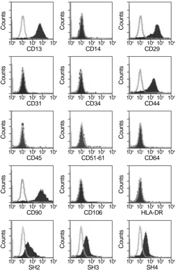
Fig. 2.Immunophenotypic characterization of the MSCs derived from hUCB. These cells were positive for antigens CD13, CD29, CD44, CD90, SH2, SH3, and SH4. These cells were negative for antigens CD14, CD31, CD34, CD45, CD51/61, CD64, CD106, or HLA-DR. These immunophenotyping was consistent with the known findings for bone marrow MSCs.
Fig. 1.Photomicrographs showing mesenchymal stem cells (MSCs) isolated from human umbilical cord blood at the day 21 (A) and at advanced passage 18 (B). hUCB-derived MSC colonies mainly consisted of bipolar fibroblast-like cells. No significant morphologi- cal changes were observed until passage 18.
A B
Counts
100 101 102103 104 CD13
Counts
100 101 102103 104 CD14
Counts
100 101 102103 104 CD29
Counts
100 101 102103 104 CD31
Counts
100 101 102103 104 CD34
Counts
100 101 102103 104 CD44
Counts
100 101 102103 104 CD45
Counts
100 101 102103 104 CD51-61
Counts
100 101 102103 104 CD64
Counts
100 101 102103 104 CD90
Counts
100 101 102103 104 CD106
Counts
100 101 102103 104 HLA-DR
Counts
100 101 102103 104 SH2
Counts
100 101 102103 104 SH3
Counts
100 101 102103 104 SH4
cell number was 2.6×10⁶(n=95, range 0.04×10⁵-22.0× 10⁶). After expansion, MSCs-like colonies from nine out of 411 hUCB harvests (2.2%) showed more than 1,000-fold
increase in cell number. These cells could be passaged up to 18 times without displaying significant changes in morpholo- gy (Fig. 1B).
ALP
1 week 2 week 3 week 4 week
1 week 2 week 3 week 4 week
M 0 wk 1 wk 2 wk 3 wk 4 wk
OP OC Col I Cbfa-1 GAPDH
453 bp 347 bp 310 bp 360 bp 398 bp 452 bp
A
B
C
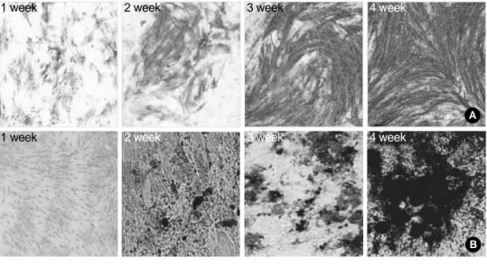
Fig. 3.Osteogenic differentiation of hUCB-derived MSCs. Cul- tured mesenchymal cells formed aggregates and increased their expression of alkaline phosphatase (A, ×100). These cells also formed extracellular calcium matrix, which was demonstrat- ed by von Kossa staining (B, ×100). hUCB-derived MSCs ex- pressed mRNA of ALP, osteopontin, osteocalcin, collagen type I, and Cbfa-I during osteogenic differentiation, which is typical of osteoblastic cells (C).
0 week
2 week 4 week 6 week
2 week 4 week 6 week
M GAPDH (452 bp) Aggrecan (392 bp) Col II(225 bp) Col II(377 bp) CoI IX (159 bp) Sox-9 (401 bp)
4 week A
B C
Fig. 4.Chondrogenic differentiation of hUCB-derived MSCs. Accumulation of sulfated proteoglycans was visualized by Safranin-O staining (A, ×200) at 2 weeks of chondrogenic differentiation. Multiple lacunae with chondrocyte-like cells were also evident in the histological sections. Extensive matrix was rich in type II collagen, which is typical of articular cartilage (B, ×200). mRNA of collagen type II, aggrecan, collagen type IX, and sox-9 was detected by RT-PCR, which is typical of chondrocytes (C).
2. Immunophenotypic characterization of hUCB -derived MSCs
Immunophenotyping of hUCB-derived MSCs with more than 1,000-fold expanding capacity demonstrated that all of them showed positive expression of the MSCs-related anti- gens, SH2, SH3 and SH4 (Fig. 2). These cells were negative for antigens CD14 (lipopolysaccharide receptor, hematopoi- etic marker), CD31 (PECAM-1), CD34 (gp105-120, hema- topoietic marker), CD45 (leukocyte common antigen), CD 51/61, CD64 (matrix receptor), CD106, or HLA-DR(HLA class II). They were found to be positive for other known antigens of MSC; CD13, CD29 ( 1 integrin), CD44 (matrix receptor for hyaluronate), and CD90(thy-1) (Fig. 2).
3. Osteogenic, chondrogenic, and adipogenic differenti- ations of hUCB-derived MSCs
The MSCs cultured in osteogenic media formed aggre- gates and increased their expression of alkaline phosphatase from the first week through forth week (Fig. 3A). These cells also formed extracellular calcium matrix, which was demonstrated by von Kossa staining (Fig. 3B). The MSCs
also expressed mRNA of bone forming cells, such as alka- line phosphatase (ALP), osteopontin (OP), osteocalcin (OC), collagen type I (Col I), and core binding factor 1 (Cbfa-I) during osteogenic differentiation (Fig. 3C).
During chondrogenic differentiation, the accumulation of sulfated proteoglycans was visualized by Safranin-O stain- ing at 2 weeks of differentiation (Fig. 4A). Multiple lacunae with chondrocyte-like cells were also evident in the histologi- cal sections. The extensive matrix was rich in type II collagen, which is typical of articular cartilage (Fig. 4B). mRNA of collagen type II (Col II), aggrecan, collagen type IX (Col IX), and SOX-9 was detected by RT-PCR at 4 weeks (Fig. 4C)
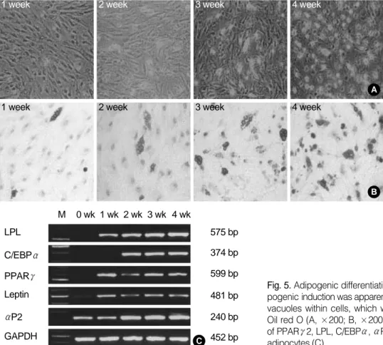
Adipogenic differentiation was apparent by the accumu- lation of lipid-rich vacuoles within cells, which were visual- ized by staining with Oil red O (Fig. 5A, B). The lipid vac- uoles continued to develop over time, coalesced, and even- tually filled the cells. These cells expressed mRNA of perox- isome proliferation-activated receptor 2 (PPAR 2), lipo- protein lipase (LPL), fatty acid-binding protein aP2, CCAAT/
enhancer-binding protein (C/EBP ), and leptin, which is typical of adipocytes (Fig. 5C).
LPL
1 week 2 week 3 week 4 week
1 week 2 week 3 week 4 week
M 0 wk 1 wk 2 wk 3 wk 4 wk
C/EBP PPAR Leptin P2 GAPDH
575 bp 374 bp 599 bp 481 bp 240 bp 452 bp
A
B
C
Fig. 5.Adipogenic differentiation of hUCB-derived MSCs. Adi- pogenic induction was apparent by the accumulation of lipid-rich vacuoles within cells, which were visualized by staining with Oil red O (A, ×200; B, ×200). These cells expressed mRNA of PPAR 2, LPL, C/EBP , P2, and leptin, which is typical of adipocytes (C).
Of the nine MSC populations that showed more than 1,000-fold expanding capacity, nine demonstrated osteogenic differentiation, eight demonstrate d chondrogenic differen- tiation, and five demonstrated adipogenic differentiation.
DISCUSSION
hUCB is known to contain a higher proportion of primi- tive hematopoietic cells, including multipotent colony form- ing cells14,20,23,28)as well as in vivo repopulating cells29), than adult bone marrow (BM). hUCB-derived HSCs are reported to have higher proliferation and expansion potentials than their adult BM counterparts9,11,24,26,27,29). In this sense, if hUCB contains MSCs, we may suppose that they have higher poten- tial for proliferation and differentiation than BM-derived MSCs. So far, it is still controversial as to whether hUCB contains MSCs. The possibility of having higher potential is still pretty much in question.
Erices et al. reported that hUCB harvests generate mes- enchymal progenitor cells with large ex vivo expansion capac- ity as well as osteogenic and adipogenic differentiation poten- tials6). On the contrary, Wexler et al.30)and Mareschi et al.16) claimed that adult BM is a reliable source of functional MSC, but hUCB is not. However, a recent study by Lee et al.13) reported that hUCB contains MSCs which can differentiate into bone, cartilage, and fat. They reported that hUCB-de- rived MSCs could even differentiated into neurogenic and hepatogenic lineage.
We performed the present study focusing on the identi- fication, expansion, and phenotypic characterization of the MSCs isolated from human hUCB. Our study demonstrat- ed that the characteristics of the hUCB-derived MSCs were very similar to the features of BM-derived MSCs15,17). Results of our present study support that hUCB does contain MSCs.
In this study, we reveald the presence of MSCs in hUCB by in vitro culture-expansion on a large scale. Our results showed that almost 25% of hUCB harvests generated MSCs-like cell populations. The immunophenotype of these cells was consistent with that reported for BM-derived MSCs15,17). The present study verified that the hUCB-derived MSCs had the capacity to differentiate into different lineages. Whether they even have the potential to differentiate into ectodermal or endomeral lineages still needs to be investigated, and will be one of our next study.
In recent years, the potential use of MSCs has been high-
lighted in tissue engineering, regeneration medicine, and cellular or gene therapy. Interestingly, MSCs have been reported to be transplantable between HLA-incompatible individuals, because they don’t elicit alloreactive lymphocyte proliferate responses and modulate immune responses1,5,12,25). MSCs are thought to be the origin of mesenchymal tissues during the process of normal growth, remodeling, and repair.
So, various tissues have been inspected as a source for MSCs, such as synovium, fat tissue, muscle as well as bone marrow with some successful results2,4,7,18,31). However, these sources are mostly not easy to collect, limited in amount of the ava- ilable tissue, or have the concern of donor site morbidity.
hUCB has the advantage of easy of collection, availability and relative abundance. It is well know that hUCB harvest is not harmful to donor baby and mother. Although the incidence of hUCB harvest producing MSCs is low in our study, some of them showed more than 1,000-fold expand- ing capacity that will be sufficient in cell number to be used for tissue regeneration strategy. So, We believe hUCB can be a useful candidate for source of MSC for future therapeutic options of tissue regeneration. Further studies will be need- ed to select optimal hUCB harvests before or during MSCs cultivation.
The limitation of this study is that the MSCs were eval- uated as a population, rather than a single colony derived cell line. Although many reported studies included this type of method by using MSC population, there has always been a critic on the possibility that the population may be a mix- ture of different progenitor cells of different lineage with or without pure MSCs.
Because we still do not have any specific markers of MSCs, it is not feasible at the present time to specifically isolate the pure MSCs from hUCB, as well as from bone marrow.
The investigation to find out methods to isolate and expand the pure MSCs will be a valuable research in the future.
CONCLUSION
Our study supports that hUCB does contain MSCs, which can be differentiated into different cell lineages. We believe hUCB will be a good source of MSCs with the advantage of availability and relative abundance. We think hUCB should not be considered as a medical waste, and it will serve as a good source of cells for tissue engineering and cellular therapy in the future.
REFERENCES
1. Arinzeh TL, Peter SJ, Archambault MP, et al: Allogeneic mesenchymal stem cells regenerate bone in a critical-sized canine segmental defect. J Bone Joint Surg Am, 85-A: 1927-1935, 2003.
2. Asakura A, Komaki M and Rudnicki M: Muscle satellite cells are multipotential stem cells that exhibit myogenic, osteogenic, and adipogenic differentiation. Differentiation, 68: 245-253, 2001.
3. Broxmeyer HE, Douglas GW, Hangoc G, et al: Human umbil- ical cord blood as a potential source of transplantable hematopoietic stem/progenitor cells. Proc Natl Acad Sci USA, 86: 3828-3832, 1989.
4. De Bari C, Dell’Accio F, Tylzanowski P and Luyten FP:
Multipotent mesenchymal stem cells from adult human synovial membrane. Arthritis Rheum, 44: 1928-1942, 2001.
5. De Kok IJ, Peter SJ, Archambault M, et al: Investigation of allogeneic mesenchymal stem cell-based alveolar bone formation:
preliminary findings. Clin Oral Implants Res, 14: 481-489, 2003.
6. Erices A, Conget P and Minguell JJ: Mesenchymal progenitor cells in human umbilical cord blood. Br J Haematol, 109: 235-242, 2000.
7. Fickert S, Fiedler J and Brenner RE: Identification, quantifica- tion and isolation of mesenchymal progenitor cells from osteoarthritic synovium by fluorescence automated cell sorting. Osteoarthritis Cartilage, 11: 790-800, 2003.
8. Gluckman E, Broxmeyer HA, Auerbach AD, et al: Hematopoi- etic reconstitution in a patient with Fanconi’s anemia by means of umbilical-cord blood from an HLA-identical sibling. N Engl J Med, 321: 1174-1178, 1989.
9. Hows JM, Bradley BA, Marsh JC, et al: Growth of human umbilical-cord blood in longterm haemopoietic cultures. Lancet, 340: 73-76, 1992.
10. Jaiswal N, Haynesworth SE, Caplan AI and Bruder SP:
Osteogenic differentiation of purified, culture-expanded human mesenchymal stem cells in vitro. J Cell Biochem, 64: 295-312, 1997.
11. Lansdorp PM, Dragowska W and Mayani H: Ontogeny-relat- ed changes in proliferative potential of human hematopoietic cells.
J Exp Med, 178: 787-791, 1993.
12. Le Blanc K, Tammik C, Rosendahl K, Zetterberg E and Ringden O:HLA expression and immunologic properties of dif- ferentiated and undifferentiated mesenchymal stem cells. Exp Hema- tol, 31: 890-896, 2003.
13. Lee OK, Kuo TK, Chen WM, Lee KD, Hsieh SL and Chen TH:Isolation of multipotent mesenchymal stem cells from umbil- ical cord blood. Blood, 103: 1669-1675, 2004.
14. Lu L, Xiao M, Shen RN, Grigsby S and Broxmeyer HE: En- richment, characterization, and responsiveness of single primitive CD34 human umbilical cord blood hematopoietic progenitors with high proliferative and replating potential. Blood, 81: 41-48, 1993.
15. Majumdar MK, Thiede MA, Mosca JD, Moorman M and Gerson SL:Phenotypic and functional comparison of cultures of marrow-derived mesenchymal stem cells (MSCs) and stromal cells.
J Cell Physiol, 176: 57-66, 1998.
16. Mareschi K, Biasin E, Piacibello W, Aglietta M, Madon E and Fagioli F:Isolation of human mesenchymal stem cells: bone marrow versus umbilical cord blood. Haematologica, 86: 1099-1100, 2001.
17. Minguell JJ, Erices A and Conget P: Mesenchymal stem cells.
Exp Biol Med (Maywood), 226: 507-520, 2001.
18. Mizuno H: [Versatility of adipose tissue as a source of stem cells].
J Nippon Med Sch, 70: 428-431, 2003.
19. Nakamura T, Shiojima S, Hirai Y, et al: Temporal gene expres- sion changes during adipogenesis in human mesenchymal stem cells. Biochem Biophys Res Commun, 303: 306-312, 2003.
20. Pettengell R, Luft T, Henschler R, et al: Direct comparison by limiting dilution analysis of long-term culture-initiating cells in human bone marrow, umbilical cord blood, and blood stem cells.
Blood, 84: 3653-3659, 1994.
21. Rubinstein P, Carrier C, Scaradavou A, et al: Outcomes among 562 recipients of placental-blood transplants from unrelated donors.
N Engl J Med, 339: 1565-1577, 1998.
22. Sekiya I, Vuoristo JT, Larson BL and Prockop DJ: In vitro cartilage formation by human adult stem cells from bone marrow stroma defines the sequence of cellular and molecular events during chondrogenesis. Proc Natl Acad Sci USA, 99: 4397-4402, 2002.
23. Sutherland HJ, Eaves CJ, Eaves AC, Dragowska W and Lansdorp PM:Characterization and partial purification of human marrow cells capable of initiating long-term hematopoiesis in vitro.
Blood, 74: 1563-1570, 1989.
24. Traycoff CM, Abboud MR, Laver J, Clapp DW and Srour EF:Rapid exit from G0/G1 phases of cell cycle in response to stem cell factor confers on umbilical cord blood CD34+ cells an enhanced ex vivo expansion potential. Exp Hematol, 22: 1264-1272, 1994.
25. Tse WT, Pendleton JD, Beyer WM, Egalka MC and Guinan EC:Suppression of allogeneic T-cell proliferation by human mar- row stromal cells: implications in transplantation. Transplantation, 75: 389-397, 2003.
26. Tsujino Y, Wada H, Misawa M, Kai S and Hara H: Effects of mast cell growth factor, interleukin-3, and interleukin-6 on human
primitive hematopoietic progenitors from bone marrow and cord blood. Exp Hematol, 21: 1379-1386, 1993.
27. van de Ven C, Ishizawa L, Law P and Cairo MS: IL-11 in combination with SLF and G-CSF or GM-CSF significantly increas- es expansion of isolated CD34+ cell population from cord blood vs.
adult bone marrow. Exp Hematol, 23: 1289-1295, 1995.
28. Verfaillie C, Blakolmer K and McGlave P: Purified primitive human hematopoietic progenitor cells with long-term in vitro repop- ulating capacity adhere selectively to irradiated bone marrow stro- ma. J Exp Med, 172: 509-502, 1990.
29. Wang JC, Doedens M and Dick JE: Primitive human hema-
topoietic cells are enriched in cord blood compared with adult bone marrow or mobilized peripheral blood as measured by the quanti- tative in vivo SCID-repopulating cell assay. Blood, 89: 3919-3924, 1997.
30. Wexler SA, Donaldson C, Denning-Kendall P, Rice C, Bra- dley B and Hows JM:Adult bone marrow is a rich source of human mesenchymal ‘stem’ cells but umbilical cord and mobilized adult blood are not. Br J Haematol, 121: 368-374, 2003.
31. Zuk PA, Zhu M, Ashjian P, et al: Human adipose tissue is a source of multipotent stem cells. Mol Biol Cell, 13: 4279-4295, 2002.
목 적: 저자들은 인간 제대혈(umbilical cord blood)에서 간엽 줄기 세포를 분리 및 확인할 수 있었고, 이들 세포군에 대하 여 골, 연골 및 지방세포로의 분화가 가능하였기에 이를 보고하고자 한다.
대상 및 방법: 제대혈(n=411)로부터 단핵세포군을 원심분리 방법을 이용하여 분리한 후 배양하였으며, 5-8회 개대배양 시 에 1,000배 이상의 증식을 보인 세포군에 대하여 간엽줄기세포 및 타 세포계열 표면항원 검사를 시행하였다. 분리된 간엽 줄기 세포군에 대하여 골, 연골 및 지방세포로의 분화를 시도하였다.
결 과: 총 411개의 제대혈 중 95개(23.1%)에서 첫 배양 시 간엽 줄기 세포양 세포군(MSC-like cell population)이 확인되었 으며, 5-8회 개대배양 시 1,000배 이상의 증식을 보인 세포군은 9개(2.2%)였다. 이들 세포군은 기존에 알려진 모든 간엽줄 기세포 표면항원검사에 양성, 타 세포계열 표면항원 검사에는 모두 음성이었다. 분리된 간엽 줄기 세포군은 골, 연골 및 지방세포로의 분화가 가능하였다.
결 론: 본 연구의 결과는 인간 제대혈이 서로 다른 세포 계열로 분화할 수 있는 간엽줄기세포를 포함하고 있다는 사실을 지 지하는 것으로 판단된다. 인간 제대혈은 상대적으로 많은 양의 간엽줄기세포를 비교적 용이하게 얻을 수 있다는 장점이 있는 세포 공급원이 될 수 있을 것으로 사료되며, 향후 조직 공학 및 세포 치료를 위한 세포 공급원의 하나가 될 수 있을 것 으로 기대한다.
색인 단어: 제대혈, 간엽 줄기 세포, 골세포 분화, 연골세포 분화, 지방세포 분화
인간 제대혈에서 간엽줄기세포의 분리, 확인 및 골, 연골 및 지방세포로의 분화
양성은*ㆍ양윤선*ㆍ하철원
성균관대의과대학 삼성서울병원 정형외과, 메디포스트(주) 생명공학연구소*