Salicylate Can Enhance Osteogenic Differentiation of Human Periosteum- derived Mesenchymal Stem Cells
Bo Gyu Kim1, A ram Lee1, Bo Young Lee1, Sungbo Shim2, Dong kyu Moon3, Sun-Chul Hwang3, June-Ho Byun4* and Dong Kyun Woo1*
1College of Pharmacy and Research Institute of Pharmaceutical Sciences, Gyeongsang National University, Jinju 52828, Korea
2Department of Biochemistry, Chungbuk National University, Cheongju 28644, Korea
3Department of Orthopedic Surgery and Institute of Health Sciences, School of Medicine and Hospital, Gyeongsang National University, Jinju 52727, Korea
4Department of Oral and Maxillofacial Surgery and Institute of Health Sciences, School of Medicine and Hospital, Gyeongsang National University, Jinju 52727, Korea
Received October 10, 2018 /Revised October 22, 2018 /Accepted October 22, 2018
Due to a rapidly expanding aging population, the incidence of degenerative bone disease has in- creased, and efforts to handle the issue using regenerative medicine have become more important. In order to control various bone diseases such as osteoarthritis and osteoporosis, regenerative medicine utilizing adult stem cells has been extensively studied. And it is now clear that the mitochondrial en- ergy metabolism, oxidative phosphorylation, is important for the process of stem cell differentiation.
Interestingly, a recent study reported that salicylate promotes mitochondrial biogenesis by regulating the expression of PGC-1α in murine cells. However, the possible effects of salicylate on osteogenic dif- ferentiation through increased mitochondrial biogenesis in stem cells remain unknown. Thus, here we investigated whether salicylate could influence osteogenic differentiation and mitochondrial biogenesis of periosteum-derived mesenchymal stem cells (POMSCs). We found that salicylate treatments of POMSCs undergoing osteogenic differentiation increased the activity of alkaline phosphatase, a well-known early marker of bone cell differentiation. In addition, we observed that mitochondrial mass was increased by salicylate treatments in POMSCs. Together, these results indicate that salicylate can enhance osteogenic differentiation and mitochondrial biogenesis in POMSCs. Therefore, the find- ings in this study suggest that small molecules augmenting mitochondrial function such as salicylate can be a novel modulator for osteogenic differentiation and regenerative medicine.
Key words : Differentiation, mitochondria, osteogenesis, regenerative medicine, stem cell
*Corresponding authors
*Tel : +82-55-772-2428, Fax : +82-55-772-2429
*E-mail : [email protected] (Dong Kyun Woo) [email protected] (June-Ho Byun)
This is an Open-Access article distributed under the terms of the Creative Commons Attribution Non-Commercial License (http://creativecommons.org/licenses/by-nc/3.0) which permits unrestricted non-commercial use, distribution, and reproduction in any medium, provided the original work is properly cited.
Journal of Life Science 2018 Vol. 28. No. 12. 1455~1460 DOI : https://doi.org/10.5352/JLS.2018.28.12.1455
서 론
최근 들어 급속한 고령화 사회가 진행되고 있으며 이로 인 해 골관절염과 골다공증 등의 퇴행성 골질환 환자수도 동반하 여 증가하고 있다. 그리고 고령화에 따른 골관련 질환의 새로 운 제어와 치료법 개발을 위해 재생의학도 활발히 연구되고 있다. 재생의학이란 생체에 적합한 재료나 줄기세포를 단독 혹은 복합적으로 이용하여 새로운 조직이나 장기를 만들어 손상되거나 결함이 있는 조직과 장기를 대체하는 치료방법이 다[13]. 중간엽 유래 성체줄기세포(mesenchymal stem cells,
MSCs)는 자가증식이 가능하며 골세포, 연골세포 그리고 지방 세포 등으로 다양하게 분화할 수 있기 때문에 퇴행성 골질환 을 타겟으로한 재생의학의 소재로써 활발히 연구되고 있다 [14, 17, 18, 22]. MSC로부터 조골세포 또는 골세포로의 분화유 도를 활용한 골재생 연구에는 전통적으로 골수 기원의 성체줄 기세포(bone marrow-derived mesenchymal stem cells, BMMSCs)가 이용되어 왔다. 하지만 BMMSCs의 경우에는 골 수 흡입에 의한 줄기세포 추출 시에 동반되는 통증과 감염의 위험이 있다는 단점을 갖는다. 최근에는 이러한 단점들을 보완 함과 동시에, 골재생에 중요한 골전구세포들(osteoprogenitor cells)을 포함하는 골막 기원의 성체줄기세포(periosteum-de- rived mesenchymal stem cells, POMSCs)도 재생의학의 연구 재료로 사용되고 있다. POMSCs는 치과적으로 국소마취하에 매복치 발치 등을 포함한 일반적인 시술을 통하여 쉽게 채취 할 수 있는 골막에서 추출할 수 있으며 골재생에 중요한 역할 을 하는 것으로 밝혀지고 있다[1, 4, 7, 9, 17, 20].
흥미롭게도, 최근 줄기세포의 분화와 신진대사(metabo- lism) 관련 연구에서, 줄기세포의 분화가 진행되는 과정에서
해당작용(glycolysis)은 점차 감소하는 반면에 분화가 이루어 질수록 산화적인산화(oxidative phosphorylation)가 증가되는 것으로 보고되고 있다[11, 24]. 산화적인산화는 미토콘드리아 에서 일어나는 생화학적 반응으로 생물학적 에너지인 ATP 생산에 중요한 과정이다[3, 10]. 이러한 산화적인산화는 미토 콘드리아 내막에 존재하는 5개의 산화적인산화 단백질복합체 들(oxidative phosphorylation complexes, OXPHOS com- plexes)에 의해 이루어진다. 포유류에서 OXPHOS complexes 기능에 핵심적인 13개의 막관통 단백질 소단위(transmem- brane protein subunit)는 미토콘드리아 DNA에 암호화되어 있으며, 미토콘드리아 내에서 전사 및 번역되어 미토콘드리아 내막에 삽입된다. 따라서 미토콘드리아 DNA는 산화적인산화 에 의한 ATP 생산에 필수적이라고 할 수 있다. 하지만 POMSCs 의 골세포 분화과정에서 미토콘드리아 생합성 및 산화적인산 화의 증가나 미토콘드리아가 분화에 미치는 영향에 관한 연구 결과는 아직 미비한 실정이다.
Salicylate는 오래된 의약품 중 하나인 아스피린의 주성분이 며 항염증제, 진통제 그리고 해열제로 널리 사용되어 왔다[2, 12, 23]. 최근 연구에서, salicylate처리가 동물세포에서 PGC-1 α 발현을 조절하여 미토콘드리아 생합성을 유도시키는 것으 로 보고되었다[26]. 이러한 연구결과는 본 연구진으로 하여금, salicylate에 의한 미토콘드리아 생합성 증가가 줄기세포의 골 세포 분화과정을 촉진할 수 있는 지에 대한 의문을 갖게 하였 다. 또한, POMSCs를 이용하여 줄기세포의 골세포 분화과정 을 규명하는 많은 연구가 진행되어 왔지만, 미토콘드리아 생 합성 및 에너지대사 관점에서 골세포 분화과정을 분석하는 시도는 아직 미비하다[8, 15, 16, 21]. 따라서, 본 연구에서는 미토콘드리아 생합성을 증진시키는 효과가 있는 salicylate가 POMSCs의 골세포 분화과정에 미치는 영향을 연구하였다.
재료 및 방법
POMSCs의 세포배양 및 골세포 분화유도
경상대학교병원 윤리위원회(the Ethics Committee of Gyeongsang National University Hospital, GNUH 2014-05- 012)의 규정에 따라 환자의 동의 하에 골막조직 샘플을 획득하 였고 POMSCs 분리하였다[15]. 세포배양은 10% fetal bovine serum과 1% penicillin/streptomycin이 첨가된 Dulbecco's modified Eagle's medium (DMEM배양액)을 사용하여 통상 적인 37℃, 5% CO2의 배양조건에서 이루어졌다. 골세포 분화 유도를 위해서는 DMEM배양액에 50 μg/ml L-ascorbic acid 2-phosphate, 10 nM dexamethasone, 그리고 10 mM β-glyc- erophosphate를 첨가한 골세포분화유도배양액(osteogenic differentiation induction medium, OM배양액)에서 세포배양 하였다. 골세포 분화를 위한 세포배양에서 OM배양액은 3일 을 주기로 교체해주었다.
POMSCs의 증식능력 및 생존능력 분석
세포증식과 생존능력에 대한 salicylate의 영향을 분석하기 위해 2×10⁴ cells/well의 농도로 POMSCs를 24-well plate에 seeding한 후에 salicylate (Thermo Fisher Scientific, Wal- tham, USA)를 200 μM과 1 mM 농도로 처리한 OM배양액에 서 각각 세포배양하였다. 세포배양 5일과 10일에 hemocy- tometer를 이용한 cell counting으로 세포의 증식능력이 분석 되었고, MTT assay로 세포의 생존능력이 분석되었다.
Alkaline phosphatase (ALP) 활성 분석
POMSCs의 골세포분화에 대한 salicylate의 영향을 알아보 기 위해 골세포분화의 초기 표지자인 ALP의 활성을 분석하였 다. POMSCs를 2×10⁴ cells/well의 농도로 24-well plate에 seeding한 후에 salicylate를 200 μM과 1 mM 농도로 각각 처 리한 OM배양액에서 세포배양하였다. 골세포 분화유도 세포 배양 5일과 10일에 ALP 활성을 분석하였다. 간략히 기술하면, 먼저 NP40 Cell Lysis Buffer (Life Technologies, Carlsbad, USA)를 사용하여 세포용해(cell lysis)를 실시하였다. 여기서 얻어진 일부 cell lysates에 완충액과 ALP의 기질(substrate)인 p-nitrophenyl phosphate (Thermo Fisher Scientific, Wal- tham, USA)를 첨가한 후 10분간 효소반응시켰다. 이 후 mi- croplate reader (Molecular Devices, San Jose, USA)를 이용하 여 405 nm 파장의 흡광도를 측정하여 ALP 활성을 분석하였 다. 다른 일부 cell lysates에 대해서는 Bradford assay로 단백 질 농도를 분석하였다. 세포수에 따른 흡광도 보정을 위해서, cell lysates에서 같은 양의 단백질 당 흡광도를 계산하여 최종 적인 ALP 활성을 결정하였다.
미토콘드리아 생합성 분석
POMSCs의 골세포 분화과정에서 salicylate가 미토콘드리 아 생합성에 미치는 영향을 알아보기 위하여 2×10⁴ cells/well 의 농도로 POMSCs를 24-well plate에 seeding한 후에 salicy- late (200 μM과 1 mM)가 함유된 OM배양액에서 세포배양하 였다. 2주일간의 골세포 분화유도 세포배양후에 세포내 미토 콘드리아를 특이적으로 염색하는 형광 dye인 Mitotracker® Green FM (Life Technologies, Carlsbad, USA)을 200 nM 농도 로 20분간 분화된 POMSCs에 처리하였다. 다음으로 trypsin 처리를 통하여 세포를 수득한 뒤에 PBS에 부유시키고 flow cytometry로 분석하였다. 또한 POMSCs를 위와 동일한 조건 으로 chamber slide에서 세포배양한 후에, Mitotracker® Green FM 형광 dye로 세포내 미토콘드리아를 염색하여 Observer.
Z1 (ZEISS, Oberkochen, Germany) 현광현미경으로 관찰하였 고, 획득된 형광현미경 사진을 Image J program (NIH, Bethe- sda, USA)을 이용하여 정량적으로 분석하였다.
통계분석
모든 실험은 최소한 3회 이상의 독립적인 반복을 실시하였
A B
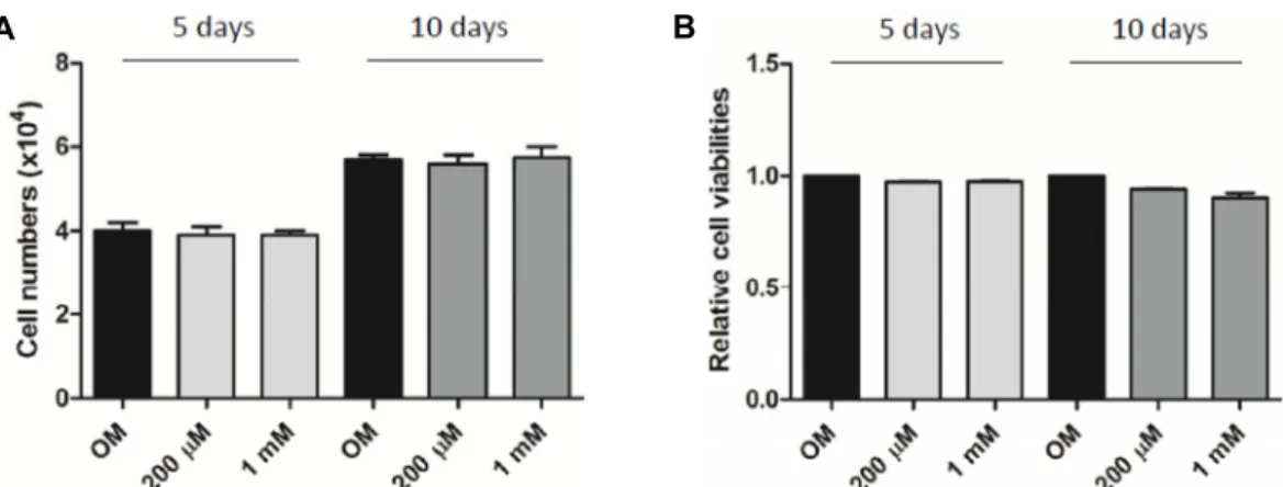
Fig. 1. Effects of salicylate on the proliferation and viability of POMSCs. Salicylate treatments (200 μM and 1 mM) do not affect neither POMSC proliferation nor its viability in cell cultures with an osteogenic differentiation induction medium (OM).
(A) Cell proliferations of 5- and 10-day POMSC cultures were determined by cell counting. (B) Cell viabilities of 5- and 10-day POMSC cultures were assessed by MTT assay.
Fig. 2. Effects of salicylate on osteogenic differentiation of POMSCs. POMSCs were cultured and induced to osteo- genic differentiation with OM for 5 and 10 days. Treat- ments of 1 mM salicylate increased ALP activities of both 5- and 10-day POMSC cultures in a time-depend- ent manner. Treatments of 200 μM salicylate enhanced ALP activities of 10-day POMSC cultures but not of 5-day cultures. One asterisk (*) indicates p<0.05.
고, 반복실험에서 얻은 결과는 Graphpad Prism 7 software (GraphPad, La Jolla, USA)를 이용하여 분산분석을 수행하였 고, 평균±표준편차로 나타내었다. 각 실험군의 평균값 차이를 통계분석하여 p<0.05인 경우에 통계학적으로 유의하다고 판 단하였다.
결과 및 고찰
POMSCs의 세포증식과 생존능력에 대한 salicylate의 영 향 분석
POMSCs의 세포증식과 생존능력에 대한 salicylate의 영향 을 알아보기 위하여 POMSCs에 salicylate를 200 μM과 1 mM 의 농도로 각각 처리하고 골세포분화 OM배양액에서 5일간 그리고 10일간 세포배양하였다. 이후 cell counting 그리고 MTT assay를 수행하였다. Salicylate를 처리하지 않은 대조군 과 비교하였을 때, 200 μM과 1 mM salicylate를 처리한 그룹에 서 5일 및 10일 간의 세포배양 기간에 증식된 세포의 수는 비슷한 것으로 나타났다(Fig. 1A). MTT assay 실험결과에서도 salicylate (200 μM과 1 mM) 처리는 POMSCs의 생존능력에 관하여 별다른 영향을 주지 않는 것으로 나타났다(Fig. 1B).
이러한 실험결과들은 salicylate가 최소한 200 μM과 1 mM의 농도에서는 POMSCs의 증식능력과 생존능력에 영향을 미치 지 않은 것으로 판단된다.
POMSCs의 골세포 분화유도에 대한 salicylate의 영향 분석
POMSCs의 골세포 분화유도에 대한 salicylate의 영향을 알 아보기 위해, POMSCs를 200 μM 그리고 1 mM salicylate가 첨가된 골세포분화 OM배양액으로 세포배양하였다. 5일 그리 고 10일간의 골세포분화 유도 후에 초기 골세포분화 표지자로 잘 알려진 ALP의 활성을 측정하였다. Fig. 2에서 보여지는 바
와 같이, 골세포분화 유도효과가 없는 통상적인 DMEM배양 액을 사용한 대조군에 비해, 골세포분화를 유도하는 OM배양 액 그룹은 ALP 활성이 5일간의 배양에서는 약 2배 그리고 10일간의 배양에서는 약 4배 정도 증가하였다. 이러한 결과는 본 실험에 사용되는 POMSCs가 OM배양액에 반응하여 골세 포분화의 초기단계인 조골세포로 분화하는 능력이 있음을 분 명하게 제시해 준다. 또한 OM배양액에 1 mM salicylate 처리 는 5일간 그리고 10일간의 세포배양 모두에서 salicylate를 처 리하지 않은 OM배양액 그룹에 비해 통계적으로 유의하게 ALP 활성을 증가시켰다. 상대적으로 낮은 200 μM salicylate 처리는 5일간의 배양에서는 차이가 없었으나 10일간의 배양
A
B
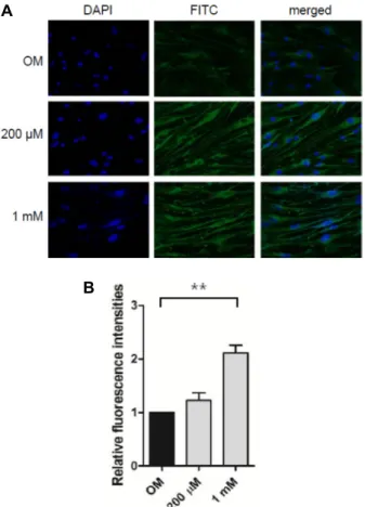
Fig. 3. Effects of salicylate on mitochondrial biogenesis during osteogenic differentiation of POMSCs. (A) POMSCs were cultured and induced to osteogenic differentiation with OM for two weeks. The resulting differentiated POMSCs were stained with a fluorescent dye, Mitotrack- er Green, which is localized in mitochondria in the cell.
The stained cells were assessed for mitochondrial mass by flow cytometry. Salicylate treatments (200 μM and 1 mM) increased mitochondrial contents in a dose-de- pendent manner. (B) Flow cytometric data from three independent experiments are plotted. * indicates p<0.05 and ** indicates p<0.01.
에서는 다소 ALP 활성이 증가됨이 관찰되었다. Fig. 2에 제시 되지는 않았지만, 보다 높은 농도인 2 mM salicylate 처리는 OM배양액 그룹에 비해 골세포 분화유도 증가가 관찰되지 않 았다. 종합하여, 이러한 결과는 salicylate 처리가 POMSCs의 골세포분화의 초기과정을 촉진시킬 수 있다는 것을 제시한다.
POMSCs의 골세포분화 과정에서 salicylate의 미토콘드 리아 생합성에 대한 영향 분석
POMSCs의 골세포분화 과정에서 salicylate가 미토콘드리 아 생합성에 미치는 영향을 알아보기 위하여 POMSCs를 sali- cylate (200 μM과 1 mM)가 첨가된 골세포분화 OM 배양액으 로 2주일 동안 세포배양하였다. 이렇게 배양된 세포에 Mito- tracker® Green FM 형광 dye를 처리하여 세포내 미토콘드리 아를 염색하였다. 염색된 POMSCs를 flow cytometry 기법으 로 세포내 미토콘드리아의 양을 정량적으로 분석하였다. Fig.
3A에서 salicylate를 처리하지 않은 대조군과 비교하여 200 μM 그리고 1 mM salicylate 처리는 두 농도 모두에서 형광값이 증가하였고 이러한 결과는 세포내 미토콘드리아의 양이 증가 한 것을 제시한다. Fig. 3B에서 flow cytometry 실험결과를 정량적으로 제시하였다. 한편, chamber slides에서 위와 동일 한 조건으로 POMSCs의 골세포 분화유도와 동반된 salicylate 처리 후에, Mitotracker® Green FM 형광 dye로 배양된 POMSCs를 염색하여 형광현미경으로 관찰하였다(Fig. 4A).
Salicylate (200 μM 그리고 1 mM)를 처리한 두 그룹에서 대조 군에 비해 뚜렷이 증가된 녹색형광을 보였고 이는 Mitotracker®
Green FM 형광 dye에 염색된 세포내 미토콘드리아의 증가를 의미한다. Fig. 4B에서 형광현미경사진의 녹색형광을 Image J program로 정량하여 그래프로 나타내었다. 종합하면, 골세 포분화 유도 후에 세포내 미토콘드리아의 양을 분석한 이러한 실험결과에서, salicylate 처리는 POMSCs의 골세포 분화과정 에서 미토콘드리아 생합성을 증가시킨다는 것을 제시한다.
미토콘드리아 생합성 증가와 POMSCs 골세포 분화촉진 최근 들어 MSCs의 조골세포/골세포 분화능력을 이용한 재 생의학이 골질환 치료연구에서 주목받고 있다[17, 25]. MSCs 의 분화과정은 매우 복잡하고 다양한 작용을 통해 조절된다[6, 27]. 이중 골세포분화에는 bone morphogenetic protein를 매 개로 한 신호전달 그리고 collagen type I이나 osteocalcin과 같은 골형성 단백질들이 관여한다는 것이 잘 알려져 있다[5, 19]. 한편, 줄기세포의 증식과정과 비교하였을 때, 분화과정이 진행되는 동안에는 해당과정이 줄어들고 산화적인산화가 증 가하는 것이 최근 연구결과에서 보고되고 있다[11, 24]. 산화적 인산화는 미토콘드리아에서 조절되며 ATP 생산에 중요한 과 정이다. 흥미롭게도, 최근 연구에서 의약품으로 널리 사용되 고 있는 salicylate가 동물세포에서 미토콘드리아 생합성을 증 가시키는 효과가 있다고 알려졌다[26]. 하지만 salicylate를 이
용한 미토콘드리아 생합성 증진을 골세포분화와 재생의학에 적용한 연구결과는 아직 보고되지 않고 있다. 따라서 본 연구 에서는 골질환 치료를 위한 재생의학의 관점에서 salicylate가 성체줄기세포의 골세포 분화과정에 미치는 영향에 대해 분석 하였으며, 골막유래 성체줄기세포인 POMSCs가 골세포로 분 화하는 과정에서, salicylate의 골세포분화 증진효과 및 미토콘 드리아 생합성 증진효과를 확인할 수 있었다. 이러한 결과들 은 salicylate가 POMSCs의 미토콘드리아 생합성에 영향을 주 어 골세포분화를 촉진시킬 수 있음을 암시한다. 미토콘드리아 의 산화적인산화를 통한 ATP 생성은 줄기세포 분화과정의 진행에서 중요하다고 알려지고 있다. 그러므로 salicylate에 의 한 미토콘드리아 생합성 증진과 골세포분화 촉진과의 관련성 을 보여주는 본 연구는 에너지대사를 중심으로 한 골 재생의 학의 새로운 시도라고 할 수 있다. 따라서 향후 추가적인 관련 연구가 계속되어 골 재생의학의 조절물질로서 salicylate가 응
A
B
Fig. 4. Visualization and quantitation of mitochondrial bio- genesis during osteogenic differentiation of POMSCs.
(A) POMSCs were cultured on chamber slides and in- duced to osteogenic differentiation with OM for two weeks. The resulting differentiated POMSCs were stained Mitotracker Green to visualize mitochondria by fluorescent microscopy. From fluorescence microscope images shown, salicylate treatments (200 μM and 1 mM) increased mitochondrial mass in a dose-dependent manner. (B) Fluorescence microscopy images from three independent experiments are plotted. ** indicates p<0.01.
용될 수 있길 기대한다.
감사의 글
본 연구는 교육부와 미래창조과학부의 재원으로 한국연구 재단의 지원(과제번호: NRF-2014R1A1A2A16055714, NRF-2016 R1D1A1B03931722, NRF-2017R1D1A1B03035996 및 2018EG 021010105)을 받아 이루어졌으며 이에 대해 감사드립니다.
References
1. Agata, H., Asahina, I., Yamazaki, Y., Uchida, M., Shinohara, Y., Honda, M. J., Kagami, H. and Ueda, M. 2007. Effective
bone engineering with periosteum-derived cells. J. Dent. Res.
86, 79-83.
2. Baroni, M. D., Colombo, S. and Martegani, E. 2018. Anta- gonism between salicylate and the cAMP signal controls yeast cell survival and growth recovery from quiescence.
Microb. Cell 5, 344-356.
3. Bergman, O. and Ben-Shachar, D. 2016. Mitochondrial oxi- dative phosphorylation system (OXPHOS) deficits in schizo- phrenia: Possible interactions with cellular processes. Can.
J. Psychiatry 61, 457-469.
4. Breitbart, A. S., Grande, D. A., Kessler, R., Ryaby, J. T., Fitzsimmons, R. J. and Grant, R. T. 1998. Tissue engineered bone repair of calvarial defects using cultured periosteal cells. Plast. Reconstr. Surg. 101, 567-574.
5. Chen, G., Deng, C. and Li, Y. P. 2012. TGF-β and BMP sig- naling in osteoblast differentiation and bone formation. Int.
J. Biol. Sci. 8, 272-288.
6. Chung, J. E., Park, J. H., Yun, J. W., Kang, Y. H., Park, B.
W., Hwang, S. C., Cho, Y. C., Sung, I. Y., Woo, D. K. and Byun, J. H. 2016. Cultured human periosteum-derived cells can differentiate into osteoblasts in a perioxisome prolifer- ator-activated receptor gamma-mediated fashion via bone morphogenetic protein signaling. Int. J. Med. Sci. 13, 806-818.
7. Ferretti, C. and Mattioli-Belmonte, M. 2014. Periosteum de- rived stem cells for regenerative medicine proposals: Boost- ing current knowledge. World J. Stem Cells 6, 266-277.
8. Hah, Y. S., Joo, H. H., Kang, Y. H., Park, B. W., Hwang, S. C., Kim, J. W., Sung, I. Y., Rho, G. J., Woo, D. K. and Byun, J. H. 2014. Cultured human periosteal-derived cells have inducible adipogenic activity and can also differentiate into osteoblasts in a perioxisome proliferator-activated re- ceptor-mediated fashion. Int. J. Med. Sci. 11, 1116-1128.
9. Hutmacher, D. W. and Sittinger, M. 2003. Periosteal cells in bone tissue engineering. Tissue Eng. 9, 45-64.
10. Huttemann, M., Lee, I., Samavati, L., Yu, H. and Doan, J.
W. 2007. Regulation of mitochondrial oxidative phosphor- ylation through cell signaling. BBA 1773, 1701-1720.
11. Ito, K. and Suda, T. 2014. Metabolic requirements for the maintenance of self-renewing stem cells. Nat. Rev. Mol. Cell Biol. 15, 243-256.
12. Kakehata, S. and Santos-Sacchi, J. 1996. Effects of salicylate and lanthanides on outer hair cell motility and associated gating charge. J. Neurosci. 16, 4881-4889.
13. Mao, A. S. and Mooney, D. J. 2015. Regenerative medicine:
Current therapies and future directions. Proc. Natl. Acad. Sci.
USA. 112, 14452-14459.
14. Oryan, A., Kamali, A., Moshiri, A. and Eslaminejad, B. M.
2017. Role of mesenchymal stem cells in bone regenerative medicine: What is the evidence? Cells Tissues Organs 204, 59-83.
15. Park, B. W., Hah, Y. S., Kim, D. R., Kim, J. R. and Byun, J. H. Osteogenic phenotypes and mineralization of cultured human periosteal-derived cells. Arch. Oral Biol. 52, 983-989.
16. Park, B. W., Hah, Y. S., Kim, D. R., Kim, J. R. and Byun, J. H. 2008. Vascular endothelial growth factor expression in cultured periosteal-derived cells. Oral Surg. Oral Med. Oral
초록:Salicylate가 성체줄기세포의 골분화에 미치는 영향
김보규1․이아람1․이보영1․심성보2․문동규3․황선철3․변준호4*․우동균1*
(1경상대학교 약학과, 2충북대학교 생화학과, 3경상대학교병원 정형외과, 4경상대학교병원 구강악안면외과)
최근 들어 급속한 고령화 사회가 진행되고 있으며 이로 인해 골관절염과 골다공증 등의 퇴행성 골질환 환자수 도 동반하여 증가하고 있다. 따라서 고령화에 따른 골관련 질환의 새로운 제어와 치료법 개발을 위해 성체줄기세 포의 골세포 분화유도를 활용한 재생의학도 활발히 연구되고 있다. 또한 관련 연구에서 줄기세포의 분화과정에서 미토콘드리아의 산화적인산화가 중요하다고 알려지고 있다. 흥미롭게도 최근 연구에서 아스피린의 주성분인 sali- cylate가 동물세포의 미토콘드리아 생합성을 증진시키는 효과가 보고되었다. 그러나 성체줄기세포에서 salicylate 가 골세포분화나 미토콘드리아 생합성을 유도할 수 있는지에 대한 연구결과는 미비한 실정이다. 본 연구에서는 인체 골막 유래의 성체줄기세포를 이용하여 골세포분화나 미토콘드리아 생합성에 대한 salicylate의 영향을 분석 하였다. 골막 유래 성체줄기세포의 골세포 분화유도 과정에 동반한 salicylate 처리는 잘 알려진 골세포분화 표지 자인 alkaline phosphatase의 활성을 증가시키는 결과를 본 연구에서 얻었다. 이러한 연구결과는 salicylate가 줄기 세포로부터 골세포로의 분화를 조절할 수 있는 물질이 될 수 있음을 제시한다. 또한 이러한 골세포 분화과정에서 미토콘드리아 생합성도 salicylate 처리에 의해 증가됨이 관찰되었다. 따라서, 미토콘드리아 생합성이나 기능을 조 절하는 물질이 성체줄기세포의 골세포 분화과정에도 영향을 줄 수 있으며, 이러한 물질이 골세포분화나 재생의학 의 새로운 조절 물질로 응용될 수 있음을 제시한다.
Pathol. Oral Radiol. Endod. 105, 554-560.
17. Park, H. C., Son, Y. B., Lee, S. L., Rho, G. J., Kang, Y. H., Park, B. W., Byun, S. H., Hwang, S. C., Cho, I. A., Cho, Y.
C., Sung, I. Y., Woo, D. K. and Byun, J. H. 2017. Effects of osteogenic-conditioned medium from human periosteum- derived cells on osteoclast differentiation. Int. J. Med. Sci.
14, 1389-1401.
18. Park, J. H., Park, B. W., Kang, Y. H., Byun, S. H., Hwang, S. C., Kim, D. R., Woo D. K. and Byun, J. H. 2017. Lin28a enhances in vitro osteoblastic differentiation of human peri- osteum-derived cells. Cell Biochem. Funct. 35, 497-509.
19. Rammelt, S., Neumann, M., Hanisch, U., Reinstorf, A., Pompe, W., Zwipp, H. and Biewener, A. 2005. Osteocalcin enhances bone remodeling around hydroxyapatite/collagen composites. J. Biomed. Mater. Res. A. 73, 284-294.
20. Ringe, J., Leinhase, I., Stich, S., Loch, A., Neumann, K., Haisch, A., Häupl, T., Manz, R., Kaps, C. and Sittinger, M.
2008. Human mastoid periosteum-derived stem cells: prom- ising candidates for skeletal tissue engineering. J. Tissue Eng.
Regen. Med. 2, 136-146.
21. Rhy, Y. M., Hah, Y. S., Park, B. W., Kim, D. R., Roh, G. S., Kim, J. R., Kim, U. K., Rho, G. J., Maeng, G. H. and Byun, J. H. 2011. Osteogenic differentiation of human periosteal- derived cells in a three-dimensional collagen scaffold. Mol.
Biol. Rep. 38, 2887-2894.
22. Schafer, R., Spohn, G. and Baer, P. C. 2016. Mesenchymal stem/stromal cells in regenerative medicine: Can precondi- tioning strategies improve therapeutic efficacy? Transfus.
Med. Hemother. 43, 256-267.
23. Steinberg, G. R., Dandapani, M. and Hardie, D. G. 2013.
AMPK: mediating the metabolic effects of salicylate-based drugs? Trends Endocrinol. Metab. 24, 481-487.
24. Shum, L. C., White, N. S., Mills, B. N., Bentley, K. L. and Eliseev, R. A. 2016. Energy metabolism in mesenchymal stem cells during osteogenic differentiation. Stem Cells Dev.
25, 114-122.
25. Shyam, H., Singh, S. K., Kant, R. and Saxena, S. K. 2017.
Mesenchymal stem cells in regenerative medicine: a new paradigm for degenerative bone diseases. Regen. Med. 12, 111-114.
26. Yan, Y., Yang, X., Zhao, T., Zou, Y., Li, R. and Xu, Y. 2017.
Salicylates promote mitochondrial biogenesis by regulating the expression of PGC-1α in murine 3T3-L1 pre-adipocytes.
Biochem. Biophys. Res. Commun. 491, 436-441.
27. Yoon, D. K., Park, J. S., Rho, G. J., Lee, H. J., Sung, I. Y., Song, J. H., Park, B. W., Kang, Y. H., Byun, S. H., Hwang, S. C., Woo, D. K., Cho, Y. C. and Byun, J. H. 2017. The involvement of histone methylation in osteoblastic differ- entiation of human periosteum-derived cells cultured in vitro under hypoxic conditions. Cell Biochem. Funct. 35, 441-452.