70% 간절제술을 받은 쥐에서 피브린젤을 이용한 중간엽줄기세포의 생체내 간세포 분화
Hepatic Differentiation of Bone Marrow Derived Mesenchymal Stem Cell Using Fibrin Gels in 70% Hepatectomized Rat
Purpose: To investigate the differentiation of rat bone marrow-derived mesenchymal stem cells (MSCs) into hepatocytes by cell transplantation using fibrin gels in a 70% hepatectomized rat model.
Methods: MSCs were isolated from Sprague-Dawley rats. MSCs (1.5×10
7cells) were mixed with fibrin gels and injected immediately into the abdominal cavity of 70% hepatectomized rats.
Fibrin-gels consisted of 500 IU/ml of thrombin and 90 mg/ml of fibrinogen. Transplanted MSCs in the fibrin scaffold were retrieved from surgically opened peritoneal cavities of rats on days 5, 10, 15, and 21 after the operation. The specimens were analyzed histologically and immunohistochemically.
Results: On H&E staining, MSCs from hepatectomized rats had changed to a round shape, while MSCs of the control group kept their spindle shape. When the fibrin matrix was biodegraded at day 15, the morphology of the MSCs had changed to hepatocyte-like cells without sinusoids and the hepatocyte-like cells had formed a three-dimensional tissue permitting cell-to-cell contacts within the matrix. On immunohistochemistry, MSCs expressed the hepatocyte markers cytokeratin 18, albumin, and alpha-fetoprotein, after 15 days of transplantation.
Conclusion: When bone marrow-derived MSCs are transplanted using fibrin gels in the 70%
hepatectomized rat, MSCs differentiate into hepatocyte-like cells and are conglomerated so that they form three-dimensional tissue-like hepatocytes without sinusoids.
신우영, 정성은1
, 민혜숙
2, 이해원
1,
조응호1, 이남준
1, 서경석
1, 이건욱
1전남대학교 의과대학 외과학교실, 서울대학교 의과대학
1외과학교실,
2
병리학교실
Woo Young Shin, M.D., Sung Eun Jung, M.D.
1, Hye Sook Min, M.D.
2, Hae Won Lee, M.D.
1, Eung-Ho Cho, M.D.
1, Nam-Joon Yi, M.D.
1, Kyung-Suk Suh, M.D.
1, Kuhn Uk Lee, M.D.
1Departments of Surgery, Chonnam National University College of Medicine, Departments of
1Surgery and
2Pathology, Seoul National University College of Medicine
책임저자 정 성 은
서울시 종로구 연건동 28 서울대학교 의과대학 외과학교실 우편번호 110-744
Tel: 02-2072-2927 Fax: 02-766-3975
E-mail: [email protected]
*본 연구는 서울대학교병원 일반 연구과제(04-2005-053-0) 지원하여 시행되었으며, 2006년 제 25차 한국간담췌외과학회 추계학술대회에서 구연발표되었음.
Key Words : Mesenchymal stem cell, Hepatocyte, Cell differentiation, Fibrin 중심단어 : 중간엽줄기세포, 간세포, 세포분화, 피브린
Received: 2010. 4. 7 Accepted: 2010. 5. 20
화할 수 있다고 보고되었다.2-5 또한 MSCs는 생체내외에서 간 세포로도 분화할 수 있다.6-10
현재까지 MSC의 생체내 간세포분화를 위해 MSC의 세포이 식 수단은 간에 직접 주입하거나, 정맥 내로 주입하는 것이
다.11,12 이들 방법은 이식된 MSCs의 분화여부를 확인할 수는
있으나, 이식된 세포를 적출하여 직접 관찰하기 어렵다. 최근 MSCs가 피브린지지체(fibrin scaffold)내에서 서로 접촉하면서 증식할 수 있음이 보고되었다.13 또한 피브린젤과 간세포를 혼 합하여 생쥐의 복강 내에 주입하였을 때, 수술 후 1주일까지 간세포가 응집되면서, 기능을 유지하는 것이 확인되었다.14 피 브린젤을 이용한 세포 이식법은 조작하기 쉽고, 이식된 세포 를 적출하기에도 용이하다. 하지만, 피브린젤 내에서 MSCs가 간세포로 분화가 가능한지에 대한 연구는 아직 이루어 지지 않았다. 따라서, 본 연구에서 피브린젤을 이용하여 골수 유래 의 MSCs가 쥐의 복강 내로 이식되었을 때 간세포로 분화가 가능한지를 연구하였다.
방 법
모든 동물관련 연구 과정은 서울대학교병원 임상의학연구 소의 실험동물운영위원회(Institutional Animal Care and Use Committee)의 승인을 받았다(No. 06174).
골수 유래의 MSCs를 분리 배양하기 위해 200∼250 g의 Sprague-Dawley 쥐를 이용하였으며, 분리 과정은 다음과 같 다. 쥐를 이산화탄소를 이용하여 질식사 시킨 후, 대퇴골과 경골을 분리하였다. 수질강(medullary cavity)을 노출시킨후 3,000 rpm에서 30초간 원심분리하여 얻어진 세포를 Low glucose Dulbecco’s modified Eagle’s medium containing 1 g/L glucose (DMEM-LG, WelGENE Inc. Daegu, South Korea)로 세척하였다. Phosphate buffer solution (PBS)로 세 정 후 골수 세포를 20 ml 1.077 g/L-Histopaque® (Sigma-
물을 제거하고, media에 부착된 세포만 계속해서 배양하였다.
배양액은 매 3일마다 교체하였고, 약 4주 후 5계대 째 배양세 포를 분화를 위한 실험에 이용하였다.
골수에서 분리 배양된 세포가 다분화능을 가진 MSCs인 것 을 확인하기 위해 5계대의 골수 세포를 골세포와 지방세포로 분화를 유도하였다. Chemicon (Temecula, CA, USA)과 Bio- Whittaker (Walkersville, MD, USA)의 분화 유도 배지를 이용 하였다.
골수 유래의 MSCs의 복강 내 이식을 위해 300∼350 g의 SD 쥐를 Isoflurane으로 마취하였다. 간세포성장인자(hepato- cyte growth factor; HGF)는 많은 연구에서 가장 많이 이용되 는 간세포 분화 유도 인자 중 하나로, 간절제술 후에 생체 내 에서 증가하는 것으로 알려져 있다.15,16 HGF의 증가를 통해 MSCs의 간세포로의 분화를 유도하기 위해 70% 간절제술을 시행하였다. 실험군(n=12)은 70% 간절제술 후, 피브린젤 (Greenplast kit, GreencrossPD Co. YongIn, South Korea)을 이용하여 세포이식을 시행하였다. 1.5×10⁷개의 5계대 MSCs 를 트롬빈 500 IU/ml에 부유한 후 피브리노젠 90 mg/ml에 섞고, 간절제면에 주입하였다. 대조군(n=3)은 간절제술 없이 동일한 방법으로 세포를 이식하였다. 쥐들은 이식 후 20oC 하 에서 12시간의 주야간격으로 사육되었고, 물은 자유롭게 이용 하게 하였다.
혈중 HGF의 증가 유무를 확인하기 위해 실험군과 대조군에서 수술 후 Enzyme-Linked Immunosorbent Assay kit (Institute of Immunology, Tokyo, Japan)를 이용하여 혈중 HGF 농도를 측정 하였다. 두 군간의 HGF농도의 차이는 반복 측정 ANOVA로 분석 하였다. 통계적 분석은 SPSS 12.0을 이용하였다.
피브린지지체를 수술 후 5, 10, 15, 21일 째 수술적으로 적 출하여, 포르말린에 고정 후 4 μm 두께로 슬라이드를 제작하 였다. Hematoxylin & eosin (H&E)염색으로 조직학적 검사를 시행하였다. 간세포로의 분화를 확인하기 위해 Periodic acid-
Fig. 1. (A) Cells 7 days after isolation. Spindle-shaped cells could be seen (×40). (B) Rat MSCs at passage 5 are induced to differentiate into osteocytes and stain positive for Alizarin Red S (×200). (C) Under adipogenic conditions, rat MSCs differentiated into adipocytes, which are positively stained for Oil Red O (×200).
Fig. 2. The levels of serum hepatocyte growth factor of hepa- tectomized and control groups. The level of hepatectomized group was significant higher than control group (p<0.001).
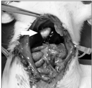
Fig. 3. Fibrin scaffold was seen in abdominal cavity (white arrow).
Schiff 염색을 이용하여 분화된 세포의 glycogen의 유무를 확 인하였고, 간세포의 특이적인 항체인 알부민(polyclonal rab- bit anti-human albumin antibody, DakoCytomation, Kyoto, Japan), cytokeratin 18 (mouse anti-human CK18 antibody, Abcam, Cambridge, MA, USA), 알파태아단백(polyclonal rabbit anti-human AFP antibody, DakoCytomation)에 대한 면역조직염색을 시행하였다. 모든 조직학적 검사는 해부병리 과 전문의의 판독을 거쳤다.
결 과
골수 세포를 분주한 후 배지 바닥에 방추형의 세포가 관찰
되었다(Fig. 1A). 5계대 배양 후 골세포들을 골세포와 지방세 포로 분화를 유도하였고, 이들 세포들은 각각 Alizarin Red S 염색과 Oil Red O 염색에서 각각 염색이 되어 골수 세포의 다분화능을 확인할 수 있었다(Fig. 1B, 1C).
쥐의 혈중 HGF 농도는 실험군과 대조군 모두에서 수술 후 2일째 최고치를 보인 후 감소하여, 수술 후 10일째 수술 전 농도로 돌아왔다. 그러나 최고 HGF 농도의 평균값은 70% 간 절제술을 받은 실험군에서 17.1±3.6 ng/ml로 대조군의 2.9±0.5 ng/ml에 비해 유의하게 높았다(Fig. 2,
p
<0.001).이식된 MSCs를 얻기 위해 재개복하였을 때 피브린지지체 는 복강 내에서 관찰되었고, 수술적으로 쉽게 적출 가능하였
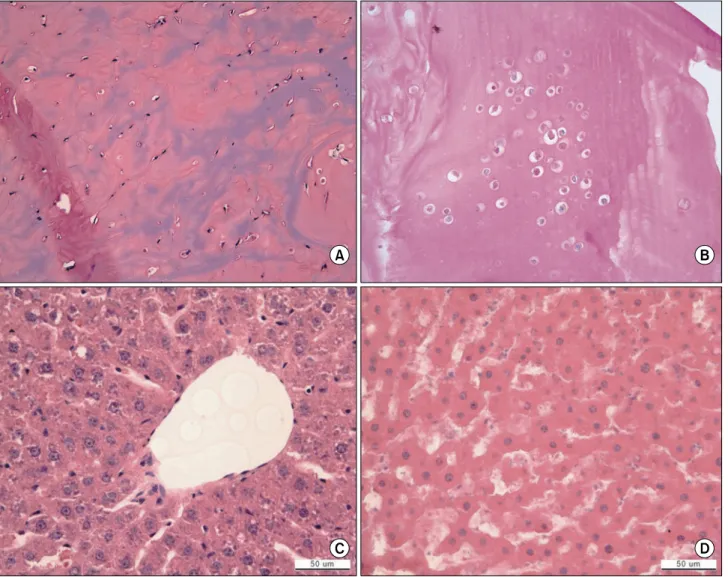
Fig. 4. (A) MSCs in control group at 15 days. MSCs kept spindle shape (×200). (B) MSCs in experimental group at 15 days.
MSCs changed to round shape (×200). (C) Normal rat hepatocytes at 21 days (×400). (D) Hepatocyte-like differentiated cells at 21 days (×400).
다(Fig. 3). 피브린지지체는 생체 내에서 분해되기 시작하여 이식 15일째부터 적출 시 크기의 감소가 관찰되었다.
H&E 염색 상 실험군의 MSCs는 원형으로 모양이 바뀌었으 나, 대조군의 MSCs는 여전히 방추형을 유지하였다(Fig. 4A, B). 수술 21일째 실험군의 MSCs는 굴모양혈관(sinusoid)이 없 는 간세포의 형태로 변하였다. 3차원적인 조직의 형태를 갖추 었다(Fig. 4C, B). PAS 염색상, 수술 후 21일째 분화 유도된 MSCs에서 glycogen이 양성으로 확인되었다(Fig. 5A). 면역조 직염색상 CK-18은 10일째부터, 알부민과 알파태아단백은 15 일째부터 양성으로 염색되었다(Fig. 5B∼D).
고 찰
본 연구의 목적은 피브린젤을 이용하여 골수유래의 MSCs 를 복강 내 이식하였을 때 간세포로 분화가 되는지를 확인하 는 것이다.
In vitro
하에서 HGF, 상피세포성장인자(epider- mal growth factor), 종양성장인자(tumor growth factor), 알 파-섬유아세포성장인자(α-fibroblast growth factor)가 간세 포의 분화를 촉진하는 데 주로 이용되고 있다.In vivo
하에서 는 수 많은 싸이토카인과 성장인자가 간세포의 성장 및 분화 에 영향을 준다.15 본 연구에서는 간세포의 분화를 유도할 수Fig. 5. (A) The cells stained with PAS suggested glycogen storage at 21 days (white arrows). Immunohistochemistry of rat MSCs retrieved after 15 days. There were MSCs positively stained with CK 18 (B), albumin (C), and alpha-fetoprotein (D) (×400).
있는 조건을 만들기 위해 간절제술을 시행하였다. 간절제술 시행군에서는 혈중 HGF의 증가와 함께 MSCs의 분화가 관찰 되었으나, 대조군에서는 그렇지 않았다. 기존의
in vivo
분화 연구는 MSCs를 간 내에 직접 주입하거나, 말초 혈관 주입을 통해 간 내에 유입시켜 분화를 유도한 것으로 간절제술과 같 은 전처치가 필요하지 않았다.11,12 하지만, 본 연구의 복강내 주입법에서 간절제술을 받지 않은 대조군에서는 MSCs는 분화 되지 않았다. 간내 분화의 경우 혈액을 통해 분화에 필요한 성장인자 등이 직접 전달되지만, 복강내에서 분화를 유도할 경우 이들 인자들이 확산에 의해 세포로 전달되어야 한다. 따 라서 분화에 필요한 최소 농도를 조성하기 위해서, 더 높은혈중 농도가 필요할 것이다.
In vitro
하에서 MSCs의 간세포 분화를 위해 최소 20 ng/ml이 필요한 것으로 알려져 있지만,15in vivo
하에서는 알려진 바 없다. 본 연구에서 HGF의 평균 최대 농도는 17.1±3.6 ng/ml이고, 복강 내 분화법이기 때문 에 실제 MSCs에 전달되는 HGF는 이보다 낮을 것으로 예상된 다. 하지만,in vivo
조건이므로 혈장 내의 다양한 싸이토카인 의 협력작용에 의해 MSCs가 간세포와 유사한 세포로 분화가 가능하게 되었을 것이다.피브린젤은 수술적 지혈과 조직을 메우는데 사용되는 물질 로 정상적인 상처 치유과정에 중요한 것으로 알려져 있다.17-19 세포이식에 있어 피브린젤은 지지체로서 몇 가지 장점이 있
비율이라고 보고하였다.13 아직 간세포 분화에 대하여 피브린 젤의 적정 농도비에 대한 연구는 없다. 본 연구에서는 MSCs 의 증식에 초점을 맞춰 Benesaïd 등의 비율을 이용하여 트롬 빈 500 IU/ml+피브리노젠 90 mg/ml을 사용하였다. 셋째로 피브린젤을 이용한 세포 이식은 MSCs가 세포간의 접촉을 가 능하게 하여 3차원적인 입체 구조를 형성할 수 있게 해준다는 것이다.20 이는 분화된 세포들이 조직의 형태를 갖추게 해준 다. 간세포의 배양에 있어 세포-세포간의 상호작용은 간세포 의 생존을 증가시키고, 간 기능을 유지하게 하는 필수 요소로 생각되고 있다.22 따라서 피브린젤을 이용한 세포이식법은 세 포간의 접촉을 가능하게 함으로써 세포-세포 상호작용을 유도 할 수 있는 이점이 있다. 넷째, 생체 내 이식된 세포를 직접 적출하여 세포에 대한 직접 관찰을 가능하게 해준다. 하지만, 수술 후 시간이 경과함에 따라 피브린지지체는 생체내에서 분 해된다. 본 연구에서는 15일경부터 생체내분해로 인해 피브린 지지체의 크기가 감소하는 것이 관찰되었고, 21일 째 적출한 피브린지지체의 크기는 눈에 띠게 작아져 있었다. 따라서, 추 후 연구 진행 시 이식 후 4주 이내에는 적출을 요할 것으로 생각된다.
결 론
골수 유래의 중간엽 줄기세포를 피브린젤을 이용하여 복강 내 이식을 하였을 때, 이식된 세포들을 쉽게 적출하여 관찰할 수 있었다. 이식된 MSCs는 간세포로 분화가 가능하였고, 피브 린젤 내에서 이식된 MSCs는 서로 응집하여 3차원적인 조직의 형태를 갖추었다. 하지만, 간세포로의 분화를 유도하기 위해 간절제술과 같은 전처치가 필요하였다.
참 고 문 헌
1. Caplan AI. Mesenchymal stem cells. J Orthop Res 1991;
Regulation of neural markers nestin and GFAP expression by cultivated bone marrow stromal cells. J Cell Sci 2003;116:
3295-3302.
5. Orlic D. Adult bone marrow stem cells regenerate myocar- dium in ischemic heart disease. Ann N Y Acad Sci 2003;
996:152-157.
6. Schwartz RE, Reyes M, Koodie L, et al. Multipotent adult pro- genitor cells from bone marrow differentiate into functional hepatocyte-like cells. J Clin Invest 2002;109:1291-1302.
7. Lee KD, Kuo TK, Whang-Peng J, et al. In vitro hepatic di- fferentiation of human mesenchymal stem cells. Hepatology 2004;40:1275-1284.
8. Hong SH, Gang EJ, Jeong JA, et al. In vitro differentiation of human umbilical cord blood-derived mesenchymal stem cells into hepatocyte-like cells. Biochem Biophys Res Commun 2005;330:1153-1161.
9. Kang XQ, Zang WJ, Song TS, et al. Rat bone marrow me- senchymal stem cells differentiate into hepatocytes in vitro.
World J Gastroenterol 2005;11:3479-3484.
10. Shi XL, Qiu YD, Wu XY, et al. In vitro differentiation of mouse bone marrow mononuclear cells into hepatocyte-like cells. Hepatol Res 2005;31:223-231.
11. Sato Y, Araki H, Kato J, et al. Human mesenchymal stem cells xenografted directly to rat liver are differentiated into human hepatocytes without fusion. Blood 2005;106:756-763.
12. Lagasse E, Connors H, Al-Dhalimy M, et al. Purified hema- topoietic stem cells can differentiate into hepatocytes in vivo.
Nat Med 2000;6:1229-1234.
13. Bensaïd W, Triffitt JT, Blanchat C, Oudina K, Sedel L, Petite H. A biodegradable fibrin scaffold for mesenchymal stem cell transplantation. Biomaterials 2003;24:2497-2502.
14. Gwak SJ, Choi D, Paik SS, Lee EY, Lees KS, Kim BS. Stable hepatocyte transplantation using fibrin matrix. Biotechnol Lett 2004;26:505-508.
15. Heng BC, Yu H, Yin Y, Lim SG, Cao T. Factors influencing stem cell differentiation into the hepatic lineage in vitro. J Gastroenterol Hepatol 2005;20:975-987.
16. Liu ZC, Chang TM. Transdifferentiation of bioencapsulated bone marrow cells into hepatocyte-like cells in the 90%
hepatectomized rat model. Liver Transpl 2006;12:566-572.
17. Albala DM. Fibrin sealants in clinical practice. Cardiovasc
Surg 2003;11(Suppl 1):5-11.
18. Jackson MR. Fibrin sealants in surgical practice: an overview.
Am J Surg 2001;182(Suppl 2):1S-7S.
19. Mankad PS, Codispoti M. The role of fibrin sealants in hemostasis. Am J Surg 2001;182(Suppl 2):21S-28S.
20. Bruns H, Kneser U, Holzhuter S, et al. Injectable liver: a novel approach using fibrin gel as a matrix for culture and intrahepatic transplantation of hepatocytes. Tissue Eng 2005;
11:1718-1726.
21. Catelas I, Sese N, Wu BM, Dunn JC, Helgerson S, Tawil B.
Human mesenchymal stem cell proliferation and osteogenic differentiation in fibrin gels in vitro. Tissue Eng 2006;12:
2385-2396.
22. Lee DH, Yoon HH, Park JK. Hepatocyte culture technology and its application to bioartificial liver. Korean Chem Eng Res 2004;42:129-138.