Enhanced hepatic differentiation of
human cord blood-derived
mesenchymal stem cells
using activin A-based protocol
Sinyoung Kim
Department of Medicine
Enhanced hepatic differentiation of
human cord blood-derived
mesenchymal stem cells
using activin A-based protocol
Directed by Professor Hyun Ok Kim
The Doctoral Dissertation
submitted to the Department of Medicine,
the Graduate School of Yonsei University
in partial fulfillment of the requirements for the degree
of Doctor of Philosophy
Sinyoung Kim
This certifies that the Doctoral
Dissertation of Sinyoung Kim is
approved.
---
Thesis Supervisor: Hyun Ok Kim
---
Kwang-Hyub Han: Thesis Committee Member#1
---
Yoo Hong Min: Thesis Committee Member#2
---
Dong-Wook Kim: Thesis Committee Member#3
---
Kyung Sik Kim: Thesis Committee Member#4
The Graduate School
Yonsei University
ACKNOWLEDGEMENTS
While pursuing my doctoral degree with various research projects, a
number of generous individuals guided and supported me. Firstly, I owe
much gratitude to my dissertation advisor, Professor Hyun Ok Kim, who
has not only advised me through the scientific endeavors of my projects,
but also encouraged me with enthusiasm and inspired me in every situation.
Let me take this opportunity to also express my sincere gratitude to other
members of the dissertation committee, Professor Kwang-Hyub Han,
Professor Yoo Hong Min, Professor Dong-Wook Kim, and Professor
Kyung Sik Kim, for all of their professional advice in leading me through
my thesis completion. And I want to thank Professor Han-Soo Kim for the
direction he has provided and the focus he allowed me to achieve.
And most of all, I would like to thank my parents for their patience, my
wife, Yunsook Oh, for her unrelenting support and inspiration at times
when I needed it most, and my lovely daughter for making everyday a
pleasure.
9 May 2009
Sinyoung Kim
<TABLE OF CONTENTS>
ABSTRACT··· 1
I. INTRODUCTION··· 4
II. MATERIALS AND METHODS··· 7
1. Isolation and primary culture of cord blood-derived mesenchymal stem
cells (CBMSCs) ··· 7
2. Immunophenotyping of CBMSCs··· 8
3. In vitro differentiation assays··· 8
A. Osteogenic differentiation··· 8
B. Chondrogenic differentiation··· 9
C. Adipogenic differentiation···10
4. Hepatic differentiation protocol···10
5. RNA isolation and reverse transcriptase-polymerase chain reaction
···12
6. Treatment of animals··· 14
7. Biochemical analysis··· 15
8. Immunohistochemical analysis··· 15
III. RESULTS··· 17
1. Isolation and expansion of CBMSCs··· 17
2. Immunophenotypes of CBMSCs··· 17
3. In vitro differentiation assays··· 19
4. In vitro hepatic differentiation potential··· 19
5. In vivo hepatic differentiation of CBMSCs in cirrhotic rat model··· 23
6. Changes of liver function after CBMSC infusion··· 26
IV. DISCUSSION··· 30
V. CONCLUSION··· 34
REFERENCES··· 35
LIST OF FIGURES
Figure 1. Photomicrographs of isolated CBMSCs···17
Figure 2. Immunophenotypes of CBMSCs···18
Figure 3. Multi-lineage in vitro differentiation capacity of
CBMSCs···20
Figure 4. Hepatic differentiation of CBMSCs in vitro···22
Figure 5. Immunohistochemical detection of human
hepatocytes···24
Figure 6. Immunohistochemical detection of human cells···25
Figure 7. Representative figures showing fibrotic area in
placebo and CBMSC infused group···28
Figure 8. Comparison of fibrotic area between the placebo
and CBMSC infused group···29
LIST OF TABLES
Table 1. Primer pairs used for polymerase chain reactions to
detect hepatocyte-specific gene expression···13
Table 2. Comparison of serum markers related to liver function
between placebo and CBMSC infused group···26
Table 3. Quantitative measurement of picro-sirius red stained
<ABSTRACT>
Enhanced hepatic differentiation of human cord blood-derived
mesenchymal stem cells using activin A-based protocol
Sinyoung Kim
Department of Medicine
The Graduate School, Yonsei University
(Directed by Professor Hyun Ok Kim)
Recent studies have shown that mesenchymal stem cells (MSCs), which are isolated from various sources such as bone marrow, cord blood, adipose tissue, amniotic fluid, and the placenta, could differentiate into hepatocyte-like cells in vitro. However, these in vitro differentiation protocols are not practical for clinical usage, as more than 28 days is required to differentiate MSCs into hepatic lineages and the differentiation efficiency is low. In addition, the in vivo hepatic differentiation of MSCs and therapeutic efficacy of MSC administration in chronic liver injury models remains controversial. The goal of the present study was to improve and modify the hepatic differentiation protocol of cord blood-derived MSC (CBMSC), based on the current knowledge of liver ontogeny. Also, we investigated the therapeutic effects of systemic CBMSC infusion in cirrhotic rat model.
adherence method, and its surface phenotype and multi-lineage differentiation potential were examined. Isolated CBMSCs were differentiated into hepatic lineages using the activin A-based protocol or previously published two-step protocol. The activin A-based protocol was modified from the two-step protocol to reflect cytokine expression during embryonic hepatogenesis (activin A plus fibroblast growth factor (FGF)-4, followed by a combination of hepatocyte growth factor, FGF-4, oncostatin M and dexamethasone). Differentiated hepatocyte-like cells were examined for hepatocyte-specific markers, such as alpha-fetoprotein (AFP) and albumin. Ex vivo expanded CBMSCs were infused in Wistar rats with thioacetamide-induced chronic liver injury. Biochemical markers, liver fibrosis and engraftment of CBMSCs were assessed 1 or 4 weeks after cell or placebo infusion.
After induction under each protocol, cuboidal morphology, which is characteristic of hepatocytes, was observed. These hepatocyte-like cells, which were induced by the two-step protocol, did not express mRNA of hepatocyte-specific AFP and albumin at 4 weeks of induction. However, using the activin A-based protocol, CBMSCs (five among 12 cell lines from different donors) revealed expression of AFP, albumin and cytokeratin 19 as early as at 2 weeks of induction. Four weeks after CBMSC infusion into rats with chronic liver injury, there was no difference in serological markers of liver injury and area of liver fibrosis between the cell and placebo infusion groups. Infused CBMSCs were detected in the perivascular and fibrous region of the liver and did not
acquire mature hepatic phenotypes.
CBMSCs have the potential to differentiate into hepatic lineages in vitro and activin A is hereby essential to promote differentiation of CBMSCs towards functional hepatocyte-like cells. However, improvement of liver function and liver fibrosis after infusion of CBMSCs could not be found in our experimental cirrhotic rat model.
--- Key words : cord blood, mesenchymal stem cell, hepatocyte, differentiation
Enhanced hepatic differentiation of human cord blood-derived
mesenchymal stem cells using activin A-based protocol
Sinyoung Kim
Department of Medicine
The Graduate School, Yonsei University
(Directed by Professor Hyun Ok Kim)
I. INTRODUCTION
Liver transplantation is currently the only successful treatment for acute hepatic failure or ends-stage liver disease. Several problems are associated with liver transplantation, which include: lack of donors, surgical complications, rejection, and high cost. Hepatocyte transplantation has been the alternative option for bridging patients to liver transplantation. However, primary hepatocytes are only available from a restricted number of donor organs, which are usually not allocated for organ transplantation1. Also primary hepatocytes have a poor proliferative potential, and probably are not sufficient to effectively repopulate the host liver and it is difficult to maintain in vitro cultures without loss of functions2. Additionally, their usage does not solve the problem of immune rejection.
have to be established. Stem cells have been identified in a variety of tissues, where they play critical roles in the tissues’ maintenance and repair. But, identification and characterization of hepatic stem cells has been technically difficult because sensitive and stable single cell based assays have not yet been developed. Several studies have shown that bone marrow-derived hematopoietic stem cells (HSCs) have the capacity to differentiate into a variety of non-hematopoietic cells such as hepatocytes3, 4, myocardium5, and neurons6. However, HSCs contribute little to hepatocyte formation under either physiological or pathological conditions, although they provide cytokines and growth factors that promote hepatocyte functions by the paracrine mechanism7.
The other potential candidate stem cells are mesenchymal stem cells (MSCs), which could be obtained from various sources such as bone marrow8, cord blood9, amniotic fluid10, placenta11, or adipose tissue12. Cord blood has a number of advantages over the other sources of MSCs in terms of cell procurement, which include abundance, lack of donor attrition, and a low risk of viral transmission for disease such as the herpes family viruses. Furthermore, MSCs have a reduced risk of allogenic transplant rejection, because they are immuno-privileged with a low major histocompatibility complex (MHC) I and no MHC II class expression13, 14. For these reasons, cord blood-derived MSCs (CBMSCs) could be a prominent source of cells for transplantation for various diseases.
15-17
. Multipotent adult progenitor cells, which were discovered by Schwartz et al., were the first plastic cells found within bone marrow that gained the ability to undergo hepatic differentiation under the fibroblast growth factor (FGF)-4 and hepatocyte growth factor (HGF)18. Thus far, several studies have revealed the hepatic differentiation potential of MSCs using different protocols. Proposed protocols included stimulation of MSCs with hepatogenic factors, added either as a mixture (FGF + HGF19; FGF + HGF + oncostatin M (OSM)20) or separately (HGF / OSM21; FGF / HGF / OSM15, FGF + HGF / OSM22). However, the resultant populations were far from homogeneous and the hepatocyte-like cells differentiated by the abovementioned protocols showed hepatocyte-specific mRNA expression at over 30 days of induction. Such a long period of induction time did require large quantities of cytokines and may have problems related to long term in vitro cultivation.
Activin A, a member of the TGF-β superfamily, has been identified as a potential regulator for the induction of mesoderm and endoderm in embryonic stem cells23, 24. Activin A has also demonstrated to provide an efficient signal into human embryonic stem cells for definitive endodermal differentiation via activin/nodal signaling pathway25. Cai et al. differentiated human embryonic stem cells into functional hepatic cells after sequential treatment with activin A and FGF4/bone morphogenetic protein-226. This process generally mimicked natural embryonic liver development observed in vivo. In the present study, we investigated to modify and improve the hepatic differentiation protocol of
CBMSC using activin A, based on the current knowledge of liver ontogeny. The administration of MSCs has been shown to improve liver function in animal model of chronic liver injury27, 28. However, evidence supporting the in vivo ability of transplanted human MSCs to enter liver parenchyma and to acquire markers of hepatocyte-like differentiation is still lacking29. Also, we investigated the therapeutic effect of systemic CBMSC infusion in the cirrhotic rat model.
II. MATERIALS AND METHODS
1. Isolation and primary culture of CBMSCs
Umbilical cord blood samples from normal full-term deliveries were collected after obtaining written informed consent and were stored at 22℃ before processing. Mononuclear cells (MNCs) in cord blood were separated within 6 hours of collection by Ficoll-Hypaque (density 1.077; Pharmacia Biotech, Uppsala, Sweden) using density gradient centrifugation at × 435g for 25 minutes. MNCs were removed from the interface and washed three times with phosphate buffered saline (PBS). MNCs were set in culture at a density of 3 × 10⁶ cells per cm2 into fibronectin (Sigma-Aldrich, St. Louis, MO, USA) coated 6-well plates (Nunc, Rochester, NY, USA) in Endothelial Growth Medium-2 (EGM-2; Gibco, Grand Island, NY, USA) or MSCGM medium (MSCGM BulletKit; CellSystems, St. Katharinen, Germany). The
media was changed every 3 days. After colonies formed between days 14 through 20, cells were harvested using 0.1% trypsin-EDTA solution (Gibco) and cultured thereafter in Dulbecco’s modified Eagle medium (DMEM; Gibco) containing 10% fetal bovine serum (FBS) and Penicillin-Streptomycin solution (Gibco).
2. Immunophenotyping of CBMSCs
For flow cytometric analysis, the cells were harvested using 0.1% trypsin-EDTA treatment, and detached 5 × 10⁵ cells were resuspended in 200 µL PBS and incubated for 20 min at room temperature with the following antibodies; CD14, CD29, CD31, CD34, CD44, CD45, CD73, CD90, and CD105, all of which were conjugated with either fluorescein isothiocyanate (FITC) or phycoerythrin (PE) (all from BD Sciences, San Jose, CA, USA). An isotype control that was FITC- or PE-labeled was included in each experiment, and specific staining was measured from the cross-point of the isotype with specific antibody graphs. The fluorescence intensity of the cells was evaluated by flow cytometry using a flow cytometer (Cytomics FC500; Beckman Coulter, Hialeah, FL, USA).
3. In vitro differentiation assays A. Osteogenic differentiation
of 7 × 10⁴ cells/well and cultured in twelve-chamber slides (Nunc) until they reached approximately 70% confluence. Additional culturing was performed for 14 days in Osteogenic Basal Medium (PT-3924; Cambrex Co., Walkersville, MD, USA) supplemented with hMSC Osteogenic SingleQuotsTM (PT-4120; Cambrex) containing dexamethasone, β-glycerophosphate, and ascorbate. The media was replaced every 3 days. The onset of osteoblast formation was evaluated after 2 weeks by calcium accumulation. The accumulation of mineralized calcium phosphate was assessed by von Kossa staining, as previously described30. Briefly, the cells were fixed for 15 minutes in 10% formalin, and after washing they were incubated with 5% silver nitrate (Sigma) for 20 minutes. One percent pyrogallo (Merck, Darmstadt, Germany) and 5% sodium thiosulfate were used to develop and fix the signal.
B. Chondrogenic differentiation
To induce chondrogenic differentiation, CBMSCs were seeded at a density of 7 × 10⁴ cells/well and cultured in twelve-chamber slides until they reached approximately 70% confluence. Additional culture was performed for 14 days in Chondrogenic Basal Medium (PT-3925; Cambrex) supplemented with hMSC Chondrogenic SingleQuotsTM (PT-4121; Cambrex) containing dexamethasone, ascorbate, sodium pyruvate, proline and 10 ng/mL TGFβ3 (PT-4124; Cambrex). The media was replaced every 3 days. The cells were stained with 1% safranin-O
working solution (Junsei Chemical, Tokyo, Japan). C. Adipogenic differentiation
To induce adipogenic differentiation, CBMSCs were seeded at a density of 1.5 × 10⁵ cells/well and cultured in twelve-chamber slides until they reached approximately 100% confluence. Additional culture was performed for 14 days in Adipogenic Induction Medium (PT-3102B;
Cambrex) supplemented with hMSC Adipogenic Induction
SingleQuotsTM (PT-4135; Cambrex) containing recombinant human insulin, dexamethasone, indomethacin, and 3-isobutyl-1-methyl-xanthine. The media was replaced every 3 days. The induction of adipogenic differentiation was assessed by intracellular accumulation of lipid-rich vacuoles that stained with Oil Red O (Fluka, Taufkirchen, Germany). The cells were fixed with 10% formalin, washed, and stained with a working solution of 0.18% Oil Red O for 5 minutes.
4. Hepatic differentiation protocol
Firstly, two-step hepatic differentiation of CBMSCs was performed as described in previously published study (referred to as ‘Protocol I’)15. 5th- to 10th-passage CBMSCs were serum deprived for 2 days, in Iscove’s modified Dulbecco’s medium (IMDM; Gibco) supplemented with 20 ng/mL epidermal growth factor (EGF; Peprotech, London, UK) and 10 ng/mL basic FGF (bFGF; Peprotech), prior to induction. Differentiation was induced by
treating CBMSCs with Step-1 differentiation medium, consisting of IMDM supplemented with 20 ng/mL HGF (Peprotech) and 10 ng/mL bFGF, 0.61 g/L nicotinamide (Sigma), for 7 days, followed by treatment with step-2 maturation medium, consisting of IMDM supplemented with 20 ng/mL OSM (Peprotech), 1 µmol/L dexamethasone (Sigma), and 1× ITS+ premix (Sigma), thereafter.
We modified the protocol while considering cellular confluence, cytokines related to liver ontogeny, additional nutrient support for serum free condition, and plate coating material. At passage 5 to 10, CBMSCs were plated on 1 mg/mL collagen type Ι-coated culture plate (6-well multidishes, polystyrene; Nunc) at a concentration of 4 × 10⁴ cell/cm2 in DMEM with 10% FBS. Once cells reached 100% confluence, CBMSCs were serum deprived for 1 days and hepatic differentiation induction was carried out. Initially, CBMSCs were treated for 3 days with DMEM/F12 (Gibco) (serum free) supplemented with 20 ng/mL FGF4 plus 20 ng/mL activin A (Peprotech) (referred to as ‘Protocol II-1’) or only 20 ng/mL FGF4 (Peprotech) (referred to as ‘Protocol II-2’). Afterwards, the cells were cultured in DMEM/F12 supplemented with 150 ng/mL HGF, 20 ng/mL FGF4, 20 ng/mL OSM, 0.01 mM dexamethasone, 1× ITS+ premix, 0.05 mM nicotinamide, and 0.1% dimethyl sulfoxide (DMSO; Sigma). The media was changed 3 times in a week.
5. RNA isolation and reverse transcriptase-polymerase chain reaction (RT-PCR)
Total RNA was extracted from cells using RNeasy Mini kit (Qiagen, Valencia, CA, USA). The quantity and purity of RNA was estimated after reading absorbance at 260 and 280 nm using NanoDrop (ND-3300; NanoDrop Technologies, Wilmington, DE, USA). For each sample, complementary DNA was synthesized from 1.0 µg RNA using the AccuPower CycleScript RT PreMix (Bioneer, Daejeon, Korea) according to the manufacturer’s instructions. Polymerase chain reaction (PCR) amplifications were performed using ExTaq polymerase (Takara, Shiga, Japan) at a final volume of 20 µL. The primers used are shown in Table 1. The amplified products were electrophoresed on a 2% agarose gel containing 0.5 µg/mL ethidium bromide for ultraviolet transillumination.
Table 1. Primer pairs used for polymerase chain reaction to detect hepatocyte-specific gene expression
Target Sequence (5' to 3') Annealing temperature (℃) Product length (base pairs) GTGGGGCGCCCCAGGCACCAGGGC β-actin CTCCTTAATGTCACGCACGATTTC 56 540 TGCAGCCAAAGTGAAGAGGGAAGA AFP CATAGCGAGCAGCCCAAAGAAGAA 56 217 TGAGAAAACGCCAGTAAGTGAC Albumin TGCGAAATCATCCATAACAGC 52 265 ATGGCCGAGCAGAACCGGAA CK19 CCATGAGCCGCTGGTACTCC 59 328 GAGATCGAGGCTCTCAAGGA CK18 CAAGCTGGCCTTCAGATTTC 51 357 TCATCGACAAGGTGCGGTTTC CK7 CTGCAGCTCTGTCAACTCCGTC 59 398 ACATTTACCCAAACTGTCCATT AAT GCTTCAGTCCCTTTCTCGTC 56 183 CAATGTGAGATGTCTCCAGC c-Met CCTTGTAGATTGCAGGCAGA 55 560
Abbreviations: AFP, alpha-fetoprotein; CK19, cytokeratin 19; CK18, cytokeratin 18; CK7, cytokeratin 7; AAT, α1-antitrypsin; c-Met, a specific receptor for HGF
6. Treatment of animals
Six-week-old female Wistar rats (180-220 g), obtained from Orient Bio (Seongnam, Korea), were housed in a temperature-monitored environment (20 to 22℃) with a 12-hour light-dark cycle, and fed standard rat chow ad libitum. All experiments and procedures were undertaken in accordance with a protocol approved by the Institutional Animal Care and Use Committee. The rats were administered intraperitoneal injections of thioacetamide (TAA, Sigma-Aldrich), 300 mg/kg twice weekly for 6 weeks, as previously described31. After 6 weeks of TAA injection, the rats were divided randomly into 2 groups: the (1) DMEM (400 µL) infused placebo group (n=12) and the (2) CBMSC infused experimental group (n=18). In the experimental group, 2 × 10⁶ CBMSCs were slowly injected into the lateral tail vein over 2 minutes using a 28G insulin syringe. Following CBMSC infusion, TAA administration continued twice weekly until the end of the experiment. All rats were immunosuppressed with a daily intraperitoneal injection of 10 mg/kg/day of cyclosporin A (CIPOL; Chong Kun Dang, Seoul, Korea), from 24 hours before the transplantation to the day of sacrifice.
Animals were terminated 1 or 4 weeks after transplantation. Blood was collected for evaluating parameters of liver function. Before removing the liver, each rat was perfused with saline by cardiac puncture under anesthesia to wash out circulating blood cells in the liver. The liver was dissected and fixed in 4% paraformaldehyde or 4% formalin and embedded in paraffin.
7. Biochemical analysis
Blood samples were obtained from each animal, and then plasma was immediately separated from the blood. Serological parameters related to liver function, such as ammonia, total protein, albumin, total bilirubin, aspartate aminotransferase (AST), and alanine aminotransferase (ALT), were measured on a Hitachi 7600 automated chemistry analyzer (Hitachi, Tokyo, Japan).
8. Immunohistochemical analysis
Formalin-fixed, paraffin-embedded tissues were cut into 2 µm sections and deposited on glass slides (StarFrost, Berlin, Germany). The sections were deparafinized, and rehydrated in xylene and grades of ethyl alcohol. The sections were exposed to antigen retrieval by incubating in boiling water containing 10 mM sodium citrate buffer (pH 6.0) for 15 minutes. Endogenous peroxidase was inactivated with 0.3% hydrogen peroxide for 30 minutes at room temperature. Nonspecific staining was eliminated by an additional blocking step with 1% bovine serum albumin and 0.1% Tween 20 in phosphate buffer for 1 hour at 37℃. Rehydrated slides were then incubated in the same blocking solution for 1 hour at room temperature with
primary antibodies as follows: anti-HepPar1 (1:100, M7158;
DakoCytomation, Heverlee, Belgium), anti-human cytokeratin (CK)-18 (1:100, M7010; DakoCytomation), anti-human CK-8 (1:100, MAB3414; Chemicon, Hants, England), anti-human mitochondria (1:50, MAB1273;
Chemicon), and anti-human nuclei (1:50, MAB1281; Chemicon). Tissues were washed 3 times with PBS and incubated for 1 hour at room temperature with a horseradish peroxidase polymer-conjugated secondary antibody
(EnVision detection kit peroxidase/DAB Rabbit/mouse, K5007;
DakoCytomation), according to the manufacturer’s instruction. The slides were washed in phosphate buffer (3 times for 10 min) and then stained with 3, 3’-diaminobenzidine tetrahydrochloride. Sections were counterstained with hematoxylin, and cover slipped with Permount (Fisher Scientific, Fair Lawn, NJ, USA).
For the evaluation of liver fibrosis, picro-sirius red staining was performed using 0.1% picro-sirius red solution as previously described32. The area of liver fibrosis stained with picro-sirius red was quantified using IMAGEJ software, which was developed at the National Institute of Health. The mean value of 4 randomly selected areas per slide was used as the expressed percent area of fibrosis.
9. Statistical analysis
All results are expressed as mean ± standard deviation. Statistical differences were assessed using Mann-Whitney U test, where P < 0.05 was considered statistically significant.
III. RESULTS
1. Isolation and expansion of CBMSCs
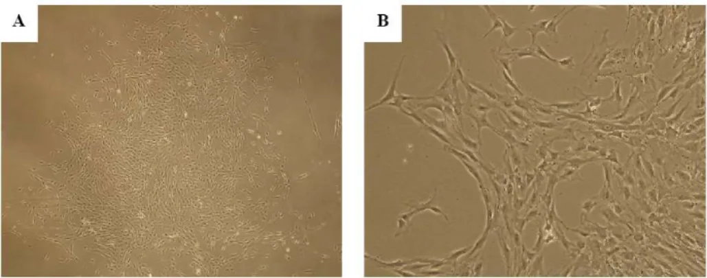
After plating the MNCs, only a few cells attached to the plastic culture dishes and formed adherent cells within 3 weeks. Upon the first passage of culture, highly proliferative fibroblast-like cells arose and became predominant over cells of other types. The adherent cells were composed almost of bipolar fibroblast-like cells that could later grow to confluence (Figure 1).
Figure 1. Photomicrographs of isolated CBMSCs. (A) CBMSCs after culturing for 2 weeks grew in colonies that contained small spindle-shaped fibroblastoid cells. Magnification 40×. (B) Magnification 100×. Abbreviation: CBMSC, cord blood-derived mesenchymal stem cell
2. Immunophenotypes of CBMSCs
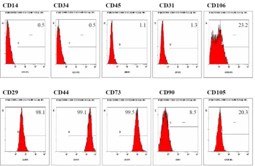
CBMSC and compared to that of 10 clones of bone marrow derived MSC (BMMSC). Neither CBMSCs nor BMMSCs expressed the hematopoietic markers such as CD14, CD34, and CD45 (Figure 2). Similarly to BMMSCs, CBMSCs were strongly positive for CD29, CD44, and CD73. However, 43.8 ± 21.0% and 21.9 ± 15.1% of CBMSC expressed CD105 and CD106, respectively.
Figure 2. Immunophenotypes of CBMSCs. Fibroblastoid cells from cord blood were trypsinized, labeled with antibodies against the indicated antigen, and analyzed by flow cytometry. A representative example is shown. Values represent the mean percentage of all assessed cells positively stained by the respective antibodies. Abbreviation: CBMSC, cord blood-derived mesenchymal stem cell
3. In vitro differentiation assays
CBMSCs from passage 2 or 3 were cultured under conditions that are favorable for an osteogenic, chondrogenic, or adipogenic differentiation, respectively. First, when induced to differentiate under osteogenic condition for 2 weeks, the spindle shape of CBMSCs flattened and broadened with increasing time of induction and resulted in accumulation of calcium deposit, as assessed by von Kossa stain (Figure 3B). Under chondrogenic conditions for 2 weeks, the accumulation of sulfated proteoglycans was visualized by safranin-O staining (Figure 3D). Adipogenic differentiation was demonstrated by the accumulation of intracellular neutral lipid vacuoles indicated by the Oil Red O stain. However, after adipogenic induction for 2 weeks, none of the CBMSCs display an adipogenic phenotype (Figure 3F). As a positive control, BMMSCs were differentiated into the cells with an adipogenic phenotype under standard adipogenic differentiation condition (data not shown).
4. In vitro hepatic differentiation potential
Because cellular confluence was the prerequisite for hepatic differentiation33, hepatogenic induction was carried out after CBMSCs reached at 100% confluence. CBMSCs were induced into hepatic lineage with the previously published two-step protocol (Protocol I)15 or newly modified activin A-based protocol (Protocol II).
Figure 3. Multi-lineage in vitro differentiation capacities of CBMSCs. CBMSCs were exposed in vitro to differentiation medium to induce osteogenic, chondrogenic, and adipogenic differentiation, respectively. (B) CBMSCs were shown to differentiate appropriately to the osteogenic lineage. Calcium mineralization detected by von Kossa stain. (D) Chondrogenic differentiation was confirmed by safranin-O staining. (F) CBMSCs did not differentiate toward adipogenic lineage, detected by Oil Red O stain. (A), (C), and (E) show non-induced controls. A representative example is shown. Abbreviation: CBMSC, cord blood-derived mesenchymal stem cell
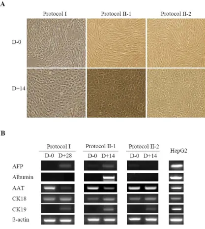
Using Protocol I, the fibroblastic morphology of CBMSCs changed into broadened, flattened morphology 1 week post-induction (step I). In the presence of OSM and dexamethasone (step II), the cuboidal morphology of hepatocyte-like cells and the appearance of abundant cytoplasmic granules
were more prominent with increasing duration of differentiation (Figure 4A). However, RT-PCR analysis failed to show the expression of AFP and albumin, although the duration of differentiation induction was prolonged from the original 4 weeks to 6 weeks (Figure 4B). In previous reports, chromatin remodeling agents, such as trichostatin A and DMSO, have been used to enhance hepatic differentiation of MSCs via epigenetic modification28, 34. However, although 1 ug/mL trichostatin A or 0.1% DMSO was added in the original 2-step protocol in this study, hepatocyte-like cells with hepatic phenotype were not detected (data not shown).
In Protocol II, FGF4 plus activin A (Protocol II-1) or FGF4 only (Protocol II-2) were used to induce hepatic differentiation. After 2 weeks of culture using both protocol, the fibroblastic morphology of CBMSCs was changed into a round or oval shape (Figure 4A). This morphological change was detected in regions of high cellular density initially, and propagated into the areas of low cell density. Undifferentiated CBMSCs did not express AFP, albumin, or CK19, but the expressed CK18, CK7, c-Met (a specific receptor of HGF), and AAT. After induction using Protocol II-1, CK18 and CK19 expression was increased, and CK7 expression was diminished. However, in the absence of supplementation with 20 ng/mL of Activin A (Protocol II-2), the more important hepatocyte-specific genes such as AFP and albumin were not expressed. HepG2 cells were used as positive controls (Figure 4B).
Figure 4. Hepatic differentiation of CBMSCs in vitro. Previously published original protocol (Protocol I), newly modified activin A-based protocol (Protocol II-1), and Protocol II-1 without activin A (Protocol II-2) were used to hepatic differentiation. (A) During differentiation, CBMSCs changed from a fibroblast-like spindle shape (undifferentiated) to a hepatocyte-like polygonal shape in all examined protocols. There is no microscopic difference in hepatocyte-like cell induced by different protocols. Magnification 100×. (B) Differentiated cells express hepatocyte-specific marker genes, by reverse transcriptase-polymerase chain reaction at the indicated time points. It is worthy
to note that AFP and albumin were expressed as early as 14 days in Protocol II-1. Abbreviations: CBMSC, cord blood-derived mesenchymal stem cell; AFP, alpha-fetoprotein
5. In vivo hepatic differentiation of CBMSCs in cirrhotic rat model
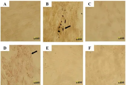
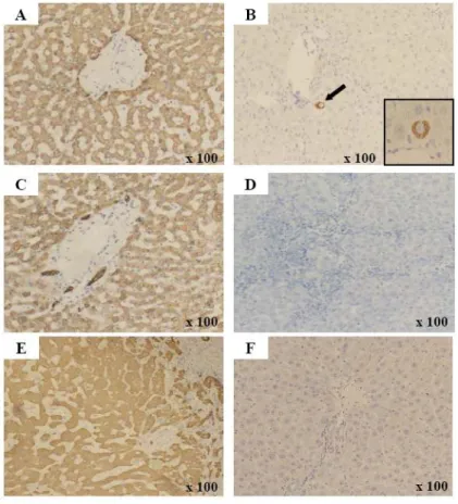
To investigate whether CBMSCs could differentiate into hepatic lineages in vivo, CBMSCs were infused in rats treated with multiple TAA for 6 weeks. The engraftment and hepatic differentiation of CBMSCs in the liver of recipient rats were analyzed at 1 and 4 weeks after infusion. We confirmed that anti-HepPar1, anti-CK18, anti-CK8, anti-human nuclei, and anti-human mitochondria antibodies are specific for human cells and do not cross-react with rat liver elements (data not shown)35. Human hepatocytes were rarely identified (positive for anti-human hepatocyte antibody), originated from CBMSCs, as single cells located near portal veins (Figure 5B). However, the HepPar1-positive cells were not stained for the human CK18 and CK8, an important marker for hepatocyte development, as shown in human liver sections used as positive controls (Figure 5D and 5F).
Figure 5. Immunohistochemical detection of human hepatocytes. Immunohistochemistry on human liver sections with HepPar1 (A), anti-CK18 (C), and anti-CK8 (E) that exclusively detect human, not mouse or rat, hepatocytes. Immunohistochemistry on rat liver sections of CBMSC infused group with anti-HepPar1 (B), anti-CK18 (D), and anti-CK8 (F). (B) Anti-HepPar1 positive cells integrated into the hepatic plate of cirrhotic rat liver. However, there is no cell positively stained with human CK18 (D) or anti-human CK8 (F). Cells were counterstained with hematoxylin. Abbreviations: CK, cytokeratin. Original magnification A-F 100×
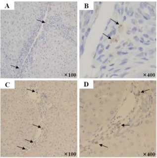
We detected infused CBMSCs by immunohistochemical stain for human nuclei and human mitochondria on liver sections from recipient rats. Cells with positive signals were observed in the region of fibrous septal, perivascular, and periportal area, with uncertain behavioral roles (Figure 6). Sequential immunohistochemical analysis revealed that anti-human nuclei or human mitochondria positive cells were not positively stained for anti-HepPar1, anti-CK18, and CK8 (data not shown).
Figure 6. Immunohistochemical detection of human cells. Infused CBMSCs were detected by immunohistochemical stain for anti-human nuclei (A, B) and anti-human mitochondria (C, D) in the region of fibrous septal, perivascular, and periportal area of CBMSCs infused group. Arrows indicate human positive cells. Cells were counterstained with hematoxylin. Abbreviation: CBMSC, cord blood-derived mesenchymal stem cell
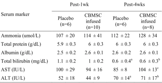
6. Changes of liver function after CBMSC infusion
Serum markers related to liver function, such as ammonia, total protein, albumin, total bilirubin, AST, and ALT were measured on the 1 and 4 weeks after CBMSC infusion. There were no statistically significant differences between the control group and MSC infused group in terms of ammonia, total protein, and albumin (Table 3).
Table 2. Comparison of serum markers related to liver function between placebo and CBMSC infused group.
Post-1wk Post-4wks Serum marker Placebo (n=6) CBMSC infused (n=10) Placebo (n=6) CBMSC infused (n=8) Ammonia (umol/L) 107 ± 20 114 ± 41 112 ± 22 128 ± 34 Total protein (g/dL) 5.9 ± 0.3 6 ± 0.3 6 ± 0.3 6 ± 0.3 Albumin (g/dL) 2.5 ± 0.2 2.6 ± 0.1 2.6 ± 0.2 2.6 ± 0.1 Total bilirubin (mg/dL) 1.1 ± 0.2 1 ± 0.2 0.6 ± 0.4a 0.6 ± 0.3b AST (IU/L) 100 ± 29 94 ± 16 85 ± 8 104 ± 13c ALT (IU/L) 52 ± 18 44 ± 9 70 ± 14a 71 ± 17b
Abbreviation: CBMSC, cord blood-derived mesenchymal stem cell
a
P < 0.05 vs. placebo group (post-1wk)
b
P < 0.05 vs. CBMSC infused group (post-1wk)
c
The concentration of total bilirubin after 4 weeks of CBMSC infusion was significantly lower than that of placebo group; on the contrary, the activity of ALT was significantly increased in CBMSC infusion group.
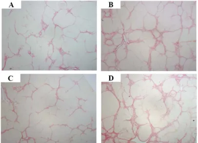
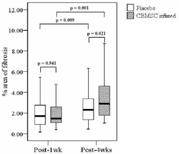
In order to quantify the amount of fibrosis over time comparing the placebo and CBMSC infused group, picro-sirius red stained areas, representing collagen deposition, were measured using IMAGEJ software. After 1 week of infusion, the CBMSC infused group had 2.25 ± 2.01% of collagen in the liver, compared to 2.05 ± 1.37% of control group. Theses results are not significantly different (P = 0.941). After 4 weeks of infusion, CBMSC infused group revealed a fibrosis area of 3.58 ± 2.24%, compared to 3.26 ± 2.76% in the control group. The liver fibrosis area after 4 weeks of CBMSCs transplantation was significantly more that that in the control group (P = 0.021, Table 3, Figure 8).
Figure 7. Representative figures showing fibrotic area in placebo and CBMSC infused group. Collagen fibers are stained in red by picro-sirius red staining. One week after placebo (A) or CBMSC (C) infusion, liver tissues showed collagen deposition. Four weeks after placebo (B) or CBMSC (D) infusion, collagen deposition was aggravated, and showed intense collagen deposition with septa interconnecting regenerative nodules.
Table 3. Quantitative measurement of picro-sirius red stained fibrotic area (% collagen/total area) Placebo group (%) CBMSC infused group (%) P value Post-1wk 2.05 ± 1.37 (n=73) 2.25 ± 2.01 (n=141) 0.941 Post-4wks 3.26 ± 2.76 (n=80) 3.58 ± 2.24 (n=81) 0.021
Figure 8. Comparison of fibrotic area between the placebo and CBMSC infused group. No differences were found between placebo and the CBMSC infusion group 1 week after infusion (P = 0.941). The fibrotic area of the CBMSC infusion group was significantly increased than that of placebo group 4 weeks after infusion (P = 0.021). Abbreviation: CBMSC, cord blood-derived mesenchymal stem cell
IV. DISCUSSION
Because of the lack of universally accepted criteria to characterize MSC, the Mesenchymal and Tissue Stem Cell Committee of the International Society for Cellular Therapy proposed minimal criteria to define human MSC. First, MSC must be plastic adherent when maintained in standard culture conditions using tissue culture flasks. Second, >/95% of the MSC population must express CD105, CD73, and CD90, as measured by flow cytometry. Additionally, these cells must lack expression of CD45, CD34, CD14 or CD11b, CD79a, or CD19 and HLA class II. Third, the cells must be able to differentiate to osteoblasts, adipocytes and chondroblasts under standard in vitro differentiating conditions36.
We successfully isolated CBMSCs, which were positive for CD73, CD44, CD29, and CD13. However, the percentage rate of CD105-expressing CBMSCs was significantly lower than that of bone marrow. These findings were generally accepted in CBMSCs, as published in previous studies37. Furthermore, we demonstrated the capacity for multi-lineage in vitro differentiation capacity toward osteogenic, chondrogenic lineages, except for adipogenic lineage. As similar to immunophenotypic characteristics, CBMSCs have a reduced capacity to differentiate into the adipogenic lineage. Actually, there are conflicting data concerning the adipogenic differentiation potential of CBMSCs37-40. The decreased adipogenic differentiation potential of CBMSCs
might be supported by the fact that adipocytes reside in adult bone marrow but are absent in fetal bone marrow and by the observation of increased adipogenesis correlated with age41.
The potential to differentiate into endodermal cells, such as hepatocytes has been investigated in vitro with human MSC from bone marrow15, 18, 39, cord blood21, 39, and adipose tissue28. Initially, Schwartz et al. induced multipotent adult progenitor cells into cells with morphological, phenotypic, and functional characteristics of hepatocytes in vitro upon exposure to well-defined hepatogenic factors18. But these culture conditions yielded a mixture of epithelioid and other cell types. Thereafter, attempts were made to improve the hepatic differentiation process through exposure to liver-specific factors in a sequential time-dependent manner, reflecting their secretion during in vivo hepatogenesis42 or epigenetic modification using histone deacetylase inhibitors, such as DMSO28, trichostatin A34, and etc. None of the previous studies produced functionally mature hepatocytes from MSCs in highly efficient ways. In this study, we tried to induce CBMSCs into hepatocyte-like cells using protocols published in previous articles, such as the two-step protocol of Lee et al.15, but could not successfully differentiate into functionally mature hepatocytes. Although the reasons underlying these controversial results are unknown, it may be partially might ascribed to subtle differences in CBMSC isolation and expansion techniques, and microenvironment (growth factors, cytokines, chemicals, medium additives, cell-cell / cell-matrix contact, etc).
Instead, we could differentiate CBMSCs into functional hepatocytes using activin A, which was reported to differentiate human embryonic stem cell to definite endoderm and finally functional hepatocyte43. At the beginning we treated CBMSCs with activin A together with FGF4, and used a number of factors essential for hepatogenic specification and hepatocyte morphology maintenance. Using this activin A-based protocol, the most important hepatocyte-specific AFP and albumin mRNA could be expressed as early as 14 days. It is important to differentiate into hepatocyte-like cells within a short period of time. Such a short induction time might reduce quantities of growth factors and be less expensive.
There are pros and cons regarding the in vivo hepatic differentiation and therapeutic efficacy of MSCs transplantation in acute or chronic liver injury. Sakaida et al. demonstrated significant fibrosis reduction and albumin increase by transplanting bone marrow MNCs in mice44. Similarly, Oyagi et al. showed that MSCs cultured with HGF are capable of improving albumin and reducing fibrosis in rats, and this effect was not observed in MSC without HGF45. On the other hand, in rat models involving bone marrow ablation, transplanted bone marrow cells were reported to engraft the liver during chronic liver diseases and to significantly contribute to liver fibrosis by differentiating into pro-fibrogenic myofibroblast-like cells, with hepatic transdifferentiation being a rare event46. Likewise, it is still controversial whether MSC transplantation can recover liver function and reduce tissue scarring in vivo.
In this study, ex vivo expanded MSCs were infused into thioacetamide treated rats to assess the in vivo hepatic differentiation potential of CBMSCs and therapeutic efficacy in chronic liver injury models. Although the experimental animal was immunosuppressed by cyclosporin A, effective repopulation by differentiated CBMSCs was not achieved. This extremely low repopulation efficiency might be a consequence of the xeno-transplantation, with the rat liver failing to provide the proper cell-cell or cell-matrix communications required for efficient integration of the human cells. In chronic liver injury, in vivo differentiation of intravenously transplanted CBMSCs into hepatocyte-like cells represents a relatively rare and quantitatively unsatisfactory rare event. Along with the low efficiency of in vivo hepatic differentiation, liver function measured by blood parameter was not significantly improved. It is widely recognized that the experimental model of liver fibrosis presents a major problem: the degree of fibrosis in each animal is highly heterogeneous. Whereas some animals show intense extracellular matrix components deposition, others are only mildly affected. So, appropriate selection of experimental animals would be important factors for future studies. We found that CBMSCs infusion did not improve or prevent liver fibrosis which is in agreement with the biochemical results.
V. CONCLUSION
We are able to isolate MSCs from umbilical cord blood, and these CBMSCs showed characteristics similar to BMMSC, except for weaker expression of CD105 and reduced capacity for adipogenic differentiation.
CBMSCs have the potential to differentiate into hepatic lineage in vitro. The newly modified activin A-based protocol is superior to the previous two-step protocol for rapid and efficient hepatic differentiation of CBMSCs. Activin A is essential to promote differentiation of CBMSCs towards functional hepatocyte-like cells.
Under our experimental conditions, infusions of CBMSCs into cirrhotic rat models did not improve biochemical markers related to liver function or liver histology.
REFERENCES
1. Lee SW, Wang X, Chowdhury NR, Roy-Chowdhury J. Hepatocyte transplantation: state of the art and strategies for overcoming existing hurdles. Ann Hepatol 2004;3:48-53.
2. Walldorf J, Aurich H, Cai H, Runge D, Christ B, Strom SC, et al. Expanding hepatocytes in vitro before cell transplantation: donor age-dependent proliferative capacity of cultured human hepatocytes. Scand J Gastroenterol 2004;39:584-93.
3. Petersen BE, Bowen WC, Patrene KD, Mars WM, Sullivan AK, Murase N, et al. Bone marrow as a potential source of hepatic oval cells. Science 1999;284:1168-70.
4. Theise ND, Nimmakayalu M, Gardner R, Illei PB, Morgan G, Teperman L, et al. Liver from bone marrow in humans. Hepatology 2000;32:11-6.
5. Orlic D, Kajstura J, Chimenti S, Jakoniuk I, Anderson SM, Li B, et al. Bone marrow cells regenerate infarcted myocardium. Nature 2001;410:701-5.
6. Eglitis MA, Mezey E. Hematopoietic cells differentiate into both microglia and macroglia in the brains of adult mice. Proc Natl Acad Sci of U S A 1997;94:4080-5.
7. Thorgeirsson SS, Grisham JW. Hematopoietic cells as hepatocyte stem cells: a critical review of the evidence. Hepatology 2006;43:2-8.
et al. Multilineage potential of adult human mesenchymal stem cells. Science 1999;284:143-7.
9. Bieback K, Kern S, Klüter H, Eichler H. Critical parameters for the isolation of mesenchymal stem cells from umbilical cord blood. Stem cells 2004;22:625-34.
10. De Coppi P, Bartsch G, Siddiqui MM, Xu T, Santos CC, Perin L, et al. Isolation of amniotic stem cell lines with potential for therapy. Nat Biotechnol 2007;25:100-6.
11. In 't Anker PS, Scherjon SA, Kleijburg-van der Keur C, de Groot-Swings GM, Claas FH, Fibbe WE, et al. Isolation of mesenchymal stem cells of fetal or maternal origin from human placenta. Stem cells 2004;22:1338-45.
12. Zuk PA, Zhu M, Mizuno H, Huang J, Futrell JW, Katz AJ, et al. Multilineage cells from human adipose tissue: implications for cell-based therapies. Tissue Eng 2001;7:211-28.
13. Di Nicola M, Carlo-Stella C, Magni M, Milanesi M, Longoni PD, Matteucci P, et al. Human bone marrow stromal cells suppress T-lymphocyte proliferation induced by cellular or nonspecific mitogenic stimuli. Blood 2002;99:3838-43.
14. Tse WT, Pendleton JD, Beyer WM, Egalka MC, Guinan EC. Suppression of allogeneic T-cell proliferation by human marrow stromal cells: implications in transplantation. Transplantation 2003;75:389-97.
vitro hepatic differentiation of human mesenchymal stem cells. Hepatology 2004;40:1275-84.
16. Wang X, Ge S, McNamara G, Hao QL, Crooks GM, Nolta JA. Albumin-expressing hepatocyte-like cells develop in the livers of immune-deficient mice that received transplants of highly purified human hematopoietic stem cells. Blood 2003;101:4201-8.
17. Hengstler JG, Brulport M, Schormann W, Bauer A, Hermes M, Nussler AK, et al. Generation of human hepatocytes by stem cell technology: definition of the hepatocyte. Expert Opin Drug Metab Toxicol 2005;1:61-74.
18. Schwartz RE, Reyes M, Koodie L, Jiang Y, Blackstad M, Lund T, et al. Multipotent adult progenitor cells from bone marrow differentiate into functional hepatocyte-like cells. J Clin Invest 2002;109:1291-302.
19. Kang XQ, Zang WJ, Bao LJ, Li DL, Song TS, Xu XL, et al. Fibroblast growth factor-4 and hepatocyte growth factor induce differentiation of human umbilical cord blood-derived mesenchymal stem cells into hepatocytes. World J Gastroenterol 2005;11:7461-5.
20. Shi XL, Qiu YD, Wu XY, Xie T, Zhu ZH, Chen LL, et al. In vitro differentiation of mouse bone marrow mononuclear cells into hepatocyte-like cells. Hepatol Res 2005;31:223-31.
21. Hong SH, Gang EJ, Jeong JA, Ahn C, Hwang SH, Yang IH, et al. In vitro differentiation of human umbilical cord blood-derived mesenchymal stem cells into hepatocyte-like cells. Biochem Biophys Res Commun 2005;330:1153-61.
22. Ong SY, Dai H, Leong KW. Hepatic differentiation potential of commercially available human mesenchymal stem cells. Tissue Eng 2006;12:3477-85.
23. Stainier DY. A glimpse into the molecular entrails of endoderm formation. Genes Dev 2002;16:893-907.
24. Kubo A, Shinozaki K, Shannon J, Kouskoff V, Kennedy M, Woo S, et al. Development of definitive endoderm from embryonic stem cells in culture. Development 2004;131:1651-62.
25. D'Amour KA, Agulnick AD, Eliazer S, Kelly OG, Kroon E, Baetge EE. Efficient differentiation of human embryonic stem cells to definitive endoderm. Nat Biotechnol 2005;23:1534-41.
26. Cai J, Zhao Y, Liu Y, Ye F, Song Z, Qin H, et al. Directed differentiation of human embryonic stem cells into functional hepatic cells. Hepatology 2007;45:1229-39.
27. Fang B, Shi M, Liao L, Yang S, Liu Y, Zhao RC. Systemic infusion of FLK1(+) mesenchymal stem cells ameliorate carbon tetrachloride-induced liver fibrosis in mice. Transplantation 2004;78:83-8.
28. Seo MJ, Suh SY, Bae YC, Jung JS. Differentiation of human adipose stromal cells into hepatic lineage in vitro and in vivo. Biochem Biophys Res Commun 2005;328:258-64.
et al. Human mesenchymal stem cells as a two-edged sword in hepatic regenerative medicine: engraftment and hepatocyte differentiation versus profibrogenic potential. Gut 2008;57:223-31.
30. Cheng SL, Yang JW, Rifas L, Zhang SF, Avioli LV. Differentiation of human bone marrow osteogenic stromal cells in vitro: induction of the osteoblast phenotype by dexamethasone. Endocrinology 1994;134:277-86.
31. Nakamura T, Torimura T, Sakamoto M, Hashimoto O, Taniguchi E, Inoue K, et al. Significance and therapeutic potential of endothelial progenitor cell transplantation in a cirrhotic liver rat model. Gastroenterology 2007;133:91-107.
32. Sakaida I, Terai S, Yamamoto N, Aoyama K, Ishikawa T, Nishina H, et al. Transplantation of bone marrow cells reduces CCl4-induced liver fibrosis in mice. Hepatology 2004;40:1304-11.
33. Aurich I, Mueller LP, Aurich H, Luetzkendorf J, Tisljar K, Dollinger MM, et al. Functional integration of hepatocytes derived from human mesenchymal stem cells into mouse livers. Gut 2007;56:405-15.
34. Snykers S, Vanhaecke T, De Becker A, Papeleu P, Vinken M, Van Riet I, et al. Chromatin remodeling agent trichostatin A: a key-factor in the hepatic differentiation of human mesenchymal stem cells derived of adult bone marrow. BMC Dev Biol 2007;7:24.
35. Newsome PN, Johannessen I, Boyle S, Dalakas E, McAulay KA, Samuel K, et al. Human cord blood-derived cells can differentiate into hepatocytes in the mouse liver with no evidence of cellular fusion. Gastroenterology
2003;124:1891-900.
36. Dominici M, Le Blanc K, Mueller I, Slaper-Cortenbach I, Marini F, Krause D, et al. Minimal criteria for defining multipotent mesenchymal stromal cells. The International Society for Cellular Therapy position statement. Cytotherapy 2006;8:315-7.
37. Kern S, Eichler H, Stoeve J, Klüter H, Bieback K. Comparative analysis of mesenchymal stem cells from bone marrow, umbilical cord blood, or adipose tissue. Stem cells 2006;24:1294-301.
38. Erices A, Conget P, Minguell JJ. Mesenchymal progenitor cells in human umbilical cord blood. Br J Haematol 2000;109:235-42.
39. Lee OK, Kuo TK, Chen WM, Lee KD, Hsieh SL, Chen TH. Isolation of multipotent mesenchymal stem cells from umbilical cord blood. Blood 2004;103:1669-75.
40. Chang YJ, Shih DT, Tseng CP, Hsieh TB, Lee DC, Hwang SM. Disparate mesenchyme-lineage tendencies in mesenchymal stem cells from human bone marrow and umbilical cord blood. Stem cells 2006;24:679-85.
41. Moerman EJ, Teng K, Lipschitz DA, Lecka-Czernik B. Aging activates adipogenic and suppresses osteogenic programs in mesenchymal marrow stroma/stem cells: the role of PPAR-gamma2 transcription factor and TGF-beta/BMP signaling pathways. Aging cell 2004;3:379-89.
42. Snykers S, Vanhaecke T, Papeleu P, Luttun A, Jiang Y, Vander Heyden Y, et al. Sequential exposure to cytokines reflecting embryogenesis: the key
for in vitro differentiation of adult bone marrow stem cells into functional hepatocyte-like cells. Toxicol Sci 2006;94:330-41.
43. Hay DC, Fletcher J, Payne C, Terrace JD, Gallagher RC, Snoeys J, et al. Highly efficient differentiation of hESCs to functional hepatic endoderm requires ActivinA and Wnt3a signaling. Proc Natl Acad Sci U S A 2008;105:12301-6.
44. Sakaida I, Terai S, Nishina H, Okita K. Development of cell therapy using autologous bone marrow cells for liver cirrhosis. Med Mol Morphol 2005;38:197-202.
45. Oyagi S, Hirose M, Kojima M, Okuyama M, Kawase M, Nakamura T, et al. Therapeutic effect of transplanting HGF-treated bone marrow mesenchymal cells into CCl4-injured rats. J Hepatol 2006;44:742-8.
46. Russo FP, Alison MR, Bigger BW, Amofah E, Florou A, Amin F, et al. The bone marrow functionally contributes to liver fibrosis. Gastroenterology 2006;130:1807-21.
<ABSTRACT (IN KOREAN)>
Activin A를 이용한
인간 제대혈 유래 중간엽줄기세포의 효율적 간세포 분화
<지도교수 김현옥>
연세대학교 대학원 의학과
김신영
최근 골수, 제대혈, 지방조직 및 태반 등의 다양한 조직에서 분리한 중간엽줄기세포로부터 간세포로의 생체외 분화가 시도되고 있다. 하지만 기존의 연구에 의하면 간세포로의 분화는 28일 이상의 장기간 분화유도가 소요되며 분화 효율이 매우 낮아 추가적인 동물 실험이나 임상으로의 적용이 어렵다. 또한 중간엽줄기세포의 생체내 간세포로의 분화 여부 및 만성 간손상 동물모델에서 중간엽줄기세포 이식의 치료효과는 연구마다 상이한 결과를 보이고 있다. 따라서 본 연구에서는 인간 제대혈 유래 중간엽줄기세포로부터 간세포로의 분화여부를 확인하고, 보다 효율적이며 신속한 분화 유도를 위한 분화조건을 탐색하고자 하였다. 또한, 인간 제대혈 유래 중간엽줄기세포를 간경변 유도 동물모델에 주입하여 생체 내에서의 간세포 분화능 및 간기능 개선 효과를 분석하고자 하였다. 제대혈로부터 중간엽줄기세포를 분리하여 골세포, 연골세포, 및 지방세포로의 생체외 분화능과, 유세포분석을 통한 세포 표면 항원 발현을 분석하였다. 기존의 2단계 분화방법과 새로이 개발한 activin A 기반의 분화방법을 이용하여 제대혈 유래 중간엽줄기세포로부터 간세포로의 생체외 분화를 유도하였다. Activin A 기반의 분화방법은 간의 발생시에 표현되는 시토카인을 반영하여, 1차로 activin A와
fibroblast growth factor (FGF)-4를 처리한 이후 간세포 특이
성장인자인 hepatocyte growth factor, FGF-4, oncostatin M 및
dexamethasone을 이용하여 간세포로의 분화를 유도하였다.
백서(Wistar rat)에서 thioacetamide를 6주간 투여하여 간경변을
유도하였으며, 제대혈 유래 중간엽줄기세포를 꼬리정맥으로 주입하여 간조직에서의 생착여부 및 간기능 개선효과를 면역조직화학염색, picro-sirius red 염색 및 생화학적 검사를 통하여 분석하였다. 제대혈 유래 중간엽줄기세포는 골수 유래 중간엽줄기세포와 유사한 특징을 보였지만, CD105 세포 표면 항원의 발현이 감소되어 있었으며, 지방세포로의 분화능도 감소되어 있었다. 기존의 2단계 분화방법을
이용하여 간세포와 유사한 입방형 세포가 형성되었으나, 4주 이상의 분화 유도에도 알파태아단백 및 알부민과 같은 간세포 특이 mRNA의 발현은 관찰되지 않았다. 하지만, activin A 기반의 분화방법을 이용할 경우 서로 다른 12개의 세포주 중 5개의 세포주에서 분화 유도 후 약 2주에 알파태아단백 및 알부민 등의 mRNA 발현이 관찰되었다. 간경변이 유도된 백서로 주입된 인간 제대혈 유래 중간엽줄기세포는 이식 4주 후 간의 혈관주위 및 섬유화 부위에서 관찰이 되었으나 성숙한 간세포의 특징을 표현하지 않았다. 대조군 및 실험군에서 간기능 관련 생화학적 검사 결과 및 간섬유화 정도의 차이는 관찰되지 않았다. 결론적으로 인간 제대혈 유래 중간엽줄기세포로부터 생체외 간세포 분화능은 확인할 수 있었으며, 특히 activin A 기반의 분화방법을 사용할 경우 보다 효율적이며 신속한 분화가 유도되었다. 반면, 본 실험에서 사용한 간경변 유도 동물모델에서의 인간 제대혈 유래 중간엽줄기세포의 생체내 간세포 분화능은 매우 감소되어 있음을 확인하였다. --- 핵심되는 말 : 제대혈, 중간엽줄기세포, 간세포, 분화