Sustained release of
Ascorbate-2-phosphate and Dexamethasone from
porous PLGA scaffolds for osteogenic
differentiation of mesenchymal stem cells
Hyongbum Kim
Department of Medicine
Sustained release of
Ascorbate-2-phosphate and Dexamethasone from
porous PLGA scaffolds for osteogenic
differentiation of mesenchymal stem cells
Directed by Professor Hwal Suh
The Master’s Thesis submitted to the Department of
Medicine, the Graduate School of Yonsei University in
partial fulfillment of the requirements for the degree of
Master of Medicine
Hyongbum Kim
This certifies that the Master’s Thesis of Hyongbum Kim
is approved.
_______________________________________
Thesis Supervisor : Hwal Suh
_______________________________________
Seong Hwan Moon
_______________________________________
Hyun Woo Kim
The Graduate School
Yonsei University
감사의 글 의과대학을 마치고 희망과 설렘으로 시작한 대학원 생활이 벌써 2년이 지났고 이제 그간의 결실로서 이 논문을 완성하게 되었습니다. 이는 저를 아끼고 사랑해 주신 많은 분들의 도움이 있었기에 가능한 일이라고 생각합니다. 무엇보다도 먼저 의과대학 시절과 대학원 과정 동안 한없이 부족한 저에게 항상 연구자로서 의사로서 그리고 교육자로서의 자세에 대해 지도해 주시고 좋은 환경에서 공부에 전념할 수 있도록 해 주신 서활 교수님께 고개 숙여 깊은 감사의 말씀을 올립니다. 바쁘신 중에도 저의 석사 논문에 대해 많은 지도 편달을 아끼지 않으신 정형외과학 교실의 문성환 교수님과 김현우 교수님께 깊이 감사 드립니다. 저의 예과 시절부터 줄곧 지도를 아끼지 않으신 김덕원 교수님께 감사 드립니다. 대학원 입학 때부터 지도와 조언을 주신 박종철 교수님께 감사의 말씀을 드리고 싶습니다. 학문의 길에 있어서 디딤돌이 되어 주신 의학공학교실의 김남현 교수님과 유선국 교수님께 감사 드립니다. 격려와 조언을 주시면서 저를 지켜봐 주신 의과대학 성형외과학 교실의 나동균 교수님, 피부과학 교실의 이민걸 교수님과 이광훈 교수님, 흉부외과학 교실의 박영환 교수님, 약리학 교실의 안영수 교수님, 김동구 교수님 그리고 이민구 교수님, 미생물학 교실의 최인홍 교수님, 내과학 교실의 송시영 교수님, 소아과학 교실의 박국인 교수님, 병리학 교실의 김호근 교수님, 해부학 교실의 이종은 교수님께 깊은 감사의 말씀을 올립니다. 또한 대학원 강의를 통해 많은 것을 가르쳐 주신 연세대 화공 생명 공학부의 김중현 교수님과 생물학과의 이준호 교수님께 감사의 말씀을 드립니다. 실험실 선배님들로서, 학문적으로 인도해 주신 동희형, 친누나처럼 잘 해 주신 종은이 누나와 고참이 되신 시내 누나, 선배로서 조언을 해 주신 구현철 선배님, 항상 열심히 하시는 봉주형, 진짜 사나이 동욱이 형, 실험이 무엇인지 말해 주신 유식이 형, 그리고 미국에 계신 학준이 형과 민정이에게 감사 드립니다. 그리고 하나뿐인 나의 동기 종훈이 형에게도 감사하다고 말하고 싶습니다. 같이 입학한 혜원이와 고분자 방의
성욱에게도 고마움을 전하고 싶습니다. 그리고 귀여운 후배들인 민섭, 유석, 재민, 태윤에게도 고맙다는 말을 하고 싶습니다. 시험 감독과 조교 생활을 같이 한 창용이형, 수찬이형, 기창이형, 재성, 선희, 준희에게 고마움을 전합니다. 옆 실험실에서 여러모로 도와 주신 김윤희, 김수향, 김은정, 김향, 경희, 언혜 선생님과 마지막으로 늦게 알았지만 과학자의 자세를 보여 주시고 마무리 실험을 도와 주신 김형태 선생님 고맙습니다. 그리고 상규 형, 김 설 선생님, 이미라 선생님, 백현숙 선생님, 김연희 선생님과 길미화 선생님께도 감사의 말씀을 올립니다. 의과대학 입학 및 졸업 동기로서 같이 기초의학을 시작하여 각자 서로 다른 분야에서 물심양면으로 도와 준 혜미와 현기에게 고맙다고 말하고 싶습니다. 그리고 기초 의학의 길을 먼저 시작하여 조언을 아끼지 않으신 약리학의 우인이 형과 진우 형, 해부학의 희준이 형, 기생충학의 심서보 선배님, 생화학 교실의 김하일 선배님과 상규 형, 그리고 미생물학 교실의 박상면, 최윤희 선배님 고맙습니다. 그리고 저보다 1년 늦게 기초를 시작한 약리학의 지하에게도 고맙다고 말하고 싶습니다. 또한 친구 경석이와 태인이 그리고 해중이에게도 고맙다는 말을 하고 싶습니다. 무엇보다도 항상 옆에서 힘이 되어준 나의 귀여운 여자친구 진영이에게 진심으로 감사드립니다. 마지막으로 저에게 힘이 되어 주시고 아낌없는 사랑과 격려를 보내주신 부모님께는 어떤 감사하다는 표현도 부족할 것 같습니다. 사랑하는 어머니, 아버지 고맙습니다. 그리고 떨어져 있으면서도 자상히 챙겨주신 누나와 씩씩한 우리 남동생 형용이에겐 각각 좋은 동생과 든든한 형이 되고자 노력하겠습니다. 저에게 힘이 되어 주신 분들의 믿음을 저 버리지 않고 저 또한 그 분들께 힘이 될 수 있도록 앞으로 성실히 노력하겠습니다. 2003 년 1 월 김형범 올림
TABLE OF CONTENTS
TABLE OF CONTENTS ...i
LIST OF FIGURES ... iii
LIST OF TABLES ...iv
LIST OF TABLES ...iv
ABSTRACT...1
I. INTRODUCTION ...3
II. MATERIALS AND METHODS...5
1. Measurement of AsAP and Dex concentration: HPLC ...5
2. AsAP particles in chloroform ...5
A. Minimizing the size of Ascorbate-2-phosphate particles in chloroform...5
B. Characterization of AsAP particles in chloroform ...6
(A) Size and morphology of the AsAP particles ...6
(B) Measurement of concentration of AsAP in chloroform ...6
3. Fabrication and chracterization of the scaffolds...6
A. Fabrication of AsAP and Dex incorporated scaffolds...6
B. Scanning electron microscopy (SEM) analysis of the scaffolds ...8
C. Measurement of the AsAP and Dex incorporated into the scaffolds ...8
4. Drug release study of AsAP and Dex from the scaffolds ...9
5. In vitro degradation study...9
A. Gel permeation chromatography...9
6. Isolation and culture of mesenchymal stem cells ...10
A. Isolation of MSCs from bone marrow ...10
(A) Isolation of human MSCs ...10
(B) Isolation of rabbit MSCs...11
(C). Confirmation of differentiation potential of rabbit MSCs ...11
B. Seeding of MSCs in the scaffolds ...11
A. Alkaline phosphatase activity assay...12
B. Calcium assay ...13
8. In vivo osteogenesis of MSCs in the AsAP and Dex incorporated scaffolds : Reverse Transcription–Polymerase Chain Reaction...13
III. RESULTS AND DISCUSSION...15
1. AsAP particles in chloroform ...15
2. Fabrication of Dex and AsAP incorporated scaffolds ...18
A. Incorporation efficiency...18
3. Degradation, water uptake and drug release study...22
A. Water uptake and degradation test ...22
B. Release study...25
4. Isolation of human and rabbit MSCs...30
5. Increased osteogenesis of MSCs in the Dex and AsAP releasing scaffolds in vitro ...34
6. Osteogenic differentiation of MSCs seeded in the Dex and AsAP releasing scaffolds in vivo ...36
IV. CONCLUSION ...37
REFERENCES ...38
LIST OF FIGURES
Figure 1. Size distribution of AsAP particles in chloroform... 16
Figure 2. Morphology of AsAP particles by SEM... 17
Figure 3. Scanning electron micrograph of cross section of PLGA foams. ... 19
Figure 4. Section of scaffold walls... 20
Figure 5. Incorporation efficiency of Dex and AsAP in porous PLGA foams... 21
Figure 6. Water uptake test... 23
Figure 7. Weight average molecular weight of the PLGA scaffolds... 24
Figure 8. Dex release from porous PLGA scaffolds ... 28
Figure 9. AsAP release from porous PLGA scaffolds... 29
Figure 10 Human MSCs ... 30
Figure 11. In vitro osteogenic differentiation... 31
Figure 12. In vitro chondrogenesis of mesenchymal stem cell... 32
Figure 13. In vitro differentiation of mesenchymal stem cell into adipocyte ... 33
Figure 14. Calcium deposition of MSCs cultured PLGA scaffolds in vitro... 35
Figure 15. RT-PCR of MSCs transplanted into the subcutaneous area of athymic mice for 2 weeks... 36
LIST OF TABLES
Table 1. Preparation conditions for porous PLGA foams ... 7 Table 2 Zero order release parameters of 2nd phase release and total released amount of Dex and AsAP until day 35. ... 27
ABSTRACT
Sustained release of Ascorbate-2-phosphate and Dexamethasone from
porous PLGA scaffolds for osteogenic differentiation of mesenchymal
stem cells
Hyongbum Kim
Department of Medicine
The Graduate School of Yonsei University
(Directed by Professor Hwal Suh)
Mesenchymal stem cells (MSCs) are promising options for mesenchyaml tissue engineering. However, as they are multipotent, to fabricate specific mesenchymal tissue, they should be induced to specific lineage. Ascorbate-2-phosphate and dexamethasone are major inducer of osteogenic differentiation of mesenchymal stem cells in vitro. In addition, porous poly(D,L-lactide-co-glycolide) (PLGA) matrices have been used as vehicles of tissue engineering. In this experiment, we fabricated biodegradable porous scaffolds which released ascorbate-2-phosphate (AsAP) and dexamethasone (Dex) upto 35 days. AsAP is lipid insoluble and was incorporated into the PLGA scaffolds as particles. As the size of AsAP decrease, the incorporation efficiency increased. Dex was incorporated as molecularly dispersed pattern. In vitro release study of Dex and AsAP from the scaffolds showed that after day 4 and 9 respectively, release rate was zero order at least untill day 35.
When MSCs were cultured in the scaffolds in vitro, the cultures were significantly more mineralized than those in control scaffolds. When MSCs were
delivered into the subcutaneous tissue of athymic mice via the polymeric scaffolds, RT-PCR showed that MSCs in the AsAP and Dex incorporated scaffolds expressed significantly higher osteocalcin than those in control scaffolds.
In conclusion, AsAP and Dex was sustained released from the biodegradable polymeric scaffolds and MSCs were induced into osteogenic lineage in the PLGA scaffolds both in vitro and in vivo.
Key words : mesenchymal stem cells, ascorbate-2-phosphate, dexamethasone, poly(D,L-lactide-co-glycolide), osteogenesis
Sustained release of Ascorbate-2-phosphate and Dexamethasone from
porous PLGA scaffolds for osteogenic differentiation of mesenchymal
stem cells
Hyongbum KimDepartment of Medicine
The Graduate School of Yonsei University
(Directed by Professor Hwal Suh)
I. INTRODUCTION
To generate bone, mesenchymal stem cells (MSCs) should undergo differentiation into osteogenic lineage. They can be successfully induced to
differentiate into osteoblasts by ascorbate-2-phosphate (AsAP), dexamethasone (Dex) and β-glycerophosphate in vitro 1. These three reagents comprise routine ‘osteogenic media’ 1-4. However, β-glycerophosphate is a in vitro source of phosphate ions that is necessary for mineralization rather than a inducer of osteogenic differentiation1, 5-8. MSCs cultured in AsAP and Dex supplemented media generated bone tissue in vitro 1, 3 and in vivo 9, 10 but those cultured in the absence of AsAP and Dex generated little bone tissue or generated other kinds of tissue including cartilage or fibrous tissue both
in vitro 1 and in vivo 9-11.
The synthetic absorbable polymers most often utilized for 3-dimensional porous scaffolds in tissue engineering are the poly(α-hydroxy acids)12-14. These are the homopolymers of poly(L-lactide) (PLLA), poly(glycolide) (PGA) as well as poly(D,L-lactide-co-glycolide) (PLGA) copolymers. They are among the few synthetic absorbable polymers with U.S. Food and Drug Administration approval
for human clinical use. These biodegradable aliphatic polyesters have versatile biodegradation properties depending on their molecular weight and chemical compositions.
On the other hand, PLGA have been also extensively used as biodegradable carriers for drug delivery 15-18. Because they are biocompatible 19-21 and bioabsorbable, there is no needed to retrieve the carrier after the drug is depleted. Successful sustained release of drugs has been achieved by PLGA and PLLA 22.
Tissue-engineering scaffolds using poly(α-hydroxy acids) have been developed to serve as vehicles for the delivery of bioactive factors such as proteins or DNA that can direct cellular responses within or around the scaffolds. Until now, three kinds of method have been developed to deliver drugs via porous scaffolds. These include adsorption of growth factors to the surface 23, 24, incorporation of drugs during the scaffold fabrication process and incorporation of microparticles which contain the drugs. The second form of methods includes gas forming/emulsion 25, 26, gas forming/particulate leaching 27, 28, solvent casting/particulate leaching/emulsion 25 and emulsion freeze-drying process 29. The third kind of methods includes incorporation of microparticles containing growth factors into scaffolds 30, 31 and fabrication of scaffolds from drug loaded microspheres. All the drugs used in these methods are only either proteins or DNA. DNA is hydrophilic polymer and proteins are amphophilic and labile. Other kinds of drugs have not been studied in this view. Many drugs are neither polymer nor proteins. Incorporation and release pattern of drugs from polymeric scaffolds are different according to the molecular size of drugs, hydrophilicity of drugs, properties of polymers which comprise the scaffolds, porosity of the scaffolds, and other factors. Ascorbate-2-phosphate is hydrophilic but not polymer and dexamethasone is hydrophobic.
In this experiment, ascorbate-2-phosphate and dexamethasone was incorporated into and released from PLG porous scaffolds. MSCs were seeded onto the scaffolds and osteogenic functions of the MSCs were compared with those of control scaffolds.
II. MATERIALS AND METHODS
1. Measurement of AsAP and Dex concentration: HPLC
In this experiment, all concentrations of AsAP and Dex were measured by high pressure liquid chromatography (HPLC). The sample solutions were filtered through 0.45um filter and degassed by sonication before being analyzed by HPLC system. The analysis of Dex was performed using a 3.9x150-mm reverse phase Novapack C-18 column (Waters corporation, Milford, MA) flowing at 1 mL/min at 246nm and the mobile phase consisted of 58:42 2-mM acetate buffer (pH 4.8) to acetonitrile32. The analysis of AsAP was performed using the same column flowing at 1ml/min at 257nm and the mobile phase consisted of 50mM KH2PO4(pH 2.2) containing 5%(v/v) acetonitrile and 0.0475% n-octylamine33.
Chemical stability of Dex and AsAP through scaffold fabrication was confirmed by comparing high pressure liquid chromatography spectra obtained from Dex and AsAP samples eluted from scaffolds with those derived from freshly prepared Dex and AsAP solution 34.
2. AsAP particles in chloroform
A. Minimizing the size of Ascorbate-2-phosphate particles in chloroform
We expected that AsAP would be incorporated into the septa or walls of polymeric scaffolds as particles and the incorporation efficiency will increase as the size of AsAP particles decrease. We confirmed this hypothesis by preliminary studies and tried to minimize the size of AsAP particles in chloroform. Three kinds of methods were tried.
Method A: Ascorbate-2-phosphate was dissolved in distilled water (DW) at the concentration of 1mg/ml. The solution was quick-frozen by quenching in liquid nitrogen and lyophilized. The remaining fine powder was suspended in chloroform.
The AsAP suspension was mildly sonicated for 1 hr.
Method B: AsAP was suspended in chloroform and sonicated for 2 hrs. Method C: AsAP was suspended in chloroform and stirred for 2hrs.
B. Characterization of AsAP particles in chloroform
(A) Size and morphology of the AsAP particles
10ul of properly diluted AsAP suspension in chloroform was dropped onto clean slide glasses. In a few seconds, chloroform was evaporated and AsAP powder was left on the slide glass. Their sizes and morphologies was observed by light microscopy through CCD camera and analyzed by MetaMorph image analyzer (Universal Imaging Corporation, Downingtown, PA, USA). Their morphologies were analyzed by scanning electron microscopy (SEM).
(B) Measurement of concentration of AsAP in chloroform
Aliquots of AsAP suspension in chloroform was placed into glass vials and they were left overnight under vacuum for the chloroform to evaporate. The remaining power was dissolved in DW and their concentration was measured using HPLC system.
3. Fabrication and chracterization of the scaffolds
A. Fabrication of AsAP and Dex incorporated scaffolds
Poly(D,L-lactide-co-glycolide)(PLGA) was from Purac (Purasorb, lot number:0010000072, Netherlands). Scaffolds were fabricated by an established solvent-casting, particulate-leaching technique with NaCl as the porogen 35, 36. Briefly, 2 g PLGA and various amount of Dex were dissolved in AsAP-suspended
chloroform. 20 g NaCl particles (Sigma) sieved to 250um-350um were added to the solution. The dispersion was then cast in a 10 cm glass Petri dish. The samples were air-dried for 48 h and subsequently vacuum-dried for 24 h to remove any remaining solvent. The resulting PLGA/Dex /AsAP/salt composite membranes were then immersed in distilled deionized water (ddH2O) for 10 h (water changed every 3 h) with mild stirring to leach out the salt, and freeze-dried. The produced porous membranes were cut into disks of diameter of 6 mm and height of 1.5mm and stored in a desiccator under vacuum at –20°C until use.
Table 1. Preparation conditions for porous PLGA foams
Foam codes Dex concentration (Dex/PLGA wt/wt ppm)
AsAP concentration (AsAP/PLGA wt/wt ppm) D0/A0 0 0 D20/A0 20 0 D80/A0 80 0 D320/A0 320 0 D1100/A0 1100 0 D0/A1100 0 1100 D0/A3300 0 3300 D80/A3300 80 3300 D320/A3300 320 3300
B. Scanning electron microscopy
(SEM
)analysis of the scaffolds
The polymer constructs were quick-frozen in liquid nitrogen and sectioned to reveal an intact pore network. The samples were sputter coated with gold using an ion coater (Hitachi E-100, Tokyo, Japan) at 6mA for 6 min, and then observed on a scanning electron microscope (Hitachi S-800, Tokyo, Japan) at an accelerating voltage of 20kV.
C. Measurement of the AsAP and Dex incorporated into the scaffolds
Five AsAP incorporated scaffolds were dissolved in 5ml chloroform and 30ml of benzene was added and mixed by vortex. To this solution, 5ml of DW was added and mixed by vigorous stirring to extract AsAP in the organic solvent. The solution was centrifuged at 1000g for 30 minutes to separate the water and organic solvent. During this centrifugation any remaining AsAP particles in the organic solvents were spun down to the bottom layer (water phase) and dissolved in the water. AsAP
concentration in the water was measured by HPLC.
Five Dex incorporated scaffolds were dissolved in 10ml of acetonitrile and the concentration of Dex was measured by HPLC.
Incorporation efficiency was calculated as the ratio of mass incorporated in the scaffolds after leach step to the sum of mass incorporated and lost during the leach 27. Incorporation efficiency of Dex and AsAP was calculated using D1100/A0 and
D0/A1100 respectively. To investigate the effect of particle size of AsAP on incorporation efficiency of AsAP, we fabricated D0/A1100 scaffolds using three kinds of AsAP particles from method A, B, C.
4. Drug release study of AsAP and Dex from the scaffolds
In vitro release study was performed by a modification of previously described 18, 25. The dry mass of five scaffolds was measured and scaffolds were placed into polypropylene microcentrifuges tube containing 1 ml of DPBS with 0.1% (w/v) sodium azide as a bacteriostatic agent. The tubes were incubated at a constant temperature of 37°C under agitation of 15 rpm, and the DPBS buffer was changed at preset intervals. Buffers removed from the tubes were analyzed by HPLC.
5. In vitro
degradation study
Degradation study was performed as previously described 37. Scaffolds pre-wetted by immersion in ethanol were placed in glass vials containing 15 mL of Dulbecco’s phosphate buffered saline (DPBS, pH7.4, Life Technologies, Grand island, NY, USA). The samples were incubated in a 37°C incubator under mild agitation of 15 rpm. The DPBS solution was changed every week. At the end of each sampling time point, pH change in the incubation medium was monitored. The retrieved scaffold samples were subjected immediately to measurement of wet weight in a hydrated state, after surface water was removed with a Kimwipes tissue, and then they were freeze-dried. Mass erosion and water uptake of each scaffold sample during the degradation period then were determined by using the wet and dry weights of the sample.
A. Gel permeation chromatography
The molecular weight of PLGA was measured by gel permeation chromatography (GPC) (Waters Co., USA) equipped with a series of µ Styragel® columns (HR1, HR4, HR5, HR5E Å pore sizes), Isocratic HPLC pump (Waters 1515), Autosampler (Waters 717), Refractive Index detector (Waters 2410) and
integrator at 40℃. Tetrahydrofuran (THF) was used as an eluent at 1.0ml/min of flow rate and 1.0×10³ Pa of the pump pressure.
6. Isolation and culture of mesenchymal stem cells
A. Isolation of MSCs from bone marrow
(A) Isolation of human MSCs
Human MSCs (hMSCs) were isolated from the marrow of the osteotomy specimens which were obtained as results of routine hip surgery in children (4-8 years old) after informed consent. All animal experiment procedures were managed in accordance with the Guidelines and Regulations for Use and Care of Animals in Yonsei University3. Briefly, about 0.5ml to 2 ml of marrow aspirate was collected into a heparinized syringe to prevent clotting. The marrow sample was washed with Dulbecco's phosphate-buffered saline (DPBS), cells were recovered after centrifugation at 900g, and the process was repeated once more. Cells were resuspended in 4ml of Tyrode’s salt solution and loaded onto 7 ml of Percoll of a density of 1.073 g/ml in a 15-ml conical tube. Cell separation was accomplished by centrifugation at 1100g for 30 min at 20°C. The nucleated cells were collected from the interface, diluted with three volumes of DPBS, and collected by centrifugation at 900g. The cells were resuspended, counted, and plated at 2 x 10⁶ nucleated cells/cm2. The human and rabbit MSCs were cultured in complete media which consisted of Dulbecco's modified Eagle's medium (DMEM) (low glucose) containing 10% fetal bovine serum (FBS, Invitrogen corporation, Grand island, N.Y., USA). Medium was replaced at 24 and 72 hours and every third or fourth day thereafter. hMSCs grew as symmetric colonies and were subcultured at 10 to 14 days by treatment with 0.05% trypsin and 0.53 mM EDTA for 5 min, rinsed from the substrate with serum-containing medium, collected by centrifugation at 200g for 5 min, and seeded into fresh flasks at 5*103 cells/cm2. With each treatment of
trypsin-EDTA and replating, the passage number was increased and represented approximately three population doublings.
(B) Isolation of rabbit MSCs
All animal experiment procedures were managed in accordance with the Guidelines and Regulations for Use and Care of Animals in Yonsei University. Rabbit MSCs were obtained from adult female white New Zealand rabbits aged between 8 months and 1 year, weighing 2.5kg to 3.3kg, using a modification of the method previously described 38-40. The rabbits were anesthetized with intramuscular administration of ketamine (50mg/kg) and xylazine (10mg/kg). Under general anesthesia, bone marrow was aspirated from the tibia with a 10ml syringe containing 0.1mL heparin (3000U/mL saline solution), with a 16-gauge needle. The marrow aspirates were suspended in DPBS, centrifuged, and resuspended in the complete medium. Rabbit MSCs reached confluency at 7 to 10 day and were subcultured following the method for hMSCs.
(C). Confirmation of differentiation potential of rabbit MSCs
The differentiation potential of rabbit MSCs was confirmed by differentiation induction in vitro. They were induced for osteogenic, chondrogenic and adipogenic differentiation as previously described10. For identification of osteogenic differentiation, MSCs cultures were stained with Alizarin Red S and von Kossa. For confirmation of chondrogenic differentiation, they were stained with Alcian blue. For identification of adipogenic differentiation, they were stained with Oil red O.
B. Seeding of MSCs in the scaffolds
Before mesenchymal stem cells seeding, scafffolds were prewet and sterilized by immersion in 100% ethanol for 1hr, 70% ethanol for 1 hr and washed with PBS 41. Then they were coated with serum protein by sinking in FBS supplemented by 10%
antibiotic for 2hrs and washed with sterile DW. The MSC cultures were trypsinized at 3rd passasge and cells were suspended at the concentration of 4x10⁷ cells/ml and
25ul of the cells suspension were poured onto the scaffolds. The size of the scaffolds was 6 mm diameter and 1.5mm height. The cells were allowed to adhere to the scaffolds for 3 h. Rabbit MSCs were used for in vitro study and human MSCs were used for in vivo study.
For in vitro study, the cell/polymer constructs were placed in 12-well plates and covered with 2 mL of media. Culture media for in vitro culture were complete media supplemented by 10mM β-glycerophosphate and were changed every fourth day.
For in vivo study, the cell/polymer constructs were transplanted into subcutaneous area of the dorsal surface of 8-week-old nude mice. The transplants were recovered after 2 wks and RT-PCR was performed.
7. In vitro osteogenesis of MSCs in the AsAP and Dex incorporated
scaffolds
A. Alkaline phosphatase activity assay
Alkaline phosphatase (ALP) acitivity was measured using a modification of the previously described method 2. Briefly, scaffold cultures were rinsed with Tyrode’s salt solution (Sigma, Saint Louis, Mo, USA) twice. The scaffolds were homogenized in 1 ml of alkaline buffer solution (Sigma Diagnostics, Inc.) for ALP assay using homogenizer (IKA Werke GMBH&Co., Staufen, Germany) and centrifuged at 5000 rpm for 5 min. 100ul of the supernatants were added to 96 well plate which contain 100ul of 5mM p-nitrophenyl phosphate and the optical density was measured at 405nm in dynamic mode and the slope was calculated and converted into enzyme activity based on the standard curve. Standard curve was obtained from the successive dilutions of p-nitrophenol standard solution (Sigma Diagnostics, Inc.).
B. Calcium assay
Deposited calcium was measured by a modification of the previously described 1. Scaffolds were rinsed with calcium free-DPBS and fixed with 1% (v/v) glutaraldehyde in Tyrode’s for 30 min. Following fixation, scaffolds were mildly rinsed twice with DW, calcium was extracted with 1ml of 0.6N HCl per scaffold in epppendorf tubes. They were placed on a orbital shaker (Boekel industries inc.) overweekend and centrifuged at 10000 rpm for 5 minutes. Aliquots of the supernatants were properly diluted and assayed using a commercial calcium assay kit (Sigma Kit #587) according to the manufacturer’s instruction. The optical density was read at 575 nm with a microplate reader. Calcium concentration was calculated with a standard curve generated from a series dilution of calcium standard solution (Sigma Diagnostics, Inc.).
8. In vivo osteogenesis of MSCs in the AsAP and Dex incorporated
scaffolds : Reverse Transcription–Polymerase Chain Reaction
Total RNA was prepared from 2-week-old MSC implants by using Rneasy Mini Kit(Qiagen). First-strand cDNA was synthesized from 1.5ug of total RNA in a 20-ul reaction mix using AMV reverse transcriptase XL (Takara bio incorporation, shiga, Japan) and an oligo-dT primer. PCR reactions were carried out in a mixture of 20 µl containing1.5 mM MgCl2; 0.2 mM each of dATP, dGTP,and dTTP; 5 µM dCTP; 1 µCi of [ -32P]dCTP (3000 Ci/mmol;DuPont New England Nuclear, Boston, MA); 4ul of cDNA;1x PCR buffer; 1.25 units of Taq DNA polymerase Gold (Perkin–Elmer) ; and 20 pmol of human specific primer sets: osteocalcin (sense,5’-CATGAGAGCCCTCACA-3’; antisense, 5’-AGAGCGACACCCTAGAC-3’) and glyceraldehyde-3-phosphatedehydrogenase (GAPDH: sense,
AGCCGCATCTTCTTTTGCGTC-3’; antisense 5’-TCATATTTGGCAGGTTTTTCT-39). After denaturation at 95°C for10 min, DNA amplification was performed for 29 cycles (GAPDH) or 37 cycles (osteocalcin) consisting of denaturation at 94°C for 30 s, primer annealingat 56°C (osteocalcin) or 57°C (GAPDH) for 30 s, and elongation at 72°Cfor 30 s. PCR products were separated in 5% polyacrylamide gels containing 5.6 M urea, followed by autoradiography.
III. RESULTS AND DISCUSSION
1. AsAP particles in chloroform
Method A resulted in significantly smaller AsAP particles compared to the other methods (Fig.1) and sonication of AsAP particles (method B) resulted in slight decrease of AsAP particles compared to those of method C. The average diameter of AsAP from method A, B, C was 6.0, 11.2, 12.8um respectively. Method D caused aggregation of AsAP rather than diminishing the size of AsAP and was not further studied. When AsAP solution was frozen slowly in –20°C instead of quick freezing in liquid nitrogen during the method A, the size of AsAP was similar to those of method B. Slow decrease of temperature of AsAP solution can cause aggregation of AsAP particle in the solution during the freezing process. However, in quick-freezing by quenching in liquid nitrogen, the time for the AsAP particles to aggregate is too short.
The morphology of AsAP of method A was also quite different from those of method B and C (Fig.2). AsAP particles from method A were like dispersed meshes with void area within their particles. However, those from methods B and C were more condensed than those of method A without void area within the particles. The difference in morphology of particles of method B and C is their surface character. Sonication caused disintergration of small AsAP particles from the large particles resulting cleaner surface than those from method C.
0 5 10 15 20 25 30 1 10 100 1000 Diameter(um) Distribution(%) A B C
Figure 1. Size distribution of AsAP particles in chloroform.
Sonication(method B) diminished the size of AsAP particles compared to stirring (method C). Quick freezing and lyophilization followed by mild sonication (method A) caused smallest particles among the three methods.
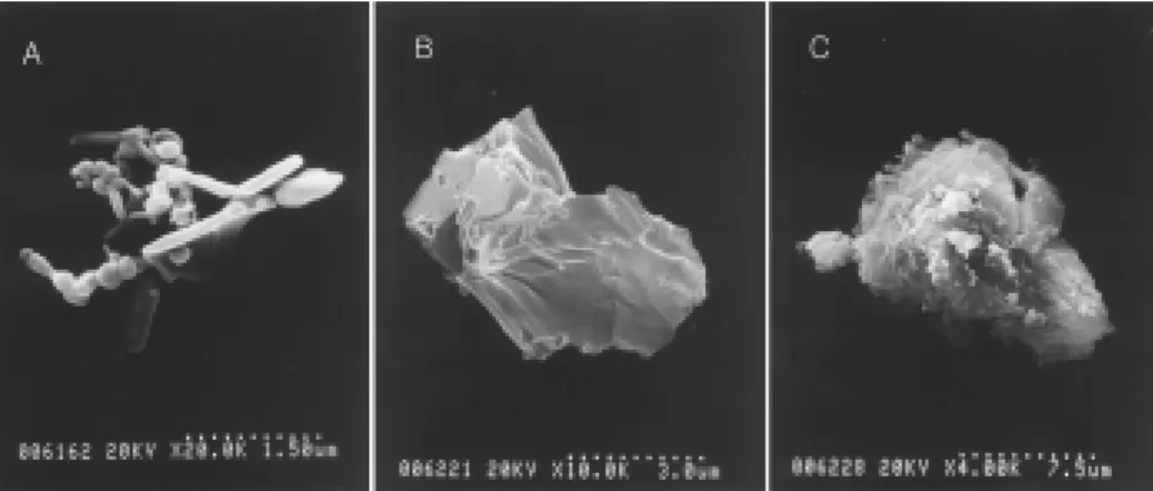
Figure 2. Morphology of AsAP particles by SEM.
A,B,C are the particle of method A, B,C respectively. Quick freezing and lyophilization followed by mild sonication (A:method A) resulted in dispered mesh shape of AsAP with void area in the particles. Particles from method B and C are more condensed morphology when compared to method A.
2. Fabrication of Dex and AsAP incorporated scaffolds
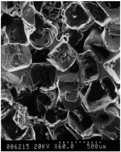
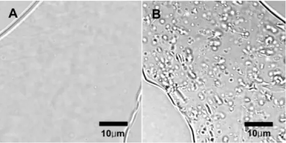
Dex and AsAP incorporated porous PLGA scaffolds were fabricated by the solvent casting/particulate leaching method (Fig.3). There was no difference in porosity and pore size between the Dex or AsAP incorporated scaffolds and the control through observation using SEM. Cross sectional view of the scaffold walls showed that AsAP incorporated scaffold walls had numerous small porous networks, which lacked in Dex incorporated scaffold walls (Fig. 4) and the control scaffold. This suggests that hydrophilic AsAP was incorporated into the hydrophobic PLGA scaffold walls as particles. There was no morphological difference between the Dex ncorporated and control scaffold through a light microscopic observation. This assumes to be related to the hydrophobicity of Dex that incorporates with the hydrophobic PLGA42.
A. Incorporation efficiency
Incorporation efficiency of AsAP increased as AsAP size decreased (Fig.5). To be incorporated into the scaffolds, in other words, not to be leached out during the particulate leaching step, the AsAP particulate should be totally entrapped by PLGA and should not exposed to the solvent of AsAP or water during the leach process. The entrapped AsAP is located in the septum or wall of porous scaffolds. The range of wall thickness was approximately 5-50um and the average was approximately 10um (Fig 3). To be incorporated into the wall, the diameter of particles should be smaller than the thickness of wall and small particles have more probability to be incorporated into the wall than large ones. PLGA polymer walls encapsulating smaller AsAP particles seemed to have a higher percentage of small pores distributed more evenly throughout the polymer walls. The incorporation efficiency of Dex was higher than those of AsAP because they are almost molecularly dispersed in the PLGA polymer, in other words, they are much more smaller than AsAP in the polymer. The higher hydrophobicity of Dex will be another reason for
higher incorporation efficiency than those of hydrophilic AsAP (Fig 5).
The chemical stability of AsAP and Dex through was confirmed by graph comparing freshly perpared HPLC graph. There was no difference in HPLC graph between the graphs of freshly prepared AsAP and Dex solution and those of incorporated AsAP and Dex respectively.
Figure 3. Scanning electron micrograph of cross section of PLGA foams.
The PLGA foam was fabricated by solvent casting/particulate leaching method and Dex and AsAP was incorporated into the foam during the fabrication process of the foam. The pore size was in the range of 250 to 350um and the pores were connected each other. Size bars and original magnification are shown on photomicrographs.
Figure 4. Section of scaffold walls
Porous scaffolds D320/A0(A) and D0/A3300(B) were frozen sectioned to 10um thickness and observed by a light microscopy at x1000 magnification to reveal the structure of scaffold walls. AsAP incorporated scaffolds(B) had small pores or porous networks in their walls, which lacked in Dex incorporated scaffolds(A).
0 10 20 30 40 50 60 70 80 90 100 Dex AsAP(Method A) AsAP(Method B) AsAP(Method C) Incorporation efficiency
Figure 5. Incorporation efficiency of Dex and AsAP in porous PLGA foams.
Incorporation efficiency of Dex was higher than those of AsAP. Particle size of AsAP decreased in order of method A, B and C. Larger AsAP particles (method C) are poorly incorporated into the PLGA foams.
3. Degradation, water uptake and drug release study
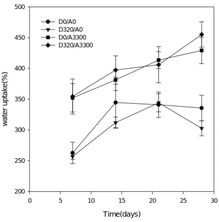
A. Water uptake and degradation test
Water uptake profiles, which measure the swelling extent of scaffolds, showed that AsAP incorporated scaffolds uptaked more water than control scaffolds(Figure 5). Incorporation of Dex into the scaffolds had no influence on the water uptake profile. Because AsAP is much more hydrophilic than PLGA and Dex, incoporation of AsAP into the PLGA increase hydrophilicity resulting in higher water uptake. In addition, water solvates the AsAP particles close to the surface and micropores are generated. These micropores became to be filled with surrounding water and the penetration of water into the PLGA polymer wall is increased through these pores. But, Dex is molecularly dispersed in the polymer and hydrophobic, Dex incorporated scaffolds does not have these micropores and consequently resulting in lower water uptake and penetration than those of which AsAP was incorporated.
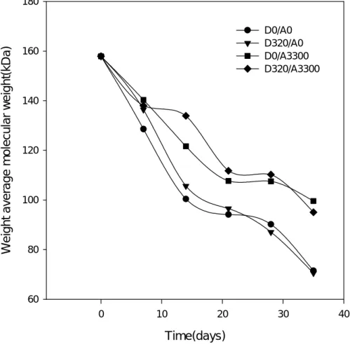
AsAP incorporated scaffolds degraded more slowly than Dex incorporated scaffolds or control scaffolds (Figure 6). Because hydrophilic AsAP was incorporated into the scaffolds as particles, as stated above, AsAP incorporated scaffolds have more micropores as AsAP is dissolved by surrounding or penetrating water than those of control or Dex incorporated scaffolds. These micropore slow down the degradation of AsAP. Similar results were previously reported 36, 43. The faster degradation of foams without these micropore was due to the greater extent of autocatalytic effect. The intermediate degradation products were trapped inside the wall structure before their molecular weights decreased to a critical value of about 1100 to be soluble in water. The accumulation of carboxylic groups led to faster degradation of PLGA that constititute the scaffold walls. In addition to these micropore effect, AsAP is basic and neutralize PLGA degradation products 44 and inhibits the autocatalysis.
Time(days) 0 5 10 15 20 25 30 w a ter up take( % ) 200 250 300 350 400 450 500 D0/A0 D320/A0 D0/A3300 D320/A3300
Figure 6. Water uptake test.
Porous PLGA scaffolds were placed in DPBS at 37°C and 15 rpm and the water uptake was calculated from wet weight and dry weight. AsAP incorporation increased water uptake of the PLGA scaffolds. Incorporation of Dex had no influence on the water uptake profile of the scaffolds. Results were shown by average±standard deviation (n=6).
Time(days) 0 10 20 30 40 We ig ht av era ge mol e cul a r w e ig ht (k D a ) 60 80 100 120 140 160 180 D0/A0 D320/A0 D0/A3300 D320/A3300
Figure 7. Weight average molecular weight of the PLGA scaffolds.
Porous PLGA scaffolds were place d in DPBS at 37°C and 15 rpm. Their Mw was measured by gel permeation chromatography.
B. Release study
The Dex of PLGA scaffolds had an initial burst release period for 4 days followed by sustained release at least until day 36(Figure 8). The second period release has almost zero order following the equation given below.
Ct=kt+b (1)
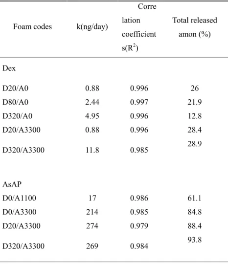
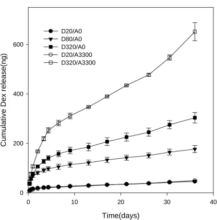
where Ct is the amount of drug that has been released by time t, k is a kinetic constant and b is an constant determined by the amount of drugs released during the first period. During the burst period, 6-13 percent of incorporated Dex was released. After day 4, the release pattern followed the above equation with high correlation coefficients (Table 2). Initial burst release followed by zero order release is ideal release pattern for many biomedical application. This release pattern increase the concentration of drug quickly by burst release for the optimal concentration and maintain the optimal, constant concentration by zero order release. This release pattern was seen all the Dex concentration in the PLGA used for this experiment suggesting that we can effectively control the concentration of Dex within or near the PLGA scaffolds by adjusting the concentration of Dex in the PLGA polymer. Until day 35, only 10 to 28 percent of the incorporated amount was released (Table 2).
The releases of AsAP from the PLGA scaffold also have initial burst release followed by zero order release(Table 2). However, the burst effect of AsAP release was severer than those of Dex. The duration of burst release was 9 days and during the burst release 52-72 percent of total incorporated drugs has been released.
By comparing the release of Dex from D320/A0 and D20/A0 to those from D320/A33000 and D320/A0 respectively, it can be inferred that release of Dex was faster when AsAP particles were co-incorporated (Figure 8 and Table 2). As stated above, water uptake and penetration were increased by incorporating AsAP into the scaffolds. This increased water penetration into the polymer wall resulted in increased release of Dex from the scaffolds. This effect of AsAP incorporation to
the release of incorporated drugs was further potentiated as the dose of AsAp increase. As shown by Table 2 and figure 9, release of incorporated drug, AsAP from D0/A3300 was faster than those from D0/A1100. As mentioned above, incorporation of AsAP into the PLGA scaffold walls results in micropores. Incorporation of higher amount of AsAP into the scaffolds results in a larger network of interconnecting channels and, therefore, more access to the surrounding medium than the lower loaded scaffolds 45. In contrast, incorporation of higher dose of Dex resulted in slower release pattern and decrease water uptake as shown by the release pattern of D20/A0, D80/A0 and D320/A0. Dex is hydrophobic and this molecule fills the space in the PLGA molecule, especially the amorphous area, through which water diffuse and penetrate in the polymer, resulting in decreased water uptake, water diffusion and consequently slower release of the incorporated drugs. When AsAP is coincorporated, this effect of Dex was not observed in water uptake and drug release profile.
Table 2 Zero order release parameters of 2nd phase release and total released amount of Dex and AsAP until day 35.
2nd phase zero order release of Dex and AsAP was initiated from day 4 and 9, respectively.
Foam codes k(ng/day)
Corre lation coefficient s(R2) Total released amon (%) Dex D20/A0 0.88 0.996 26 D80/A0 2.44 0.997 21.9 D320/A0 4.95 0.996 12.8 D20/A3300 0.88 0.996 28.4 D320/A3300 11.8 0.985 28.9 AsAP D0/A1100 17 0.986 61.1 D0/A3300 214 0.985 84.8 D20/A3300 274 0.979 88.4 D320/A3300 269 0.984 93.8
Time(days) 0 10 20 30 40 Cum u lativ e Dex re lease( ng) 0 200 400 600 D20/A0 D80/A0 D320/A0 D20/A3300 D320/A3300
Time(days) 0 10 20 30 40 Cumu lat ive AsAP r e leas e( ug) 0 5 10 15 20 25 30 35 D0/A1100 D0/A3300 D20/A3300 D320/A3300
4. Isolation of human and rabbit MSCs
Human MSCs were isolated (Figure 9). They were spindle shaped and reached confluency on the culture day of 9 to 14 days. Isolated rabbit MSCs reached confluency on the culture day of 7 to 10 days. They underwent osteogenic, chondrogenic and adipogenic differentiation depending the culture condition (Figure 10,11,12).
Figure 10 Human MSCs
Figure 11. In vitro osteogenic differentiation
Rabbit MSCs were cultured in control media(CM) or osteogenic supplemented media(OM) and stained with Alizarin Red S or Von Kossa.
1. Alrizarin Red S stain (6 well plate), 2. Von Kossa stain (6 well plate), 3. Alrizarin Red S stain of MSCs cultured in OM (x100), 4. Negative control (Alrizarin Red S stained MSCs cultured in CM,x100)
1
2
3
4
Figure 12. In vitro chondrogenesis of mesenchymal stem cell
Pellet culture shown by micrograph (A: Alcian blue stain, x100) and gross morphology(B)
A
Figure 13. In vitro differentiation of mesenchymal stem cell into adipocyte
Adiogenic differentiation was induced by culture in adipogenic media. The cultures were stained with Oil Red O. Magnification was x100.
A
5. Increased osteogenesis of MSCs in the Dex and AsAP releasing
scaffolds in vitro
MSCs seeded onto the scaffolds resulted in significantly increased calcium deposition than those of control scaffolds (Figure 13). Mineralization is critical indicator of osteogenic differentiation of MSCs 1-3. This strongly suggests that MSCs underwent osteogenic differentiation by the effect of Dex ans AsAP released from the scaffolds. This effect is almost equivalent to those of MSCs cultures in osteogenic media.
There was no difference in alkaline phosphatase activity between the MSC cultures on control scaffolds and those on AsAP and Dex incorporated scaffolds.
Jaiswal, N et al. suggested that the effective concentration of Dex for osteogenic differentiation of MSCs was in the range of 10nM(40ng/ml) to 100nM(400ng/ml) and this of AsAP was in the range of 50uM(16 μg/ml) to 500uM(160 μg/ml) 1. However, in case of AsAP, they did not study the effect of AsAP in lower concentration than 50uM and Park, SR et al. showed that osteogenesis of human marrow adipocytes was induced in the concentration of 10nM(3.2 ng/ml) of AsAP 46. Dex and AsAP showed toxic effect at the concentration of 1000nM(4000ng/ml) and 1000uM(1600μg/ml) respectively 1. We used 320/3330 scaffolds for in vitro study. The MSCs were cultured in 2ml media and the media was changed every third day. The estimated concentrations of Dex based on the drug release studies were 77, 32, 15 and 14 ng/ml at day 3, day 6, day9 and day 12, respectively and those of AsAP were 5, 1.8, 0.8 and 0.6μg/ml. This estimation is based on a hypothesis that Dex and AsAP are evenly distributed to the culture media by diffusion. However, the concentration around the cultured MSCs will be higher than the above estimation because the Dex and AsAP are released from the scaffolds, directly on which MSCs are attached. Accordingly, MSCs were considered to be exposed to the effective concentration of Dex at least during the
first 6 days. The first 1 week is especially important in the determination of differentiation fate of MSCs 1. In addition, considering Park’s experiment 46, the concentration of AsAP were also expected to be in the effective range.
0 20 40 60 80 100 120 D0/A0 D320/A3300 calcium(ug /s caf fo ld) p=0.0062
Figure 14. Calcium deposition of MSCs cultured PLGA scaffolds in vitro.
MSCs were cultured on two kinds of scaffolds. Calcium deposition was significantly higher in MSCs cultured on Dex and AsAP incorporated scaffolds(D320/A3300) than control scaffolds(D0/A0). (n=4)
6. Osteogenic differentiation of MSCs seeded in the Dex and AsAP
releasing scaffolds in vivo
Osteocalcin mRNA was expressed in MSCs seeded in the Dex and AsAP releasing scaffolds. However, osteocalcin mRNA was not expressed those in control scaffolds (Figure 14). Osteocalcin is a marker of osteogenic differentiation of mesenchymal stem cells 10. This suggest that the differentiation of MSCs were induced relatively specifically into osteogenic lineage by the release of Dex and AsAP in vivo.
Figure 15. RT-PCR of MSCs transplanted into the subcutaneous area of athymic mice for 2
weeks.
MSCs were transplanted by after seeding onto control scaffolds(C) or AsAP and Dex incorporated scaffolds(S). After 2 weeks, the transplanted were analyzed by RT-PCR.
IV. CONCLUSION
We successfully incorporated ascorbate-2-phosphate and dexamethasone during the porous PLGA scaffold fabrication by solvent casting/particulate leaching method. The size of AsAP particles in chloroform was significantly diminished by quick freezing of the AsAP solution followed by lyophilization and sonication. Incorporation of AsAP into the scaffolds increased water uptake and water penetration resulting faster drug release from the scaffolds. The release of Dex and AsAP was composed of two periods. The first period of Dex and AsAP release was burst release period of which duration was 4 days and 9 days. The second release period was zero order release. The second release period was at least until the duration of release study, 35 days.
When MSCs were cultured in these scaffolds, MSCs underwent osteogenic differentiation in vitro and in vivo. The osteogenic differentiation of MSCs in vitro were documented by increased calcium deposition after culture for 2 weeks. The osteogenic differentiation of MSCs in vivo were observed by increased expression of osteocalcin by RT-PCR.
REFERENCES
1.Jaiswal N, Haynesworth SE, Caplan AI, Bruder SP. Osteogenic
differentiation of purified, culture-expanded human mesenchymal stem cells in vitro. J Cell Biochem 1997;64295-312.
2.Sikavitsas VI, Bancroft GN, Mikos AG. Formation of three-dimensional cell/polymer constructs for bone tissue engineering in a spinner flask and a rotating wall vessel bioreactor. J Biomed Mater Res
2002;62136-148.
3.Pittenger MF, Mackay AM, Beck SC, et al. Multilineage potential of adult human mesenchymal stem cells. Science 1999;284143-147.
4.Lennon DP, Haynesworth SE, Arm DM, Baber MA, Caplan AI. Dilution of human mesenchymal stem cells with dermal fibroblasts and the effects on in vitro and in vivo osteochondrogenesis. Dev Dyn 2000;21950-62.
5.Maniatopolous C, Sodek J, Melcher AH. Bone formation in vitro by stromal cells obtained from bone marrow of young adult rats. Cell Tissue Res 1988;254317-330.
6.Bellows CG, Aubin JE, Heersche JNM, Antosz NM. Mineralized bone nodules formed in vitro from enzymatically released rat calvaria cell populations. Calcif Tissue Int 1986;38143-154.
7.Ecarot-Charrier B, Glorieux FH, van der Rest M, Pereira G. Osteoblasts isolated from mouse calvaria inititate matrix mineralization in culture. J Cell Biol 1983;96639-643.
8.Tenenbaum HC. Role of organic phosphate in mineralization of bone in vitro. J Dent Res 1981;60 Spec No C1586-1589.
9.Gundle R, Joyner C, Triffitt J. Human bone tissue formation in diffusion chamber culture in vivo by bone-derived cells and marrow stromal fibroblastic cells. Bone 1995;16597-601.
10.Yoshikawa T, Ohgushi H, Akahane M, Tamai S, Ichijima K. Analysis of gene expression in osteogenic cultured marrow/hydroxyapatite construct implanted at ectopic sites: a comparison with the osteogenic ability of cancellous bone. J Biomed Mater Res 1998;41568-573.
11.Haynesworth S, Goshima J, Goldberg V, Caplan A. Characterization of cells with osteogenic potential from human marrow. Bone
1992;1381-88.
12.Langer R, Vacanti J. Tissue engineering. Science 1993;260920-926. 13.Agrawal CM, Ray RB. Biodegradable polymeric scaffolds for
musculoskeletal tissue engineering. J Biomed Mater Res 2001;55141-150.
14.Freed LE, Marquis JC, Nohria A, Emmanual J, Mikos AG, Langer R. Neocartilage formation in vitro and in vivo using cells cultured on synthetic biodegradable polymers. J Biomed Mater Res 1993;2711-23.
15.Yamaguchi Y, Takenaga M, Kitagawa A, Ogawa Y, Mizushima Y, Igarashi R. Insulin-loaded biodegradable PLGA microcapsules: initial burst release controlled by hydrophilic additives. J Control Release 2002;81235-249.
16.Wang FJ, Wang CH. Sustained release of etanidazole from spray dried microspheres prepared by non-halogenated solvents. J Control Release 2002;81263-280.
17.Lam XM, Duenas ET, Cleland JL. Encapsulation and stabilization of nerve growth factor into poly(lactic-co-glycolic) acid microspheres. J Pharm Sci 2001;901356-1365.
18.Hickey T, Kreutzer D, Burgess DJ, Moussy F. Dexamethasone/PLGA microspheres for continuous delivery of an anti-inflammatory drug
for implantable medical devices. Biomaterials 2002;231649-1656. 19.Gogolewski S, Jovanovic M, Perren SM, Dillon JG, Hughes MK. Tissue
response and in vivo degradation of selected polyhydroxyacids: polylactides (PLA), hydroxybutyrate) (PHB), and poly(3-hydroxybutyrate-co-3-hydroxyvalerate) (PHB/VA). J Biomed Mater Res 1993;271135-1148.
20.Gupta RK, Alroy J, Alonso MJ, Langer R, Siber GR. Chronic local tissue reactions, long-term immunogenicity and immunologic priming of mice and guinea pigs to tetanus toxoid encapsulated in biodegradable polymer microspheres composed of poly lactide-co-glycolide
polymers. Vaccine 1997;151716-1723.
21.Conti B, Pavanetto F, Genta I. Use of polylactic acid for the preparation of microparticulate drug delivery systems. J Microencapsul 1992;9153-166.
22.Okada H, Toguchi H. Biodegradable microspheres in drug delivery. Crit Rev Ther Drug Carrier 1995;121-99.
23.Winn SR, Schmitt JM, Buck D, Hu Y, Grainger D, Hollinger JO. Tissue-engineered bone biomimetic to regenerate calvarial critical-sized defects in athymic rats. J Biomed Mater Res 1999Tissue-engineered bone biomimetic to regenerate calvarial critical-sized defects in athymic rats.
24.Ziegler J, Mayr-Wohlfart U, Kessler S, Breitig D, Gunther KP. Adsorption and release properties of growth factors from biodegradable implants. J Biomed Mater Res 2002;59422-428.
25.Hile DD, Amirpour ML, Akgerman A, Pishko MV. Active growth factor delivery from poly(D,L-lactide-co-glycolide) foams prepared in supercritical CO2. J Control Release 2000;66177-185.
vascular endothelial growth factor from mineralized poly(lactide-co-glycolide) scaffolds for tissue engineering. Biomaterials
2000;212521-2527.
27.Shea LD, Smiley E, Bonadio J, Mooney DJ. DNA delivery from polymer matrices for tissue engineering. Nat Biotechnol 1999;17551-554. 28.Sheridan MH, Shea LD, Peters MC, Mooney DJ. Bioabsorbable polymer
scaffolds for tissue engineering capable of sustained growth factor delivery. J Control Release 2000;6491-102.
29.Whang K, Goldstick TK, Healy KE. A biodegradable polymer scaffold for delivery of osteotropic factors. Biomaterials 2000;212545-2551. 30.Hu Y, Hollinger JO, Marra KG. Controlled release from coated polymer
microparticles embedded in tissue-engineered scaffolds. J Drug Target 2001;9431-438.
31.Meese TM, Hu Y, Nowak RW, Marra KG. Surface studies of coated polymer microspheres and protein release from tissue-engineered scaffolds. J Biomater Sci Polym Ed 2002;13141-151.
32.Lamiable D, Vistelle R, Millart H, et al. High-performance liquid
chromatographic determination of dexamethasone in human plasma. J Chromatogr 1986;378486-491.
33.Sakai T, Murata H, Ito T. High-performance liquid chromatographic analysis of ascorbyl-2-phosphate in fish tissues. J Chromatogr B Biomed Appl 1996;685196-198.
34.Lincoff AM, Furst JG, Ellis SG, Tuch RJ, Topol EJ. Sustained local
delivery of a dexamethasone by a novel intravascular eluting stent to prevent restenosis in porcine coronary injury model. J Am Coll Cardiol 1997;29808-816.
35.Mikos AG, Thorsen AJ, Czerwonka LA, et al. Preparation and
1994;351068-1077.
36.Lu L, Peter SJ, Lyman MD, et al. In vitro degradation of porous poly(L-lactic acid) foams. Biomaterials 2000;211595-1605.
37.Yoon JJ, Park TG. Degradation behaviors of biodegradable macroporous scaffolds prepared by gas foaming of effervescent salts. J Biomed Mater Res 2001;55401-408.
38.Im GI, Kim DY, Shin JH, Hyun CW, Cho WH. Repair of cartilage defect in the rabbit with cultured mesenchymal stem cells from bone marrow. J Bone Joint Surg Br 2001;83289-294.
39.Solchaga LA, Johnstone B, Yoo JU, Goldberg VM, Caplan AI. High variability in rabbit bone marrow-derived mesenchymal cell preparations. Cell Transplant 1999;8511-519.
40.Tsutsumi S, Shimazu A, Miyazaki K, et al. Retention of multilineage differentiation potential of mesenchymal cells during proliferation in response to FGF. Biochem Biophys Res Commun 2001;288413-419. 41.Mikos AG, Lyman MD, Freed LE, Langer R. Wetting of poly(L-lactic acid)
and poly(DL-lactic-co-glycolic acid) foams for tissue culture. Biomaterials 1994;1555-58.
42.Mu L, Feng S. Fabrication, characterization and in vitro release of
paclitaxel (Taxol) loaded poly (lactic-co-glycolic acid) microspheres prepared by spray drying technique with lipid/cholesterol emulsifiers. J Control Release 2001;76239-254.
43.Lu L, Peter SJ, Lyman MD, et al. In vitro and in vivo degradation of porous poly(DL-lactic-co-glycolic acid) foams. Biomaterials 2000;211837-1845.
44.Zhu G, Mallery SR, Schwendeman SP. Stabilization of proteins
encapsulated in injectable poly(lactide-co-glycolide). Nat Biotechnol 2000;1852-57.
45.Sandor M, Enscore D, Weston P, Mathiowitz E. Effect of protein molecular weight on release from micron-sized PLGA microspheres. J Control Release 2001;76297-311.
46.Park S, Oreffo R, Triffitt J. Interconversion potential of cloned human marrow adipocytes in vitro. Bone 1999;24549-554.
국문요약
Ascorbate-2-phosphate와 Dexamethasone를 서서히 방출하는
생분해성 다공성 지지체를 통한 중간엽줄기세포의
골화세포로의 분화
<지도교수 서활> 연세대학교 대학원 의학과 김형범 중간엽줄기세포는 근골격계 조직공학의 좋은 수단이다. 하지만 이 세포들은 여러가지 세포로 분화할 수 있는 능력을 가지고 있어 특정한 조직을 만들기 위해서는 특정한 세포로 이 세포들의 분화가 유도되어야 한다. ascorbate-2-phosphate(AsAP)와 Dexamethasone(Dex)는 생체 외에서 중간엽줄기세포가 골화세포로 분화되게 하는 역할을 한다. 한편, 다공성 poly(D,L-lactide-co-glycolide) (PLGA) 지지체는 조직공학의 지지체로 많이 사용되어 왔다. 이번 실험에서는 AsAP와 Dex를 서서히 방출하는 다공성 지지체를 제조하였다. AsAP는 유기용매에 불용성이라 PLGA지지체에 입자형태로 함유되었다. AsAP의 입자 크기가 작아질수록 함유율은 증가하였다. Dex는 분자수준에서 지지체 전반에 골고루 분포하였다. 생체외 방출실험에서 AsAP와 Dex는 각각 4일과 9일 이후에는 적어도 35일까지 0차 함수적으로 방출되었다. 중간엽줄기세포가 AsAP와 Dex가 들어간 지지체에서 배양된 경우 배양 14일째, 대조군 지지체에서 배양된 경우보다 많은 칼슘이 침착되었다. 또한 중간엽줄기세포를 이러한 지지체를 통해 면역결핍마우스의 피하에 이식한 다음2 주 이후 역전사효소를 이용한 중합효소연쇄반응(RT-PCR)을 실시한 결과 AsAP와 Dex가 들어가 있는 지지체를 통해 이식된 중간엽줄기세포에서 그렇지 않은 세포보다 보다 많은 양의 osteocalcin을 발현하였다. 결론적으로, AsAP와 Dex는 성공적으로 생분해성 다공성 고분자 지지체에 함유되었고 중간엽줄기세포는 이 다공성 고분자 지지체에서 생체내에서나 생체외에서 골화세포로 분화되었다. 핵심되는 말 : 중간엽줄기세포, ascorbate-2-phosphate, dexamethasone, 골, 생분해성 고분자 지지체