서 론
참마자, Hemibarbus longirostris는 잉어목(Cypriniformes), 잉어과(Cyprinidae), 모래무지아과(Gobioninae)에 속하며, 유 라시아에는 11속이 보고되었고(Nelson et al., 2016), 이 가운 데 누치속(Hemibarbus) 어류는 전 세계에 20종이 있는 것으 로 알려져 있다(Froese and Pauly, 2019).
우리나라에 서식하는 누치속 어류는 참마자를 비롯한 누치, H. labeo 및 어름치, H. mylodon 등 3종이 서식하고 있으며, 참 마자는 서해, 남해를 흐르는 하천과 중국, 일본 등에도 서식한 다(Kim et al., 2005).
어류의 초기생활사 연구는 종 특성을 규명하고 근연종과 의 분류학적 유연관계를 밝히기 위하여 연구되어 왔다(Balon, 1985; Shimizu et al., 1998; Sado and Kimura, 2002; Aoyama and Doi, 2011; Ko and Won, 2015). 자치어 골격발달에 대한 연구는 사육초기에 골격발달 이상을 제거하는 데 필수적이고, 종의 동정뿐만 아니라 성어의 골격특성에도 중요한 자료가 될 수 있어 구체적이며, 체계적인 연구가 필요하다. 또한 유어기 의 형태발달 단계는 기초형질 발현과 기능적인 발달경향 및 환경에 대한 선호도를 이해하는 데 도움을 주고 있어 많은 연 구가 이루어졌다(Mook, 1977; Potthoff et al., 1988; Faustino and Power, 1999; Liu, 2001; Sfakianakis et al., 2004).
참마자에 대한 연구로는 난막 미세구조(Kim et al., 2001), 수정체와 망막조직 비교(Lim et al., 2002) 등이 연구되었고, 누 치속 어류는 두부골격 형태(Lee and Kim, 2002), 세포유전학
—214 — http://www.fishkorea.or.kr
* Corresponding author: Kyeong Ho Han Tel: 82-61-659-7163, Fax: 82-61-659-7169, E-mail: [email protected]
ISSn: 1225-8598(Print), 2288-3371(online). DoI: https://doi.org/10.35399/ISK.31.4.5
accepted: november 26, 2019
한국산 참마자 , Hemibarbus longirostris (Cypriniformes: Cyprinidae) 자치어의 골격발달
문성준·박재민1·한경호2,*
경상북도청 해양수산과, 1경상북도 토속어류산업화센터, 2전남대학교 해양기술학부
Osteological Development of Larvae and Juvenile of Hemibarbus longirostris(Cypriniformes:
Cyprinidae) from Korea by Seong Jun Mun, Jae Min Park1 and Kyeong Ho Han2,* (gyeongsangbuk-Do Provincial government, Pohang 37687, republic of Korea; 1gyeongsangbuk-Do native fish Business center, uiseong 37366, republic of Korea; 2Marine technology undergraduate, chonnam national university, yeosu 59626, republic of Korea )
ABSTRACT This study suggests basic data for taxonomic research among similar species by observing osteological development of larvae of Hemibarbus longirostris. Newly hatched yolk-sac larvae were 8.10±0.02mm(n=5) in mean total length, and frontal, parasphenoid, and basioccipital in the cranium were ossified. On the 6 days after hatching, flexion-larvae were 10.9±0.26mm(n=5) in mean total length, hyomandibular, sympletic, and ectopterygoid in the cranium were ossified. On the 12 days after hatching, post-larvae were 12.2±0.55mm(n=5) in mean total length, interopercle in operculum region and postclavicle in shoulder girdle bone were ossified. On the 25 days after hatching, post-larvae were 16.1±0.27mm(n=5) in mean total length, epihyal and interhyal in hyoid arch, two epural in caudal skeleton, interneural processes, and interhaemal processes were ossified.
On the 40 days after hatching, juvenile were 27.9±3.74 mm(n=5) in mean total length, all skeletal development completed with hypural bone in the caudal skeleton region fused in three forms(1, 2+3, 4+5).
Key words: Hemibarbus longirostris, larvae, juvenile, skeleton
(Bang et al., 2008), 위내용물 분석(Choi et al., 2016) 등이 있 으나 초기발생에 따른 자치어의 골격발달 연구는 이루어지지 않았다.
같은 속 어류인 어름치는 물이 맑고, 바닥에 자갈이 깔린 하 천의 중상류에 주로 서식하는 반면(Choi and Baek, 1970), 참마 자는 바닥에 모래와 자갈이 깔린 하천의 중하류에서 누치와 함 께 서식한다. 이들은 유어기에 외부형태가 유사하여 육안으로 쉽게 구분되지 않으며, 미성어기일 때 나타나는 체측의 반점 유 무차이로 두 종을 구분할 수 있다. 따라서 이 연구에서는 참마자 의 자치어 골격발달 과정을 관찰하여 미성어기 이전에 나타나 는 분류학적 특성을 규명하고 근연종과 차이를 밝히고자 한다.
재료 및 방법
1. 친어확보
이 연구에 사용된 친어는 2015년 4월 경북 의성군 비안면 옥연리에 위치한 낙동강 지류 위천(지방 1급 하천)에서 반두 와 투망을 이용하여 포획 후 현장에서 산소비닐포장을 이용해 연구실로 운반하였다(Fig. 1). 친어는 순치사육을 위해 PVC 재질의 원형수조에 수용하여 유수식으로 사육하였으며, 수온 은 18.0~20.0℃(평균 19.0±1.0℃)를 유지하였다.
2. 자치어 사육
채집 직후 친어는 암, 수를 구분 후 별도 수용하였고, 2일 후 성숙한 개체를 선별해 알과 정액을 채취하여 습식법으로 인 공수정하였다. 수정란은 500mL 유리비커에 수용해 평균수온 19.0℃를 유지하면서 충분히 산소를 공급하였고, 부화하기까 지 매일 50% 이상 환수해 주었다. 부화된 자치어는 난황 흡수
후부터 32일까지 Artemia sp. nauplius 유생을 mL당 2~3개체 정도 공급하였고, 이후부터는 미립자 배합사료(Dry feed)를 공 급하였다. 자치어의 발육단계는 Kim et al.(2011)에 따라 난황 자어기, 중기자어기, 후기자어기, 치어기로 구분하였다.
3. 골격염색
관찰을 위해 부화 직후부터 치어기로 이행하는 70일까지 3~5일 간격으로 5마리씩 10% 중성 포르말린에 고정하였다. 샘 플된 자치어는 Walker and Kimmel(2007)의 염색법에 따라 경 골을 염색하였고, KOH 0.1% 및 Glycerol 50%에 보존하였다.
염색이 완료된 자치어는 실체현미경을 이용하여 관찰 및 스케 치하였으며, 골격의 각 부위별 명칭은 Lee and Kim(2002)을 따 랐다.
결 과
참마자 자치어의 성장 단계별 골격발달 과정은 Figs. 2, 3;
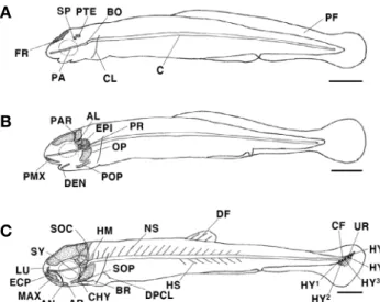
Tables 1~3과 같았다. 부화 직후 난황자어는 전장 8.08~8.14 mm(평균 8.10±0.02mm, n =5)로 두개골의 액골(frontal) 과 기저를 형성하는 부설골(parasphenoid), 기저후두골(basio- ccipital), 설이골(sphenotic) 및 익이골(pterotic)이 골화되었 다. 견대부는 쇄골(clavicle)이 골화하기 시작하였고, 척추골 (vertebra)은 추체(centrum)로 이루어져 있었다. 이 시기에는 몸 전체가 막지느러미(primordial fin)로 되어 있었다(Fig. 2A).
부화 후 3일째 전기자어는 전장 8.59~8.69mm(평균 8.64±
0.04mm, n=5)로 난황 흡수가 모두 완료되면서 먹이를 섭취 하기 시작했다. 두개골의 상이골(epiotic), 익설골(alisphenoid), 노정골(parietal), 전이골(prootic)이 골화하였고, 새개부에
Fig. 1. Map showing the sampling area in Wicheon Stream of Gyeongsangbuk-do, Uiseong-gun, Bian-myeon, Ogyeon-ri.
는 주새개골(opercle) 및 전새개골(preopercle)이 골화하였으 며, 섭이기능 역할을 하는 악골은 윗턱을 지지하는 전상악골 (premaxillary)과 아래턱을 지지하는 치골(dentary)이 골화되었 다(Fig. 2B).
부화 후 6일째 중기자어는 전장 10.7~11.3mm(평균 10.9±
0.26mm, n=5)로 두개골의 상후두골(supraoccipital) 및 안전골 (lucrymal)이 골화되었고, 구개부에는 설악골(hyomandibular), 접속골(sympletic), 외익상골(ectopterygoid)이 골화하였으며, 새개부에는 하새개골(subopercle)이 골화되었다. 설궁부에는 각 설골(ceratohyal)과 아래쪽에는 2개의 새조골(branchiostegal)이 골화되었고, 윗턱의 전상악골 아래쪽에는 주상악골(maxillary) 이 골화되었으며, 아래턱에는 각골(angular)과 관절골(articular) 이 골화되면서 치골과 결합하였다. 견대부에는 상후쇄골(dorsal post clavicle)이 골화되었고, 추체 위쪽에는 신경극(neural spine)과 아래쪽에 혈관극(hemal spine)이 골화되기 시작하였으 며, 미골부의 미부봉상골(urostyle)은 상단부로 45° 휘어지기 시 작하였다. 미부봉상골이 완전히 골화되지 않은 상태에서 하미축 골(hypural) 5개가 동시에 골화되었고, 끝부분에는 꼬리지느러 미(caudal fin) 기조가 형성되기 시작하였다. 막지느러미는 분화 하여 등과 꼬리, 배지느러미로 나누어졌으며, 등지느러미(dorsal fin)에는 3개의 기조가 발달하였다(Fig. 2C).
부화 후 12일째 후기자어는 전장 11.5~12.8mm(평균 12.2±
0.55mm, n=5)로 구개부에는 내익상골(endopterygoid), 후익 상골(metapterygoid), 방골(quadrate)이 골화하였고, 설궁부에 는 기설골(basihyal), 하설골(hypohyal)과 함께 3개의 새조골 이 골화되었으며, 새개부에는 간새개골(interopercle)이 골화 되었다. 견대부에는 상후쇄골 아래쪽에 하후쇄골(ventral post clavicle)이 골화되었고, 척추골은 처음으로 복추골(abdominal vertebrae)과 미추골(caudal vertebrae)이 골화되기 시작하였으 며, 복추골 아래쪽에는 늑골(rib)이 골화되기 시작하였다. 미골 부의 미부봉상골은 완전히 골화하였고, 아래쪽에는 준하미축골 (parhypural)이 골화되었다. 등지느러미에는 기조가 발달되기 시작하여 10개로 증가하였고, 뒷지느러미(anal fin)에는 9개의 기조가 형성되었으며, 배지느러미(ventral fin)에도 3개의 기조 가 형성되었다. 꼬리지느러미의 기조 수는 9+9=18개로 증가 하였다(Fig. 3A).
부화 후 25일째 후기자어는 전장 16.5~15.9mm(평균 16.1±
0.27mm, n =5)로 두개골의 후이골(opisthotic)과 외후두골 (exoccipital), 비골(nasal), 노정골(parietal), 안하골(suborbital) 이 골화되었고, 설궁부에는 상설골(epihyal), 간설골(interhyal) 이 골화되었으며, 견대부에는 오훼골(coracoid)이 골화되었다.
A
B
C
Fig. 2. Development stage of larvae skeleton in Hemibarbus longi- rostris. A: Newly hatched yolk-sac larvae, 8.10mm in total length (TL); B: 3 days after hatching preflexion larvae, 8.64mm in TL; C: 6 days after hatching flexion larvae, 10.9mm in TL. AL: alisphenoid;
AN: angular; AR: articular; BO: basioccipital; BR: branchiostegal; C:
Centrum; CF: caudal fin; CHY: ceratohyal; CL: clavicle; DEN: den- tary; DF: dorsal fin; DPCL: dorsal post clavicle; ECP: ectopterygoid;
EPI: epiotic; FR: flontal; HM: hyomandibular; HS: hemal spine;
HY: hypural; LU: lucrymal; MAX: maxillary; NS: neural spine; OP:
opercle; PTE: pterotic; PA: parasphenoid; PAR: parietal; PF: primor- dial fin; PMX: premaxillary; POP: preopercle; PR: prootic; SOC:
supraoccipital; SOP: subopercle; SP: sphenotic; SY: sympletic; UR:
urostyle; HY: hypural. Scale bars=1.0mm.
A
B
C
Fig. 3. Development stage of larvae and juvenile skeleton in Hemib- arbus longirostris. A: 12 days after hatching postflexion larvae, 12.2 mm in total length(TL); B: 25 days after hatching postflexion larvae, 16.1mm in TL; C: 40 days after hatching postflexion larvae, 27.9mm in TL. AF; anal fin; AV : abdominal vetebrae; BAS: basisphenoid;
BHY: basihyal; CR: coracoid; CV: caudal vertebrae; EHY: epihyal;
ENP: endopterygoid; EP: epural; EXO: exoccipital; HY: hypural;
HYE: hypohyal; IH: interhyal; INS: interneural spine; IHS: interhe- mal spine; IOP: interopercle; ME: mesethmoid MP: metapterygoid;
NA: nasal; OPI: opisthotic; PAL: parietal; PH: parhypural; Q: quad- rate; R: rib; SOR: suborbital; VF: ventral fin; VPvCL: ventral post clavicle. Scale bars=1.0mm.
미골부는 신경극과 미부봉상골 사이에 2개의 상미축골(epural) 이 골화되었고, 각 부위별 지느러미 기조 수는 배지느러미 5개, 꼬리지느러미 10+11=21개가 증가하였으며, 등지느러미에는 신경간극(interneural spine), 뒷지느러미와 배지느러미에는 혈 관간극(interhemal spine)이 골화되었다(Fig. 3B).
부화 후 40일째 치어는 전장 25.3~30.6mm(평균 27.9±
3.74mm, n=5)로 성장하였으며, 두개골에는 기저설골(basis- phenoid), 중사골(mesethmoid)이 골화되었고, 미골부는 1개의
상미축골이 추가로 골화되었으며, 하미축골은 3개(1, 2 +3, 4+5)의 형태로 융합하면서 모든 부위의 골격발달이 완성되었다 (Fig. 3C).
고 찰
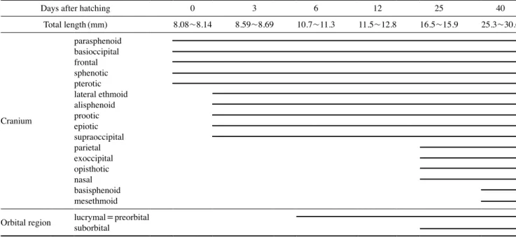
참마자는 부화 직후 평균전장 8.10mm일 때 두개골의 액 Table 1. The developmental process of cranium and orbital region of Hemibarbus longirostris
Days after hatching 0 3 6 12 25 40
Total length(mm) 8.08~8.14 8.59~8.69 10.7~11.3 11.5~12.8 16.5~15.9 25.3~30.6
Cranium
parasphenoid basioccipital frontal sphenotic pterotic lateral ethmoid alisphenoid prootic epiotic supraoccipital parietal exoccipital opisthotic nasal basisphenoid mesethmoid Orbital region lucrymal=preorbital
suborbital
Table 2. The developmental process of caudal skeleton, shoulder girdle and vertebrae of Hemibarbus longirostris
Days after hatching 0 3 6 12 25 40
Total length(mm) 8.08~8.14 8.59~8.69 10.7~11.3 11.5~12.8 16.5~15.9 25.3~30.6
Caudal skeleton
urostyle hypural(1st~5th) procurrent parhypural epural(1st~2th) 3rd
Shoulder girdle
clavicle
ventral post clavicle dorsal post clavicle coracoid
Vertebra
centrum neural spine hemal spine interhemal spine interneural spine abdominal vertebrae caudal vertebrae rib
골, 부설골, 설이골, 익이골 등이 먼저 골화되었다. 부화 후 3 일째 평균전장 8.64mm일 때는 새개부의 주새개골, 전새개골, 악골의 전상악골, 치골 등 호흡과 섭이기능 역할을 하는 부위 가 우선적으로 골화되었다. 같은 모래무지아과 어류인 모래무 지, Pseudogobio esocinus는 부화 후 5일째 평균전장 4.25mm 일 때 두개골의 부설골과 아래턱에 치골이 골화되었고(Oh, 2005), 돌고기, Pungtungia herzi는 부화 후 3일째 평균전장 6.40mm일 때 부설골, 치골, 전상악골, 쇄골 등이 골화되었으 며(Lee, 2001), 감돌고기, Pseudopungtungia nigra는 부화 후 3 일째 평균전장 6.74mm일 때 부설골, 전상악골, 치골 등이 골 화되기 시작하였다(Lee, 2004).
참마자는 부화 직후 두개골이 먼저 발달하는 반면 모래무 지, 돌고기, 감돌고기는 부화 후 3~5일부터 두개골과 악골 등 의 발달이 진행되는 것으로 보아 이들은 최초 골격발달 시기 에 참마자의 부화자어 크기보다 작았으며, 전장의 크기 차이 가 골격의 발달에 영향을 미치는 것으로 사료된다.
다른 분류군인 동사리과 어류 얼룩동사리, Odontobutis interrupta와 연어과 어류인 연어, Oncorhynchus keta는 골격의 골화가 부화 이전에 시작되어 부화 직후에는 지느러미를 지지 하는 신경간극, 혈관간극과 지느러미 기조 및 악골 등이 골화하 였다(Myoung and Kim, 1993; Park et al., 2017). 망둑어과 어류 인 미끈망둑, Luciogobius guttatus, 미끈날망둑, Chaenogobius laevis, 두줄망둑, Tridentiger trigonocephalus은 부화 직후에 골 격의 골화가 이루어지지 않았고, 상당시간이 지난 후에 악골부
터 발달하였다(Kim and Han, 1989; Kim et al., 1992; Han et al., 2018). 참마자는 부화 직후부터 골격의 발달이 관찰되었고, 부화 이전에 발달하는 얼룩동사리, 연어와 부화 후 골격이 발달 하는 미끈망둑, 미끈날망둑, 두줄망둑 사이에서 참마자의 발달 양상은 망둑어과 어류와 가까워 동사리과 및 연어과 어류와는 차이점을 보였다.
참마자의 악골은 부화 후 3일째 전상악골과 치골이 골화되 었고, 돌고기(Lee, 2001)와 감돌고기(Lee, 2004)는 부화 후 3 일째 전상악골과 치골이 골화하여 참마자의 발달시기와 동일 하였다. 반면 얼룩동사리(Park et al., 2017), 꺽지과 어류 꺽지, Coreoperca herzi, 꺽저기 Coreoperca kawamebari 등은 부화 직 후부터 악골이 골화되기 시작하였다(Han et al., 2017; Han et al., 2018). 이는 부화 직후 자어가 난황을 가짐으로 먹이 섭취의 불필요성 때문에 악골의 발달이 늦어진 것으로 판단되나 육식 성 어류인 얼룩동사리, 꺽지, 꺽저기가 악골부터 발달하는 것은 자치어기에 수서곤충 및 다른 어류의 부화자어를 포식하는 이 들의 식성과 분류군에 따라 발달정도에 차이가 있을 것으로 보 인다.
가슴지느러미를 지지하는 견대골은 어류의 유영능력에 도 움을 주며, 참마자는 부화 직후 쇄골이 골화하기 시작하여 상 후쇄골, 하후쇄골, 오훼골 순으로 골화하였다. 모래무지(Oh, 2005)는 쇄골, 상쇄골, 후쇄골, 사출골, 견갑골, 오훼골 순으로 골화하였고, 돌고기(Lee, 2001)와 감돌고기(Lee, 2004)는 쇄 골, 상쇄골, 견갑골, 오훼골, 사출골, 후쇄골 순으로 골화하였 Table 3. The developmental process of visceral skeleton of Hemibarbus longirostris
Days after hatching 0 3 6 12 25 40
Total length(mm) 8.08~8.14 8.59~8.69 10.7~11.3 11.5~12.8 16.5~15.9 25.3~30.6
Visceral skeleton
Upper jaw premaxillary maxillary Lower jaw dentary
articular angular Hyoid arch
epihyal
branchiostegal rays ceratohyal interhyal hypohyal
Palate
hyomandibular sympletic quadrate endopterygoid ectopterygoid metapterygoid Oper cular
preopercle opercle subopercle interopercle
다. 참마자의 견대골 발달은 모래무지와 유사하였으나 돌고기 와 감돌고기는 후쇄골이 가장 늦게 발달하여 차이를 보였다.
참마자와 돌고기, 감돌고기에서 나타나는 견대골의 발달과 정 차이점이 분류학적 근거자료로 제시되기 위해서는 생태학 적 연관성 등 다양한 연구가 함께 이루어져야 할 것으로 생각 된다.
다른 분류군과 비교한 결과 잉어과 어류인 버들치, Phoxinus oxycephalus는 부화 후 13일째 평균전장 6.82mm일 때 쇄골, 상쇄골, 후쇄골, 사출골, 견갑골, 오훼골 순으로 골화되었고(Han and Noh, 2001), 청어과 어류인 준치, Ilisha elongata는 평균전 장 5.02mm일 때 쇄골, 상쇄골, 후쇄골, 사출골, 견갑골 순으로 골화되었다(Han et al., 2000). 망둑어과 어류인 두줄망둑(Han et al., 2018)은 부화 후 7일째 평균전장 4.44mm일 때 쇄골, 오 훼골, 사출골 순으로 골화하였고, 큰가시고기과 큰가시고기, Gasterosteus aculeatus aculeatus는 부화 후 19일째 평균전장 8.85mm일 때 쇄골, 상쇄골, 외오훼골, 견갑골, 오훼골, 사출골 순으로 골화되었다(Han and Kim, 1989). 주둥치과 어류인 주둥 치, Leiognathus nuchalis는 평균전장 2.00mm일 때 쇄골, 후쇄 골, 상쇄골, 견갑골, 사출골, 오훼골 순으로 골화되었다(Myoung and Kim, 1984). 비교한 분류군들은 공통적으로 쇄골의 형성 이 후 상쇄골, 후쇄골 순으로 진행되었고, 사출골, 견갑골, 오훼골의 발달 순서와 정도는 종에 따라 차이를 나타냈으며, 모래무지아 과 어류와 같이 골격발달의 차이가 분류군별로 어떠한 특성을 나타내는지 추가적인 연구가 필요하다.
지속적인 유영보다 대부분을 바닥에서 주로 생활하는 어류 는 지느러미 기조보다 담기골이 먼저 골화하는 것으로 추정되 며, 유영을 많이 하는 어류는 담기골보다 지느러미 기조의 발 달이 우선적으로 이루어져 유영형태에 따라 발달정도에 차이 를 나타낸다(Park et al., 2017).
참마자의 척추골과 담기골 발달은 부화 직후 추체로 이루어 져 있었고, 부화 후 6일째 평균전장 10.9mm일 때 추체를 중 심으로 위쪽에는 신경극, 아래쪽에는 혈관극이 골화하기 시작 하였으며, 부화 후 12일째 평균전장 12.2mm일 때 복추골과 미추골이 골화하면서 부화 후 25일째 평균전장 16.1mm일 때 척추골 수는 35~36개로 골화가 완료되었다. 참마자의 척추골 은 골화하기 이전에 신경극과 혈관극이 골화하기 시작하였고, 복추골에서 미추골 순으로 골격발달이 진행되었으며, 척추골 과 담기골의 발달완료 시기는 유사하게 나타났다.
모래무지(Oh, 2005)는 척추골의 골화가 완료되기 전에 신 경극이 골화하였고, 척추골과 혈관극의 골화가 완료되면서 신경간극, 혈관간극이 골화하기 시작하였다. 척추골 수는 부 화 후 42일째 평균전장 16.7mm일 때 39개였다. 돌고기(Lee, 2001)는 척추골과 신경극이 같이 골화하기 시작하였고, 척추 골의 골화가 완료되기 전에 혈관극이 골화하기 시작하였으며, 척추골과 함께 신경간극과 혈관간극의 골화가 완료되었다. 척
추골 수는 부화 후 27일째 평균전장 10.8mm일 때 31개였다.
감돌고기(Lee, 2004)는 돌고기(Lee, 2001)의 발달양상과 유 사하였고, 척추골 수는 부화 후 78일째 평균전장 17.5mm일 때 39개였다. 참마자의 척추골과 담기골 발달은 모래무지(Oh, 2005)와 유사하였고, 참마자는 척추골의 골화 이전 신경극과 혈관극이 골화하는 반면 돌고기(Lee, 2001)와 감돌고기(Lee, 2004)는 척추골의 골화와 신경간극, 혈관간극이 함께 골화하 여 차이를 보였다. 또, 참마자의 척추골 수는 모래무지, 감돌고 기보다 적었고, 돌고기보다 많아 분류학적 차이점을 보였으며, 참마자, 모래무지, 감돌고기는 크기가 유사할 때 골화가 완료 되었으나 돌고기는 유연종 가운데 크기가 가장 작을 때 골화 가 완료되어 시기 및 크기별로 척추골의 발달정도를 알 수 있 었다.
다른 분류군과 비교해 보면 두줄망둑(Han et al., 2018)은 복 추골이 먼저 골화 후 미추골과 신경간극이 골화하였고, 꺽지 (Han et al., 2017)는 복추골과 신경극이 먼저 골화 후 미추골 과 혈관극이 골화하였다. 얼룩동사리(Park et al., 2017)는 척 추골이 골화하기 전 측돌기, 혈관극, 신경간극 및 혈관간극이 먼저 골화 후 복추골과 미추골이 골화하였다. 학공치과 어류 인 학공치, Hyporhampus sajori는 복추골과 신경극이 동시에 골화 후 복추골, 혈관극, 측돌기, 늑골 순으로 골화하였고(Lee et al., 2001), 참마자의 척추골 발달양상은 얼룩동사리(Park et al., 2017)와 유사하였고, 각 분류군마다 발달정도가 다르게 나 타나 향후 누치속 어류를 비롯해 분류군별로 척추골 발달에 대한 추가적인 연구가 필요할 것으로 생각된다.
일반적으로 농어목 어류에서 후방의 등지느러미와 뒷지느 러미의 발달은 전방의 등지느러미가 발달하기 이전에 먼저 발 달하기 시작하는 것이 일반적이다(Johnson, 1984; Faustino and Power, 1999).
참마자의 지느러미 발달은 부화 후 6일째 등과 꼬리지느러 미에 기조가 발달하기 시작하였고, 부화 후 12일째 배와 뒷지 느러미에 기조가 전방에서 후 방향으로 발달하였다. 돌고기 (Lee, 2001)와 감돌고기(Lee, 2004)는 부화 후 27일째 꼬리지 느러미 기조가 발달 이후 등지느러미가 발달하여 참마자보다 시기적으로 발달이 늦었지만 동일한 양상을 보였다. 등지느러 미가 두 개로 나누어진 두줄망둑(Han et al., 2018)은 기조가 동시에 발달하였고, 꺽저기(Han et al., 2018)는 꼬리지느러미 기조 발달 후 등과 뒷지느러미 기조가 발달하였다. 얼룩동사 리(Park et al., 2017)는 망둑어과 어류와 마찬가지로 등지느러 미가 두 개로 나누어져 있고, 제2등지느러미의 기조 발달 후 제1등지느러미와 뒷지느러미 기조가 발달하는 양상을 보였다.
참마자는 등과 꼬리지느러미가 우선적으로 발달하였고, 두 줄망둑(Han et al., 2018)과 얼룩동사리(Park et al., 2017)는 등 지느러미가 우선적으로 발달하여 농어목 어류의 일반적인 특 징을 보였다.
꼬리지느러미를 지지하는 미골부는 참마자의 경우 부화 후 6일째 5개의 하미축골이 골화하였고, 부화 후 12일째는 1개 의 준하미축골이 골화하였으며, 부화 후 40일째는 1개의 하미 축골을 제외한 2+3번째, 4+5번째가 서로 결합하면서 3개(1, 2+3, 4+5)의 골편으로 이루어졌다. 같은 모래무지아과 어류 인 모래무지(Oh, 2005), 돌고기(Lee, 2001) 및 감돌고기(Lee, 2004)의 경우 미골의 발달과정 중 5개의 하미축골이 골화하는 것이 관찰되었으나 부위별 결합과정과 골편의 개수에 대한 언 급이 없어 추가적인 연구가 필요하다.
연구결과 참마자는 유연종인 모래무지(Oh, 2005)와 견대골, 척추골, 돌고기(Lee, 2001)와 감돌고기(Lee, 2004)에서는 악골 과 지느러미의 발달과정이 유사하게 나타나 일부 모래무지아 과 어류의 골격발달 과정에서 동일한 양상을 확인할 수 있었 다. 이러한 결과를 통해 근연종과의 유연관계 규명을 위한 기 초자료가 마련되었다고 생각되며, 향후 더욱 면밀한 분류학적 특성을 확인하기 위해 모래무지아과 어류의 자치어 골격발달 연구가 지속적으로 이루어져야 할 것으로 보인다.
요 약
이 연구는 참마자 자치어의 골격발달을 관찰하여 유연종 간 의 분류학적 연구 기초자료를 제공하고자 한다. 부화 직후 난 황자어는 평균전장 8.10±0.02mm(n=5)로 두개골의 액골과 부설골, 기저후두골이 골화하였고, 부화 후 6일째 중기자어는 평균전장 10.9±0.26mm(n=5)로 두개골의 설악골, 접속골, 외익상골이 골화였으며, 부화 후 12일째 후기자어는 평균전장 12.2±0.55mm(n=5)로 새개부의 간새개골, 견대부의 하후쇄 골이 골화하였다. 부화 후 25일째 후기자어는 평균전장 16.1±
0.27mm(n=5)로 구개부의 상설골, 간설골이 골화하였고, 미 골부에는 2개의 상미축골이 골화하였으며, 신경간극과 혈관간 극이 골화하였다. 부화 후 40일째 치어는 평균전장 27.9±3.74 mm(n=5)로 미골부의 하미축골이 3개(1, 2+3, 4+5)의 형태 로 융합하면서 모든 부위의 골격발달이 완성되었다.
REFERENCES
Aoyama, S. and T. Doi. 2011. Morphological comparison of early stages of two Japanese species of eight-barbel loaches: Lefua echigonia and Lefua sp.(Nemacheilidae). Folia Zool., 60:
355-361.
Balon, E.K. 1985. The theory of fsaltatory ontogeny and life histor- ymodels revisited. In: Balon, E.K.(ed.), Early Life Histories of Fishes: New developmental, ecological and evolutionary perspective. DR W. Junk Publishers, pp. 13-30.
Bang, I.C., Y.A. Lee and W.O. Lee. 2008. Cytogenetic analysis of three Hemibarbus species(Cypriniformes) from Korea.
Aquaculture, 21: 259-264.(in Korean)
Choi, H.C., J.M. Park, G.W. Baeck and S.H. Huh. 2016. The summer diet of a juvenile barbell steed, Hemibarbus labeo, in the surf zone of the Nakdong river estuary, Korea. J. Korean Soc.
Mar. Environ. Saf., 22: 766-772.(in Korean)
Choi, K.C. and Y.K. Baek. 1970. On the life-history Gonoproktopter- us mylodon(Berg). Korean J. Limnol., 3: 21-33.(in Korean) Faustino, M. and D.M. Power. 1999. Development of the pectoral,
pelvic, dorsal and anal fins in cultured sea bream. J. Fish.
Biol., 54: 1094-1110.
Froese, R. and D. Pauly(eds.). 2019. Fishbase. World wide web electronic publication. version(08/2019). Available at: https//
fishbase.org.
Han, K.H. and B.Y. Noh. 2001. Osteological development of larvae and juveniles of Phoxinus oxycephalus(Teleostei: Cyprini- dae). Bull. Yosu Natl. Fish. Univ., 16: 369-377.(in Korean) Han, K.H., J.T. Park, D.S. Jin, D.J. Yoo and J.M. Park. 2017. Osteo-
logical development of the larvae and juvenile in Coreoperca herzi. Korean J. Ichthyol., 29: 32-40.(in Korean)
Han, K.H., J.T. Park, S.H. Lee, D.S. Jin and J.M. Park. 2018. Oste- ological development of the larvae and juveniles in aucha perch Coreoperca kawamebari(Perciformes: Centropomi- dae) in Korea. Korean J. Fish. Aqua. Sci., 51: 170-177.(in Korean)
Han, K.H., D.Y. Kim, B.Y. Noh, S.H. Oh, Y.M. Kim, D.S. Jin and Y.U.
Kim. 2000. Morphological and skeletal development of the larvae and juveniles of the slender, Ilisha elongata(Bennett) (Teleostei: Clupeidae). Korean J. Ichthyol., 12: 230-235.(in Korean)
Han, K.H., S.H. Lee, D.Y. Kim, W.I. Seo and J.M. Park. 2018b. Os- teological development of the larvae and juvenile of chame- leon goby, Tridentiger trigonocephalus. Korean J. Ichthyol., 30: 137-143.(in Korean)
Han, K.H. and Y.U. Kim. 1989. Spawning behavior, embryonic de- velopment, and morphology of larvae and juveniles of three- spined stickleback, Gasterosteus aculeatus aculeatus(Lin- naeus) reared in the laboratory. Bull. Natl. Fish. Univ. Pusan, 29: 11-36.(in Korean)
Kim, D.H., Y.K. Deung, H.Y. Kim and D.S. Reu. 2001. Ultrastructure of the fertilized egg envelope from long nose barbel, Cyprin- idae, teleost. Korean J. Electron Microscopy, 31: 85-90.(in Korean)
Kim, I.S., Y. Choi, C.L. Lee, Y.J. Lee, B.J. Kim and J.H. Kim. 2005.
Illustrated book of Korean fishes. Kyo Hak Publishing, Seoul Korean, pp. 113-125.(in Korean)
Kim, J.K., J.H. Ryu, S. Kim, D.W. Lee, K.H. Choi, T.Y. Oh, K.S.
Hwang, J.H. Choi, J.N. Kim, H.J. Kwon, H.S. Ji and J.N. Oh.
2011. An identification guide for fish eggs, larvae and juve- niles of Korea. Hanguel graphics, Busan, Korea, 275pp.(in Korean)
Kim, Y.U. and K.H. Han. 1989. 1. Egg development, larvae and juve-
niles of Chaenogobius laevis(Steindachner) - Early life his- tory of the marine animals. Bull Korean Fish. Soc., 22: 317- 331.(in Korean)
Kim, Y.U., K.H. Han, C.B. Kang and J.W. Ryu. 1992. Early life his- tory and spawning behavior of the gobiid fish, Luciogobius guttatus Gill. Korrean J. Ichthyol., 4: 1-13.(in Korean) Ko, M.H. and Y.J. Won. 2015. Egg development and early life history
of the Korean spine loach, Cobitis tetralineata(Pisces: Cobit- idae). Korean J. Ichthyol., 27: 95-103.(in Korean)
Lee, C.L. and Y.H. Kim. 2002. Skeletal morphology of the head in the genus Hemibarbus(Cypriniformes: Cyprinidae) from Korea. Korean J. Ichthyol., 14: 212-221.(in Korean) Lee, S.H. 2001. The early life history and spawning behavior of
Pungtungia herzi. Dep. Fish. Sci. Graduate School Yosu Natl. Univ., pp. 1-31.(in Korean)
Lee, S.H. 2004. Early development and reproductive ecology of Pseudopungtungia nigra. Master thesis. Graduate School Yosu Natl. Univ., pp. 1-98.(in Korean)
Lee, S.J., Y.U. Kim and K.H. Han. 2001. Osteological development of larvae and juveniles of Hyporhampus sajori(Teleostei:
Hemiramphidae). Korean J. Ichthyol., 13: 173-180.(in Kore- Lim, J.W., C.L. Lee, W.K. Lee and S.J. Jye-Gal. 2002. Comparative an)
study of lens and retinal tissues in Zacco temminckii and Hemibarbus longirostris(Cyprinidae, Cypriniformes). Kore- an J. Ichthyol., 14: 183-189.(in Korean)
Liu, C.H. 2001. Early osteological development of the yellow tail Seriola dumerili(Pisces: Carangidae). Zool. Stud., 40: 289- 298.
Mook, D. 1977. Larval and osteological development of the sheeps- head, Archosargus probatocephalus(Pisces: Sparidae).
Copeia, 1: 126-133.(in Korean)
Myoung, J.G. and Y.U. Kim. 1984. Morphology of larvae and juve- niles of Leiognathus nuchalis. Bull. Nat. Fish. Univ. Pusan,
24: 1-22.(in Korean)
Myoung, J.G. and Y.U. Kim. 1993. Morphological study of Onco- rhynchus spp.(Pisces : Salmonidae) in Korea - Ⅰ Egg devel- opment and morphology of alevin, fry and smolt of chum salmon, Oncorhynchus keta. Korean. J. Ichthyol., 5: 53-67.(in Korean)
Nelson, J.S., T.C. Grande and M.V.H. Wilson. 2016. Fishes of the world(5th ed.). John Wiley & Sons, Inc., 707pp.
Oh, G.N. 2005. Early life history of the Pseudogobio esocinus. Dep.
Aquacult. Graduate School Yosu NatL. Univ., pp. 1-38.(in Korean)
Park, J.M., J.H. Han, S.M. Yun and K.H. Han. 2017. Early osteo- logical development of larvae and juveniles in the Korean spotted sleeper Odontobutis interrupta from Korea. Korean J.
Fish. Aquat. Sci., 50: 396-405.(in Korean)
Potthoff, T., S. Kelley and L.A. Collins. 1988. Osteological develop- ment of the red snapper, Lutjanus campechanus(Lutjanidae).
Bull. Mar. Sci., 43: 1-40.
Sado, T. and S. Kimura. 2002. Descriptive morphology of the eggs, larvae, and juveniles of two cyprinid fishes belonging to the Zacco temminckii species’ group. Ichthyol. Res., 49: 245- 252.
Sfakianakis, D.G., G. Koumoundouros, P. Divanach and M. Kentou- ri. 2004. Osteological development of the vertebral column and of the fins in Pagellus erythrinus(L. 1758). Temperature effect on the developmental plasticity and morphoanatomical abnormalities. Aquaculture, 232: 407-424.
Shimizu, T., H. Sakai and N. Mizuno. 1998. Embryonic and larval de- velopment of a Japanese spinous loach, Cobitis takatsuensis.
Ichthyol. Res., 45: 377-384.
Walker, M.B. and C.B. Kimmel. 2007. A two-color acid-free cartilage and bone stain for zebrafish larvae. Biotechnic and Histo- chemistry, 82: 23-28.