서 론
꺽지(Coreoperca herzi)는 농어목(Perciformes) 꺽지과 (Centropomidae) 꺽지속(Coreoperca)에 속하는 어류로 우리 나라 강과 하천의 상류에 물살이 다소 빠르고 수질이 깨끗한 곳에 서식하는 한국고유어종이다(Han et al., 1998; Kim et al., 2005).
꺽지는 국내 내수면 산업에 중요한 종으로 손꼽히고 있으나 최근 하천의 개발과 수질오염으로 인해 서식지가 파괴되고 개
체수가 점차 감소하고 있다. 최근 내수면 양식업계에서 새로 운 어종의 개발이 요구되고 있어 우리나라 고유어종인 꺽지의 종묘생산 및 치어생산 기술개발이 크게 부각되고 있다(Han et al., 1998).
꺽지과 어류에 관한 연구로는 산란습성 및 초기생활사, 일 본산 꺽저기(C. kawamebari)의 생활사(Imai and Nakahara, 1957), 한국산 꺽저기의 산란습성 및 초기생활사(Han et al., 2000), 쏘가리(Siniperca scherzeri)의 초기생활사(Lee et al., 1997; Lee et al., 1998) 및 자치어 골격발달(Myoung et al., 2001) 등의 연구가 이루어져 왔다.
자치어의 골격발달에 대한 관심과 정확한 지식은 어류의 종
—32 — http://www.fishkorea.or.kr
* Corresponding author: Jae Min Park Tel: 82-54-830-8833, Fax: 82-54-830-8809, E-mail: [email protected] ISSn: 1225-8598(Print), 2288-3371(online)
accepted: March 18, 2017
꺽지 (Coreoperca herzi) 자치어의 골격발달
한경호·박준택1·진동수2·유동재3·박재민4,*
전남대학교 해양기술학부, 1전라남도 해양수산과학관, 2경상남도청, 3경상북도 수산자원연구소,
4경상북도 토속어류산업화센터
Osteological Development of the Larvae and Juvenile in Coreoperca herzi(Perciformes: Centropo
midae) by Kyeong Ho Han, Jun Taek Park1, Dong Soo Jin2, Dong Jae Yoo3 and Jae Min Park4,* (Marine technology under graduate, chonnam national university, yeosu 59626, republic of Korea; 1Maritime and fisheries Science Museum, yeosu 59771, republic of Korea; 2the Provincial office of gyeongsangnam-Do, changwon 51154, republic of Korea;
3gyeongsangbuk-Do fisheries resources Development Institute, yeongdeok 36405, republic of Korea; 4gyeongsangbuk- Do native fish Business center, uiseong 37366, republic of Korea)
ABSTRACT Samples were obtained from broodstok in May, 2010, while naturally fertilized embryos were maintained, and the process of skeletal development was observed from larvae and juvenile.
Prelarvae immediately after hatching showed an average total length of 6.85±0.63mm(n=5), Premaxillary and dentary were ossified, parasphenoid was ossified in the cranium, and centrum and caudal bone did not ossify. Prelarvae showed ossification with maxillary, articular, and epihyal, and branchiostegal rays of hyoid arch were ossified at 2 to 3 days after hatching with an average total length of 7.25±0.28mm(n=5). The vertebrae began to ossify in the direction of the tail, and neural spine began to ossify above the ossified vertebra. Postlarvae showed ossification of lateral ethmoid, alisphenoid, parietal, and caudal skeleton in the cranium when the average total length was 9.00±1.53 mm(n=5) in 6 days after hatching. At 40 days after hatching, postlarvae ossified maxillary in the cranium, and ossified endopterygoid and ectopterygoid, etc. in the palate, when the average length of 23.3±0.28mm(n=5). At 120 days after hatching, with the average length was 37.5±2.83mm(n=5), caudal skeleton had one additional epural bone ossification, resulting in ossification of a total of 3 epural bone to complete ossification of all spicules.
Key words: Coreoperca herzi, juvenile, larvae, osteological development
묘생산 과정 중 사육 초기에 발생하는 골격이상을 탐지하고 제거하는데 필수적이며(Koumoundouros et al., 1997a, b), 유어 기의 계통분류학적 기초형질을 제공하고 있어 이 분야에 대한 연구가 활발하다(Mook, 1977; Potthoff et al., 1987; Potthoff et al., 1988; Potthoff and Tellock, 1993; Liu, 2001; Sfakianakis et al., 2004; Coban et al., 2009). 또한 경골어류의 초기발육단계 중 형태변화는 섭이기능과 유영능력이 발달하는 후기자어에서 치어기에 가장 크게 일어나 이 시기의 골격발달은 생활방식에 따라 골화 정도와 순서에 변화를 초래해 종마다 다양한 양상 을 나타낸다(Mook, 1977; Kohno et al., 1983; Myoung et al., 2001).
꺽지를 비롯한 같은 과 어류인 꺽저기, 쏘가리 등은 자신의 영역을 지키면서 생활하며, 산란시기에는 텃세 및 영역다툼이 심한 것으로 알려져 있다(Han et al., 1998, 2000; Myoung et al., 2001). 특히 꺽지는 난황이 모두 흡수되기 전부터 먹이활 동을 시작하며, 후기자어 시기부터 개체 간의 크기 차이로 공 식현상과 기형이 발생하고 있다. 이러한 현상은 종묘생산 과정 중 자어단계에서 다량의 폐사 원인이 되어, 공식현상을 방지 하여 건강한 치어를 생산하기 위해서는 어미관리, 먹이 및 사 육환경 등에서 발생되는 골격 이상에 대한 기초자료가 필요한 실정이다. 따라서 본 연구에서는 꺽지의 발육단계별 내부형태 를 관찰하여 골격발달 과정을 살펴보고 종묘생산 및 분류학적 연구의 기초자료로 이용하고자 한다.
재료 및 방법
1. 자치어 사육
본 연구에 사용된 시료는 2010년 5월 사육 중이던 어미로부 터 자연산란 된 수정란을 확보하여 별도 관리하던 중 부화한 자어를 사육하면서 실험에 사용하였다. 부화 자어는 PVC 사각 수조(100×100×50cm)에서 수용 및 사육하였고, 사육용수는 하천수를 유수식으로 주수하였으며, 사육기간 동안 수온 범위 는 21.0~23.0℃(평균 22.0±1.0℃)를 유지시켜 주었다. 자치 어의 먹이는 부화 직후부터 15일까지 Artemia sp. nauplius 유 생을 mL 당 3~5개체 정도 공급하였고, 16일부터 30일까지 살 아있는 실지렁이(Limnodrilus gotoi)를 공급하였으며, 30일 이 후부터는 냉동 장구벌레(Blood warm Hikari, Japan)와 배합사 료(Love larva, Japan)를 혼합 공급하였다.
2. 시료확보 및 골격염색
골격발달 과정을 관찰하기 위해 부화 직후부터 120일까지 4~5일 간격으로 5마리를 무작위로 5% 중성 포르말린에 고정 하였다. 샘플된 자치어는 Walker and Kimmel(2007)의 이중염
색법에 의해 염색하였고, KOH 0.1%와 Glycerol 50%에 보존 하였다. 염색한 자치어는 실체현미경(Nikon SMZ800, Japan) 을 이용하여 관찰 및 스케치하였으며, 골격의 각 부위별 명칭 은 Kendall(1991), Okiyama(1988)에 따른 Byun et al.(2012), Kang et al.(2012)을 인용하였다.
결 과
꺽지 자치어의 골격발달 과정은 다음과 같았다(Figs. 1, 2;
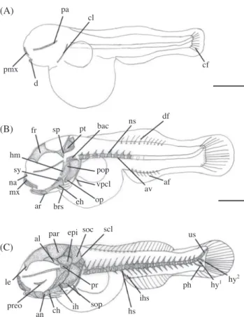
Tables 1~3). 부화 직후의 전기자어는 전장 6.45~7.34(평균 6.85±0.63)mm(n=5)로 난황을 가지고 있었고, 섭이기능과 관련되어 윗턱을 구성하는 전상악골(premaxillary)과 아래턱 을 구성하는 치골(dentary)이 골화하였으며, 두개골의 기저를 형성하는 부설골(parasphenoid)이 골화하였다. 척추골격은 척 색(notochord)으로 이루어져 있었고, 추체(centrum)와 미골부 는 골화가 전혀 일어나지 않았다. 미부봉상골(urostyle)이 상 단부로 휘어지기 시작하였으며, 견대부에는 선모양의 쇄골 (clavicle)이 골화하기 시작하였다. 이 시기에는 꼬리지느러미 에서 최초로 9개의 줄기가 발달하기 시작하였다(Fig. 1A).
부화 2~3일 후 전장 7.00~7.40(평균 7.25±0.28)mm(n=5) 일 때 전기자어는 윗턱에 주상악골(maxillary)이 골화하였고, 아래턱에는 관절골(articular)이 골화하였으며, 설궁부에는 상 설골(epihyal)과 3개의 새조골(branchiostegal rays)이 골화하 였다. 구개부에는 설악골(hyomandibular), 접속골(sympletic) 이 골화하였고, 두개골에는 기저후두골(basioccipital), 비 골(nasal), 눈의 위쪽에 액골(frontal), 설악골 위쪽에 설이골 (sphenotic), 익이골(pterotic)이 골화하였다. 새개부에는 전새개 골(preopercle), 주새개골(opercle)이 골화하였고, 쇄골의 아래 부분에는 하후쇄골(ventral post cleithrum)이 골화하기 시작하 였다. 척추골은 8개의 복추골(abdominal vertebrae)이 꼬리방 향으로 골화하기 시작하였고, 골화된 척추골의 위쪽에는 신경 극(neural spine)이 골화하기 시작하였으며, 각 부위별 지느러 미 개수는 등지느러미 11개, 뒷지느러미 8개의 줄기가 발달하 기 시작하였다(Fig. 1B).
부화 6일 후 전장 7.94~10.1(평균 9.00±1.53)mm(n=5) 일 때 후기자어는 두개골에 측사골(lateral ethmoid), 익설 골(alisphenoid), 노정골(parietal), 전이골(prootic), 상이골 (epiotic) 및 상후두골(supraoccipital)이 골화하였고, 안와 부에는 안전골(preorbital =lucrymal), 설궁부에는 각설골 (ceratohyal) 및 간설골(interhyal)이 골화하였으며, 구개부는 아 래턱에 각골(angular) 및 새개부에 하새개골(subopercle)이 골 화하였다. 미골은 미부봉상골이 상단부로 45° 휘면서 골화하였 고, 미부봉상골 아래쪽으로는 2개의 하미축골(hypural bone)과 1개의 준하미축골(parhypural bone)이 골화하기 시작하였다.
견대부에는 쇄골 상단부에 상쇄골(supracleithrum)이 골 화하였고, 척추골은 30개로 증가하였으며, 아래쪽에는 혈관 극(hemal spine), 뒷지느러미 앞쪽에는 혈관간극(interhemal spine)이 골화하기 시작하였다. 이 시기의 꼬리지느러미에는 부속줄기(procurrent)가 형성되기 시작하였다(Fig. 1C).
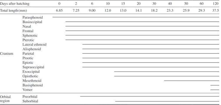
부화 15~16일 후 전장 12.5~13.9(평균 13.0±0.99)mm(n=
5)일 때 후기자어는 두개골에 외후두골(exoccipital) 및 후이 골(opisthotic)이 골화하였고, 구개부에는 방골(quadrate), 설궁 부에는 하설골(hypohyal), 새조골이 7개로 증가하였으며, 안와 부에는 안하골(suborbital), 새개부에는 간새개골(interopercle) 이 골화하였다. 주상악골의 끝쪽 상단에는 상주상악골(supr- amaxillary)이 골화하였고, 미골은 2개의 하미축골이 추가로 골
화하면서 이 시기의 하미축골 수는 4개였다. 견대부에는 상후 쇄골(dorsal post cleithrum)이 골화하였고, 배지느러미를 지지 하는 요대부에는 요대골(pelvic bone)이 골화하기 시작하였다.
등지느러미에는 신경간극(interneural spine)이 골화하기 시작 하였고, 복추골 아래쪽에는 측돌기(parapophysis)가 골화하였 으며, 꼬리지느러미의 줄기 수는 15~16개로 발달하였다(Fig.
2A).
부화 40일 후 전장 22.1~22.5(평균 23.3±0.28)mm(n=5) 일 때 후기자어는 두개골에 중사골(mesethmoid), 구개부에 내 익상골(endopterygoid), 외익상골(ectopterygoid), 후익상골 (metapterygoid), 구개골(palatine)이 골화하였다. 미골은 미부 봉상골 위쪽으로 2개의 상미축골(epural bone)이 골화하기 시 작하였고, 아래쪽에는 1개의 하미축골이 추가로 골화하면서 이 시기의 하미축골 수는 총 5개였다.
견대부는 상측두골(supraposttemporal) 및 후측두골(post- temporal)이 골화하였고, 견갑골(scapula), 오훼골(coracoid) 및 사출골(actinost)이 골화였으며, 견갑골은 망치모양으로 4개가
pa cl
pmx cf d (A)
(B)
(C) fr
hm sy namx
ar
al
le
preo an ch ih sop pr
hs ihs
us
ph hy1 hy2 par epi soc scl
brs eh op pop
vpcl av af
sp pt bac ns df
Fig. 1. Development stage of prelarvae and postlarvae of skeleton in Coreoperca herzi. A: Newly hatched larva, 6.85mm in total length (TL); B: 2~3 days after hatching, 7.25mm in TL; C: 6 days after hatching, 9.00mm in TL. af: anal fin, al: alisphenoid, an: angular, ar:
articular, av: abdominal vertebrae, bac: basioccipital, brs: branchios- tegal rays, ch: ceratohyal, cl: clavicle, cf: caudal fin, d: dentary, df:
dorsal fin, eh: epihyal, epi: epiotic, fr: frontal, hm: hyomandibular, hs:
hemal spine, hy: hypural bone, ih: interhyal, ihs: interhemal spine, le:
lateral ethmoid, mx: maxillary, na: nasal, ns: neural spine, op: oper- cle, pa: parasphenoid, par: parietal, ph: parhypural bone, pmx: pre- maxillary, pop: preopercle, pr: prootic, preo: preorbital=lucrymal, pt:
pterotic, sp: sphenotic, scl: supracleithrum, soc: supraoccipital, sop:
subopercle, sy: sympletic, us: urostyle, vpcl: ventral post cleithrum.
Scale bars=1.0mm.
sor
smx
opi
q
exo
h
ins
dpcl
iop pel
pp
me pos spt ep1ep2
hy5
pal ecpenpmp sc
cr
a vf
v
bas ep3 cdp
uro r
hy3 hy4 (A)
(B)
(C)
Fig. 2. Development stage of postlarvae and juveniles of skeleton in Coreoperca herzi. A: 15~16 days after hatching, 13.0mm in total length(TL); B: 40 days after hatching, 23.3mm in TL; C: 120 days after hatching, 37.5mm in TL. a: actinost, bas: basisphenoid, cdp:
caudal bony plate, cr: coracoid, dpcl: dorsal post cleithrum, ecp: ec- topterygoid, ep: epural bone, enp: endopterygoid, exo: exoccipital, h:
hypohyal, ins: interneural spine, iop: interopercle, me: mesethmoid, mp: metapterygoid, opi: opisthotic, pal: palatine, pel: pelvic bone, pos: posttemporal, pp: parapophysis, q: quadrate, r: rib, sc: scapula, sor: suborbital, smx: supramaxillary, spt: supraposttemporal, uro:
uroneural, v: vomer. Scale bars=1.0mm.
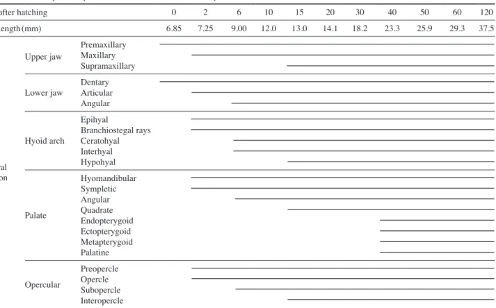
Table 1. The developmental process of cranium and orbital region of Coreoperca herzi
Days after hatching 0 2 6 10 15 20 30 40 50 60 120
Total length(mm) 6.85 7.25 9.00 12.0 13.0 14.1 18.2 23.3 25.9 29.3 37.5
Cranium
Parasphenoid Basioccipital Nasal Frontal Sphenotic Pterotic Lateral ethmoid Alisphenoid Parietal Prootic Epiotic Supraoccipital Exoccipital Opisthotic Mesethmoid Basisphenoid Vomer Orbital
region Preorbital Suborbital
Table 2. The developmental process of caudal skeleton, shoulder girdle and vertebrae of Coreoperca herzi
Days after hatching 0 2 6 10 15 20 30 40 50 60 120
Total length(mm) 6.85 7.25 9.00 12.0 13.0 14.1 18.2 23.3 25.9 29.3 37.5
Caudal skeleton
urostyle Procurrent Hypural bone 1st~2nd 3th~4th 5th
Parhypural bone Epural bone 1st~2nd 3th
Caudal bony plate uroneural
Shoulder girdle
Clavicle
Ventral post cleithrum Supracleithrum Dorsal post cleithrum Pelvic bone Supraposttemporal Posttemporal Scapula Coracoid Actinost
Vertebrae
Notochord Neural spine Hemal spine Parapophysis Interhemal spine Interneural spine rib
골화하였다. 척추골은 복추골을 따라 6개의 늑골(rib)이 골화 하였고, 각 부위별 지느러미 줄기 수는 등지느러미 극조 13개, 연조 13개, 뒷지느러미 극조 3개, 연조 8개, 배지느러미 극조 1 개, 연조 5개로 증가하였다(Fig. 2B).
부화 120일 후 전장 36.0~40.0(평균 37.5±2.83)mm (n=5)일 때 치어는 두개골에 기저설골(basisphenoid)과 서골 (vomer)이 골화하면서 두부골격의 골화가 완료되었다. 미골 은 1개의 상미축골이 추가로 골화되면서 총 3개의 상미축골이 골화하였고, 세 번째 상미축골과 미부봉상골 사이에는 미골판 (caudal bony plate)이 골화하였으며, 다섯 번째 하미축골과 미 부봉상골 사이에는 신경골(uroneural)이 골화하면서 모든 골편 들의 골화가 완성되었다(Fig. 2C).
고 찰
일반적으로 경골어류는 부화 시 골격발달에 주목할 만한 변화를 나타낸다고 알려져 있으며(Koumoundouros et al., 2001a), 농어목 어류는 부화 이후에 두개골과 지느러미의 발달 이 관찰되나 연어과 어류는 부화 이전에 골격의 발달이 시작 되는 것으로 알려져 본 종은 전자에 속하였다(Kendall et al., 1984; Matsuoka, 1985; Koumoundouros et al., 1997b, 2001a,
2001b; Faustion and Power, 1999; Sfakianakis et al., 2004, 2005).
꺽지는 평균전장 6.85mm일 때 두개골을 형성하는 골격 중 부설골, 전상악골, 치골과 견대부의 쇄골이 먼저 골화하였다.
잉어과 어류인 버들치 Phoxinus oxycephalus(Han and Noh, 2001)는 부설골이 최초로 골화하였고, 이후 외후두골 및 기 저후두골이 골화하였다. 농어목 어류인 점농어 Lateolabrax maculatus(Kang et al., 2012)는 견대부의 쇄골이 최초로 골화 하였고, 두개골의 부설골, 전상악골, 치골이 뒤이어 골화하였 으며, 잿방어 Seriola dumerili(Liu, 2001)는 전상악골, 주상악 골, 치골, 전새개골, 주새개골과 함께 안전골, 서골 등이 골화하 였다. 쏨뱅이목 어류인 쏨뱅이 Sebastiscus marmoratus(Kim et al., 1997)와 붉은쏨뱅이 S. tertius(Han et al., 2001)는 두개골 의 부설골, 악골 및 견대부의 쇄골이 먼저 골화하였고, 황점볼 락 Sebastes oblongus(Byun et al., 2012)은 두개골의 액골, 노 정골, 주상악골, 견대부의 쇄골 등이 먼저 골화하였으며, 볼락 S. inermis(Kim et al., 1993) 및 조피볼락 S. schlegelii(Kim and Han, 1991)은 악골과 견대부의 쇄골이 먼저 골화하였다. 청베 도라치과 어류인 저울베도라치 Entomacrodus stellifer lighti (Kim et al., 1992)는 쇄골과 부설골이 먼저 골화하였고, 이후 악골이 골화하였으며, 복어목 어류인 졸복 Takifugu pardalis (Han et al., 2005)은 부설골, 악골, 쇄골이 동시에 골화하였다.
Table 3. The developmental process of visceral skeleton of Coreoperca herzi
Days after hatching 0 2 6 10 15 20 30 40 50 60 120
Total length(mm) 6.85 7.25 9.00 12.0 13.0 14.1 18.2 23.3 25.9 29.3 37.5
Visceral skeleton
upper jaw Premaxillary Maxillary Supramaxillary Lower jaw Dentary
Articular Angular Hyoid arch
Epihyal
Branchiostegal rays Ceratohyal Interhyal Hypohyal
Palate
Hyomandibular Sympletic Angular quadrate Endopterygoid Ectopterygoid Metapterygoid Palatine Opercular
Preopercle Opercle Subopercle Interopercle
망둑어과 어류인 미끈날망둑 Chaenogobius laevis(Kim and Han, 1989)은 쇄골과 턱뼈, 설궁부의 내장골이 골화하여 어종 별로 다양한 골격발달 양상을 나타냈다.
한편 이들 종의 공통적인 특징으로 턱을 구성하는 악골이 먼저 골화하였는데 이는 섭이와 호흡에 필요한 중요한 골격요 소로서 골화가 우선적으로 일어나는 부위이며(Vandewalle et al., 1997), 입의 개구와 섭이기능에 sternohyoideus muscle을 지지하는 쇄골이 중요한 역할을 하기 때문이다(Wagemans and Vandewalle, 1999).
꺽지는 가슴지느러미를 지지하는 견대부 골격 중 견갑골 에 둥근 모양의 체공(foramen) 1개가 형성되는데 이는 농어 목 어류의 전형적인 형질로 알려져 있다(Koumoundouros et al., 2001b). 꺽지는 평균전장 23.3mm일 때 견갑골에 구멍이 관찰되었고, 이처럼 견갑골에 구멍이 형성되는 어류는 농어 목 어류인 참돔 Pagrus major(Matsuoka, 1987), 황돔 Dentex tumifrons(Koumoundouros et al., 2001b), 자리돔류(Emery, 1973)에서 나타났으며, 점농어(Kang et al., 2012)의 경우 견갑 골에 구멍이 형성된 후 오훼골에도 체공(foramen)이 형성되었 다 사라지는 특징을 보여 차이를 보였다. 쏨뱅이목 어류인 쏨 뱅이(Kim et al., 1997), 붉은쏨뱅이(Han et al., 2001), 조피볼락 (Kim and Han, 1991), 동갈치목 어류인 전력날치 Cheilopogon heterurus doederleini(Dasilao and Yamaoka, 1998), 복어목 어 류인 Balistes capriscus(Matsuura and Katsuragawa, 1984) 등 다른 분류군에서도 형성되는 것으로 보아 농어목 어류뿐만 아 니라 모든 어류에서 일반적으로 형성되는 전형적인 형질로 보 여 진다.
척추골은 몸의 중축을 이루는 중요한 골격 부위로서 꺽지 는 평균전장 7.25mm일 때 두개골 방향에서 복추골이 골화하 여 꼬리 방향으로 발달하기 시작하였고, 미추골과 미부봉상골 의 골화가 동시에 골화하였다. 이러한 경향은 황점볼락(Byun et al., 2012), 볼락(Kim et al., 1993), 조피볼락(Kim and Han, 1991), 붉은쏨뱅이(Han et al., 2001), 쏨뱅이(Kim et al., 1997), 점농어(Kang et al., 2012), 날치 Prognichthys agoo(Park and Kim, 1987) 및 능성어 Epinephelus septemfasciatus(Park et al., 2015b) 등에서 나타나 추체의 골화 방향은 차이가 없는 것으 로 판단된다. 꺽지는 추체가 골화한 뒤 미부봉상골이 골화되었 고, 신경극과 혈관극은 추체가 골화하면서 발달하였다. 이러한 경향은 바리과 어류인 능성어(Park et al., 2015b)의 골격발달 과 유사하였고, 실횟대 Porocottus tentaculatus(Han and Kim, 1997), 황점볼락(Byun et al., 2012), 복섬 T. niphobles(Fujita, 1992), 꽁치 Cololabis saira(Fujita and Oozeki, 1994) 및 점농 어(Kang et al., 2012), 버들치(Han and Noh, 2001) 등은 추체가 모두 골화하기 전 미부봉상골의 골화가 시작되어 차이를 보였 으며, 황해볼락 S. koreanus(Park et al., 2015a), 황점볼락(Byun et al., 2012), 복섬(Fujita, 1992), 꽁치(Fujita and Oozeki, 1994)
는 추체가 골화하기 전 신경극과 혈관극의 골화가 시작되어 다양한 골격발달 양상을 나타내었다.
지느러미의 발달은 전방의 등지느러미가 발달되기 이전 에 후방의 등지느러미와 뒷지느러미가 먼저 발달하기 시작하 는 것이 농어목 어류의 일반적인 발달 양상으로 알려져 있다 (Johnson, 1984; Faustino and Power, 1999). 꺽지는 전방의 등 지느러미가 발달되기 이전에 후방의 등지느러미와 뒷지느러 미가 발달하여 전형적인 농어목 어류의 지느러미 발달 양상을 보였다. 또한 지느러미를 지지하는 담기골의 골화는 지느러미 가 발달되면서 이루어졌고, 골화의 방향은 전방에서 후방으로 진행되었다. 담기골은 추체의 골격이 완성된 이후에 골화가 완 료되었고, 잉어 Cyprinus carpio(Itazawa, 1963), 참돔(Kohno et al., 1983), 가물치 Channa argus(Itazawa, 1963)에서 같은 경향을 보였으며, 척추골의 골화와 지느러미의 줄기 발달이 완 성된 후에 담기골의 골화가 완료되는 것은 어류가 유영하는데 있어 추진력을 증가시키는 것과 관련이 있는 것으로 보여 진다 (Lee et al., 2001).
쏘가리(Myoung et al., 2001)와 능성어(Park et al., 2015b)의 등지느러미는 전방에서 후방으로 순차적 발달이 진행되었고, 점농어는 전·후방의 등지느러미와 뒷지느러미가 동시에 발 달하여 차이를 보였다. Pagellus erythrinus(Sfakianakis et al., 2004)는 신경극의 후방에서 골화가 시작되어 이후 전, 후방 에서 신경극의 골화가 동시에 이루어졌고, Diplodus puntazzo (Sfakianakis et al., 2005) 및 Diplodus sargus(Komoundouros et al., 2001a)는 신경극과 혈관극의 골화가 전, 후방에서 거의 동시에 이루어졌으며, 참돔(Matsuoka, 1985)은 신경극과 혈관 극의 골화가 동시에 진행되나 신경극의 경우 전, 후방의 골화 가 진행된 이후 중앙 쪽으로 골화가 진행되어 어종별로 다양 한 골화과정을 나타냈다.
꼬리지느러미를 지지하는 미골부의 경우 꺽지는 평균전장 6.85mm일 때 미부봉상골의 골화는 진행되지 않았으나 상단 부를 향해 휘어지기 시작하였다. 평균전장 9.00mm일 때는 미 부봉상골은 45°로 완전히 휘어지면서 골화가 완료되었다. 또 한 평균전장 37.5mm일 때 미골부의 주요골격인 하미축골(1+
2,3 +4 +5)이 융합되어 외관상 2개의 하미축골을 형성하였 다. 이와 같이 꺽지의 하미축골 융합형태와 같은 양상을 보이 는 종은 같은 과 어류인 쏘가리와 꺽저기(Park, 2001), 방어 류(Kohno, 1997; Liu, 2001)에서 볼 수 있었다. 이 외에도 능 성어(Park et al., 2015b)는 융합 후 외관상 3개의 하미축골 (1 +2,3,4 +5)이 형성되었고, 학공치 Hyporhamphus sajori (Lee et al., 2001)와 황점볼락(Byun et al., 2012)은 하미축골 (1+2,3+4,5)이 외관상 3개였으나 융합이 다르게 나타나 차이 를 보였다. 어종마다 다양한 하미축골의 융합현상은 계통학적 연구의 기초자료로서 매우 중요하다고 판단된다. 이와 같이 어 류의 해부학적 정보는 유사종과의 식별이나 생태학적 분야에
서 유용한 자료로 활용될 수 있으며, 자치어의 골격발달 과정 은 종묘생산에서 문제가 되고 있는 기형의 발생과 원인을 분 석하는데 중요한 기초자료 될 수 있으므로 향후 이 분야의 지 속적인 연구가 필요할 것으로 생각된다.
요 약
2010년 5월 사육 중인 친어로부터 자연산란 된 수정란을 관 리하던 중 부화된 자치어를 사육하면서 발육단계에 따른 골 격발달과정을 관찰하였다. 부화 직후의 전기자어는 평균전장 6.85±0.63mm(n=5)로 전상악골과 치골이 골화하였고, 두개 골에는 부설골이 골화하였으며, 추체와 미골부는 골화가 일어 나지 않았다. 부화 2~3일 후 평균전장 7.25±0.28mm(n=5) 일 때 전기자어는 주상악골, 관절골이 골화하였고, 설궁부의 상 설골과 새조골이 골화하였다. 척추골은 꼬리방향으로 골화하 기 시작하였고, 골화된 척추골의 위쪽에는 신경극이 골화하기 시작하였다. 부화 6일 후 평균전장 9.00±1.53mm(n=5)일 때 후기자어는 두개골에 측사골, 익설골, 노정골 등이 골화하였고, 미골은 미부봉상골이 상단부로 45° 휘면서 골화하였다. 부화 40일 후 평균전장 23.3±0.28mm(n=5)일 때 후기자어는 두 개골에 중사골, 구개부에 내익상골, 외익상골 등이 골화하였다.
부화 120일 후 평균전장 37.5±2.83mm(n=5)일 때 치어는 미 골은 1개의 상미축골이 추가로 골화되면서 총 3개의 상미축골 이 골화하면서 모든 골편들의 골화가 완성되었다.
REFERENCES
Byun, S.G., C.B. Kang, J.G. Myoung, B.S. Cha, K.H. Han and C.G.
Jung. 2012. Early osteological development of the larvae and juveniles in Sebastes oblongus(Pisces: Scorpaenidae).
Kor. J. Ichthyol., 24: 67-76.(in Korean)
Coban, D., C. Suzer, H.O. Kamaci, S. Saka and K. Firat. 2009. Early osteological development of the fins in the hatchery-reared red porgy, Pagrus pagrus(L. 1758). J. Appl. Ichthyol., 25:
26-32.
Dasilao, Jr J.C. and K. Yamaoka. 1998. Osteological and functional development of the flyingfish, Cypselurus heterurus doeder- leini(Teleostei: Exocoetidae). Bull. Mar. Sci. Fish. Kochi univ., 18: 13-26.
Emery, A.r. 1973. Ecology and functional osteology damselfish (Pisces; Pomacentridae) at Alligator reef, Florida Keys.
Bull. Mar. Sci., 23: 649-770.
Faustino, M. and D.M. Power. 1999. Development of the pectoral, pelvic, dorsal and anal fins in cultured sea bream. J. Fish.
Biol., 54: 1094-1110.
Fujita, K. 1992. Development of the caudal skeleton in the tetra-
odontid fish, Takifugu niphobles. Japan. J. Ichthyol., 38:
438-440.
Fujita, K. and Y. Oozeki. 1994. Development of the caudal skeleton in the saury, Cololabis saira. Japan. J. Ichthyol., 41: 334- 337.
Han, K.H. and B.Y. Noh. 2001. Osteological development of larvae and juveniles of Phoxinus oxycephalus(Teleostei: Cyprini- dae). Bull. Yosu Natl. Fish. univ., 16: 369-377.(in Korean) Han, K.H., J.K. Cho, S.H. Lee, S.Y. Hwang, S.M. Yoon, W.I. Seo
and C.C. Kim. 2005. Osteological development of the larvae and juveniles of Takifugu pardalis(Teleostei: Tetra- odontodae). Kor. J. Ichthyol., 17: 29-35.(in Korean) Han, K.H., J.T. Park, B.M. Kim, S.H. Oh, S.H. Lee and D.S. Jin.
2000. Spawning behavior and early life history of aucha perch Coreoperca kawamebari from Korea. Kor. J. Ichthy- ol., 12: 129-136.(in Korean)
Han, K.H., J.T. Park, G.H. Jung, W.K. Lee, J.Y. Lee and I.C. Bang.
1998. Spawning behavior and early life history of Korean aucha perch, Coreoperca herzi Herzenstein. J. Aquacult., 11: 49-58.(in Korean)
Han, K.H. and Y.u. Kim. 1997. Development of larvae and juve- niles of the cottid fish, Porocottus tentaculatus(Kner). Bull.
Yosu Natl. Fish. univ., 2: 119-129.(in Korean)
Han, K.H., S.K. Lim, K.S. Kim, C.W. Kim and D.J. Yoo. 2001.
Osteological development of the larvae and juveniles of Sebastiscus tertius(Barsukov et Chen) in Korea. Kor. J.
Ichthyol., 13: 63-68.(in Korean)
Imai, S. and K. Nakahara. 1957. Life history of Coreoperca kawame- bari(Temminck et Schlegel). Suisangaku Shusei. univ.
Tokyo Press, pp. 591-601.
Itazawa, Y. 1963. The ossification sequences of the vertebral colum in the carp and snak-head fish. Bull. Jap. Soc. Sci. Fish., 29:
667-674.
Johnson, G.D. 1984. Percoidei: development and relationship. In:
Moser, H.G., W.J. richards, D.M. Cohen, M.P. Fahay, A.W.
Kendall and S.L. richardson(eds.), Ontogeny and system- atics of fishes. American Society of Ichthyologists and Her- petologists, special publication no 1, Allen Lawrence KS, pp. 464-498.
Kang, C.B., J.G. Myoung, Y.u. Kim and H.C. Kim. 2012. Early osteological development and squamation in the spotted sea bass Lateolabrax maculates(Pisces: Lateolabracidae). Kor.
J. Fish. Aquat. Sci., 45: 271-282.(in Korean)
Kendall, A.W., E.H. Ahlstrom and H.G. Moser. 1984. Early life history stages of fishes and their characters. In: Moser, H.G., W.J. richards, D.M. Cohen, M.P. Fahay, A.W. Kendall and S.L. richardson(eds.), Ontogeny and systematics of fishes.
American Society of Ichthyologists and Herpetologists, special publication no 1. Allen Lawrence KS, pp. 11-12.
Kendall, W. 1991. Systematics and identification of larvae and juve- niles of the genus Sebastes. Env. Biol. Fish., 30: 173-190.
Kim, I.S., Y. Choi, C.L. Lee, Y.J. Lee, B.J. Kim and J.H. Kim. 2005.
Illustrated book of Korean fishes. Kyo Hak Publishing,
Seoul Korean, pp. 270-271.
Kim, Y.u. and K.H. Han. 1989. Early life history of the marine an- imals 1. Egg development, larvae and juveniles of Chaeno- gobius laevis(Steindachner). Bull. Kor. Fish. Soc., 22: 317- 331.(in Korean)
Kim, Y.U. and K.H. Han. 1991. The early life history of rockfish Sebastes schlegeli. Kor. J. Ichthyol., 3: 67-83.(in Korean) Kim, Y.u., K.H. Han and C.B. Kang. 1992. Morphology and skel-
etal development of larvae and juveniles of Entomacrodus stellifer lighti(Herre). Kor. J. Ichthyol., 4: 31-43.(in Kore- Kim, Y.u., K.H. Han and S.K. Byun. 1993. The early life history of an)
the rockfish, Sebastes inermis 2. Morphological and skele- tal development of larvae and juveniles. Bull. Korean Fish.
Soc., 26: 465-476.(in Korean)
Kim, Y.u., K.H. Han, C.B. Kang, J.K. Kim and S.K. Byun. 1997.
The early life history of the rockfish, Sebastiscus marmor- atus 2. Morphology and skeletal development of larvae and juvenile. Kor. J. Ichthyol., 9: 186-194.(in Korean)
Kohno, H. 1997. Osteological development of the caudal skeleton in the carangid, Seriola lalandi. Ichthyol. res., 44: 219- 221.
Kohno, H., Y. Taki, Y. Ogasawara, Y. Shiroja, M. Taketomi and M.
Inoue. 1983. Development of swimming and feeding func- tions in larval Pagrus major. Japan. J. Ichthyol., 30: 47-60.
Koumoundouros, G., D.G. Sfakianakis, E. Maingot, P. Divanach and M. Kentouri. 2001a. Osteological development of the vertebral column and of the fins in Diplodus sargus(Tele- ostei: Perciformes: Sparidae). Mar. Biol., 139: 853-862.
Koumoundouros, G., F. Gagliardi, P. Divanach, C. Boglione, S.
Cataudella and M. Kentouri. 1997a. Normal and abnormal osteological development of caudal fin in Sparus aurata L.
fry. Aquacult., 149: 215-226.
Koumoundouros, G., G. Oran, P. Divanach, S. Stefanakis and M.
Kentouri. 1997b. The opercular complex deformity in in- tensive gilthead sea bream(Spartus aurata L.) larviculture.
Moment of apparition and description. Aquacult., 156: 165- Koumoundouros, G., P. Divanach and M. Kentouri. 2001b. Osteo-177.
logical development of Dentex dentex(Osteichthyes: Spar- idae): dorsal, anal, paired fins and squamation. Mar. Bio1., 38: 399-406.
Lee, S.J., Y.u. Kim and K.H. Han. 2001. Osteological development of larvae and juveniles of Hyporhampus sajori(Teleostei:
Hemiramphidae). Kor. J. Ichthyol., 13: 173-180.(in Kore- Lee, W.O., J.Y. Lee, S.J. Son and N.J. Choi. 1997. Early life his-an) tory and reproductive ecology of mandarin fish, Siniperca scherzeri (Pisces, Centropomidae) in Soyang lake. Kor. J.
Ichthyol., 9: 99-107.(in Korean)
Lee, W.O., S.I. Jang and J.Y. Lee. 1998. Studies on early life history of the Korean mandarine fish, Siniperca scherzeri(Perci- formes: Centropomidae). Kor. J. Ichthyol., 10: 1-10.(in
Korean)
Liu, C.H. 2001. Early osteological development of the yellow tail Seriola dumerili(Pisces: Carangidae). Zool. Stud., 40: 289- Matsuoka, M. 1985. Osteological development in the red sea bream, 298.
Pagrus major. Japan. J. Ichthyol., 32: 35-51.
Matsuoka, M. 1987. Development of the skeletal tissues and skel- etal muscles in the red sea bream. Bull. Seikai red. Fish.
res. Lab., 65: 1-14.
Matsuura, Y. and M. Katsuragawa. 1984. Osteological development of fins and their supports of larval grey triggerfish, Balistes capriscus. Japan. J. Ichthyol., 31: 411-421.
Mook, D. 1977. Larval and osteological development of the sheeps- head, Archosargus probatocephalus(Pisces: Sparidae). Co- peia, 1977: 126-133.
Myoung, J.G., J.H. Mun, J.K. Kim, K.D. Park, C.B. Kang, Y.u.
Kim and J.T. Park. 2001. Osteological development of larvae and juveniles of Korean mandarine fish, Siniperca scherzeri(Perciformes: Centropomidae). Kor. J. Ichthyol., 13: 129-135.(in Korean)
Park, J.M., J.K. Cho, H. Han and K.H. Han. 2015a. Morphological and skeletal development and larvae and juvenile of Se- bastes koreanus(Pisces: Scorpaenidae). Kor. J. Ichthyol., 27: 1-9.(in Korean)
Park, J.T. 2001. Phylogenetic study of the centropomidae(Pisces, Perciformes) in Korea. Dep. Fish. Sci. Graduate School Yosu NatL. univ., pp. 29-56.(in Korean)
Park, J.Y., C.G. Hong, J.K. Cho, M.H. Son, K.H. Han and J.M. Park.
2015b. Early osteological development of the larvae and juveniles in sevenband grouper, Epinephelus septemfascia- tus(Pisces: Serranidae). Kor. J. Ichthyol., 27: 189-198.(in Korean)
Park, Y.S. and Y.u. Kim. 1987. Studies on the larvae and juveniles of flying fish, Prognichthys agoo(Temminck et Schlegel) (Pisces, Exocoetidae) Ⅱ. Osteological development of lar- vae and juveniles. Bull. Kor. Fish. Soc., 20: 447-456.(in Korean)
Potthoff, T. and J.A. Tellock. 1993. Osteological development of the snook, Centropomus undecimalis(Teleostei, Centropo- midae). Bull. Mar. Sci., 52: 669-716.
Potthoff, T., S. Kelley and L.A. Collins. 1988. Osteological devel- opment of the red snapper, Lutjanu scampechanus(Lutjani- dae). Bull. Mar. Sci., 43: 1-40.
Potthoff, T., S. Kelly, V. Saksena, M. Moe and F. Young. 1987. De- scription of larval and juvenile damselfish Microspathodon chrysurus, Pomacentridae, and their osteological develop- ment. Bull. Mar. Sci., 40: 1-40.
Sfakianakis, D.G., C.K. Doxa, S. Kouttouki, G. Koumoundouros, E.
Maingot, P. Divanach and M. Kentouri. 2005. Osteological development of the vertebral column and of the fins in Di- plodus puntazzo(Cetti, 1777). Auqaculture, 250: 36-46.
Sfakianakis, D.G., G. Koumoundouros, P. Divanach and M. Kentou- ri. 2004. Osteological development of the vertebral column
and of the fins in Pagellus erythrinus(L. 1758). Temperature effedt on the developmental plasticity and morphoanatomical abnormalities. Auqaculture, 232: 407-424.
Vandewalle, P., I. Gluckmann, E. Baras, F. Huriaux and B. Focant.
1997. Postembryonic development of the cephalic region in Heterobranchus longifilis. J. Fish Biol., 50: 227-253.
Wagemans, F. and P. Vandewalle. 1999. Development of the carti- laginous skull in Solea solea: trends on Pleuronectiforms.
Ann. Sci. Nat., 1: 39-52.
Walker, M.B. and C.B. Kimmel. 2007. A two-color acid-free car- tilage and bone stain for zebrafish larvae. Biotechnic and Histochemistry, 82: 23-28.