서 론
붉은쏨뱅이(Sebastiscus tertius)는 쏨뱅이 (Scorpae- niform), 양볼락科(Scorpaenidae), 쏨뱅이 (Sebasticus) 에 속하는 난태생 어류로 외부형태는 쏨뱅이, Sebasticus mamoratus와 비슷하며, 1978년 Barsukov와 Chen에 의 해 으로 보고되었다. 우리나라에서는 1993년에 김 과 이가 미기록 으로 보고하였으며, 우리나라의 황해와 동중국해, 남중국해 및 일본의 남부 연안에 하고 있
다(Masuda et al., 1984; 區 究 , 1995; 한국 동물분류학회, 1997).
붉은쏨뱅이에 대한 究는 초기생활사(김 등, 1999), 형태 및 골격(한 등, 1999)이 있고, 같은 어류인 쏨뱅 이, Sebasticus mamoratus의 경우 초기생활사(김 등, 1997a, b)에 관한 연구가 있다. 우리나라에서 같은 양볼 락科에 속한 어종들에 대한 연구는 조피볼락, Sebastes schlegeli (김과 한, 1991)과 볼락, Sebastes inermis의 期 (김과 한, 1993; 김 등, 1993), 불볼락, Sebastes thompsoni과 개볼락, Sebastes pachycephalus pachyce-
─
─ 63 ──
한국산 붉은쏨뱅이 Sebasticus tertius (Barsukov et Chen) 의 골격발달
한경호∙임상구*∙김광수*∙김철원*∙유동재 여수대학교 수산생명과학부 *국립수산진흥원 완도수산종묘시험장
Osteological Development of the Larvae and Juveniles of Sebasticus tertius (Barsukov et Chen) in Korea
Kyeong-Ho Han, Sang-Ku Lim*, Kwang-Su Kim*, Chul-Won Kim* and Dong-Jae Yoo
Division of Aqua Life Science, Yosu National University, Yosu 550-749, Korea
*Wando Marine Hatchery, National Fisheries Research and Development Institute, Wando 537-800, Korea
The skeletal development of the larvae and juvenile of red marbled rockfish, Sebasticus tertius (Barsukov et Chen) was studied based on individuals that were discharged and reared in the laboratory from April to May 1997. In 8 days after bearing, the postlarvae attained 4.42 mm in total length (TL), and its parasphenoid, premaxillary, maxillary, and clavicle were ossified for the first time at this stage. In 15 days after bearing, the postlarvae attained 5.23 mm in TL, and its pterotic, basioccipital, exoccipital, opercle, and preopercle were ossif ied, with one spine on the each cranium and preopercle. In 27 days after bearing, the postlarvae attained 8.81 mm in TL, its vertebra were posteriorly ossified to the 15th centrum, and f ive spines were formed on the preopercle. In 39 days after bearing, the juveniles attained 14.21 mm in TL, and the all bones were almost completed at this stage.
Key words : Sebasticus tertius (Barsukov et Chen), larvae and juveniles, osteological develop- ment
고, 외국의 경우 황점볼락, Sebastes oblongus의 � 과 期(Fujita, 1958), 볼락 의 季 (Mizue, 1958), 年 , 및 (Mio, 1960), 交
(Shinomiya and Ezaki, 1991)에 대한 연구와 흰꼬리 볼락, Sebastes longisipinis의 (Takai and Fuku- naga, 1971), 개볼락의 期 (Shiokawa and Tsu- kahara, 1961; Shiokawa, 1962) 등이 있다.
양볼락科 어류는 자치어 단계에서 형태적으로 유사종 이 많은 분류군으로 분류, 동정이 매우 어려운 실정이 다. 이 연구는 분류학적 연구의 일환으로 붉은쏨뱅이 자 치어의 골격발달 과정을 관찰하였기에 보고한다.
재료 및 방법
이 실험에 사용된 재료는 1997년 1월부터 전라남도 완도군 연안에서 연승어업에 의해 어획된 붉은쏨뱅이 23개체를 육상수조(4.0×2.0×0.8 m)에서 사육하던 중, 4월 27일부터 5월 9일까지 친어(암컷 5마리, 전장 27.5~41.0 cm)들이 자연산출한 자어를 사육하였다. 사 육용수는 매일 1/2씩 환수하였고, 산출한 자어는 원형의 FRP수조(3 ton)에서 사육하였으며, 사육기간동안의 수 온범위는 16.0~19.5�C, 비중범위는1.023~1.025였다.
사육기간동안 먹이공급은 산출 후 1일부터 Chlorella sp. (3~6×10⁶ cells/ml), rotifer (Brachionus plicatilis, 10~15개체/ml) 및 Artemia sp. 유생을 차례로 급이하였 으며, 주수는 산출 후7일부터 분당5 l로 하였고, 사육수 조는 차광을 하여 약간 어둡게 해주었다.
자치어의 골격 관찰을 위해 자치어를 산출 직후부터 5% 중성포르말린에 고정시킨 후 Kawamura and Ho- soya (1991)의 이중염색에 의하여 내부골격을 염색한 후 입체해부현미경과 만능투영기를 사용하여 관찰, 스케치 하였고, 골격의 각 부위 명칭은 Kendall (1991) 및 Oki- yama (1993)에 따랐으며, 각 부위는 0.01 mm까지 측정 하였다.
결 과
산출 직후의 자어는 전장이 3.79~3.97 mm (평균 3.88 mm, n = 5)로 골화가 시작되지 않았으며, 산출 후 6일의 자어는 전장이4.21~4.48 mm로(평균 4.32 mm, n = 5)로 전혀 골화되지 않았다.
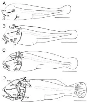
산출 후 8일의 후기자어는 전장이 4.23~4.60 mm (평 균 4.42 mm, n = 5)로 가장 먼저 肩 (shoulder girdle) 에 막대기 모양의 骨(clavicle)과 섭이 활동을 위한
骨(maxillary), 아래턱에 骨(dentary)이 骨 하기 시작하였으며, 蓋 (cranium)의 배쪽면에 가늘 고 긴 막대기 모양의 骨(parasphenoid)이 骨 되 었다(Fig. 1, A).
산출 후 11일의 후기자어는 전장이 4.38~5.02 mm (평균 4.64 mm, n = 5)로 蓋 의 등쪽면에 骨(fron- tal), 骨(parietal), 骨(supraoccipital)이 骨 하 기 시작하였고, 아래턱 骨의 뒷쪽에 關 骨(articular) 이 骨 하기 시작하였으며, 口蓋 (palate)의 骨 (hyomandibular)이 최초로 骨 하기 시작하였다. 弓 (hyoid arch)에 角 骨(ceratohyal)과 骨(epih-
Fig. 1. Skeletal ossification of Sebasticus tertius (Barsu- kov et Chen).
A: Postlarva, 8 days after bearing, 4.42 mm in total length (TL); B: Postlarva, 11 days after bear- ing, 4.64 mm in TL; C: Postlarva, 15 days after bearing, 5.23 mm in TL; D: Postlarva, 21 days after bearing, 6.54 mm in TL.
ar: articular; bo: basioccipital; br: branchiostegal ray; ch: ceratohyal; cl: clavicle; dt: dentary; eh:
epihyal; exo: exoccipital; f: frontal; hm: hyomandi- bular; iop: interopercle; mx: maxillary; ns: neural spine; oot: opisthotic; op: opercle; pa: parietal;
pmx: premaxillary; po: preopercle; pro: prootic; ps:
paraspenoid; soc: supraoccipital; sop: subopercle;
suo: supraoccipital spine; pto: pterotic. Scale bars
= 1.00 mm.
yal)이 骨 하기 시작하였으며, 2개의 새조골(branchio- stegal ray)이 骨 하여 角 骨에 관절하였다(Fig. 1, B).
산출 후 15일의 후기자어는 전장이 4.81~5.51 mm (평균 5.23 mm, n = 5)로 蓋 에 骨(pterotic), 基
骨(basioccipital), 骨(exoccipital)이 骨 하기 시작하였고, 에 1개의 棘이 형성되었으며, 아가미뚜껑 부분을 지지하는 새개부(opercular)에 삼각 형 모양의 주새개골(opercle)과 가늘고 긴 선모양에1개 의 棘을 가진 전새개골(preopercle)이 최초로 骨 하기 시작하였다(Fig. 1, C).
산출 후 21일의 후기자어는 전장이 5.83~7.10 mm (평균 6.54 mm, n = 5)로 蓋 에 새개부(opisthotic)과 骨(prootic)이 骨 하였고, 蓋 에 초생달 모양의 하새개골(subopercle)과 간새개골(interopercle)이 骨 하였으며, 전새개골의 뒤쪽면에 날카로운 3개의 가시가 형성되었다. 弓 에5개의 새조골이 骨 하였고, 骨(vertebra)은 앞쪽에서부터 5개의 經棘(neural spine)이 骨 하였다(Fig. 1, D).
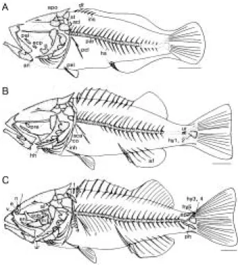
산출 후 27일의 후기자어는 전장이 7.94~9.85 mm (평균8.81 mm, n = 5)로 전새개골에5개의 棘이 형성 되 었고, 아래턱 부분에 角骨(angular)이 骨 하여 턱부분 골격의 骨 가 완성되었으며, 口蓋 의 口蓋骨(palate), 骨(ectopterygoid), 骨(quadrate)이 骨 하기 시작하였다. 蓋 에 骨(epiotic)이 骨 하였고, 骨에 棘이 형성되었으며, 骨은 머리쪽에서 꼬 리쪽으로 骨 하여 17개의 (centrum)가 骨 되었 다. 배지느러미를 지지하는 骨(pelvic girdle)이 최초 로 骨 하기 시작하였고, 管間棘(interhemal spine)과 經間棘(interneural spine)이 骨 하기 시작하였으며, 肩 에 骨의 위쪽 부분의 骨(supratem- poral), 骨(supraclavicle)과 2개의 骨(postcla- vicle)이 骨 하기 시작하였다. 弓 에 6개의 새조골 이 骨 하여 새조골의 骨 가 완성되었다(Fig. 2, A).
산출 후 33일의 치어는 전장이 9.80~12.36 mm (평균 11.24 mm, n = 5)로 이 시기에 눈주위를 구성하는 골격 인 骨(preobital)이 최초로 骨 하기 시작하였고, 弓 의 間 骨(interhyal)과 骨(hypohyal)이 骨 하였으며, 肩 는 骨(coracoid)과 肩胛骨(sca-
pula)이 骨 하였다. 骨은21개의 가 骨 하였
고, 骨(urostyle bone)과 제1, 2 骨(hy- pural bone)이 骨 하기 시작하였다. 骨의 骨 가 완성되었으며, 11개의 經間棘과 6개의 管間棘이 骨 하였다(Fig. 2, B).
산출 후 39일의 치어는 전장이 14.14~14.27 mm (평 균 14.21 mm, n = 5)로 蓋 의 骨(vomer), 骨(eth-
moid), 骨(nasal), 骨(sphenotic), 骨(lateral ethmoid), 骨(alisphenoid)이 骨 되어 蓋 가 완 성되었고, 의 骨(suborbital), 弓 의 骨 (urohyal), 口蓋 의 內 骨(ectopterigoid), 骨 (metapterygoid), 骨(symplectic)이 骨 되어 內 骨의 骨 가 완성되었다. 骨은 25 (10±15)개의
와 8개의 骨(rib)이 모두 骨 하였고, 제3, 4, 5 骨과 骨(parhypural), 骨(epural bone) 이 骨 하였으며, 肩 는 骨(actinost)이 骨 하 여, 모든 골격이 완성되었다(Fig. 2, C).
Fig. 2. Skeletal ossif ication of Sebasticus tertius (Barsukov et Chen).
A: Postlarva, 27 days after bearing, 8.81 mm in total length (TL); B: Juvenile, 33 days after bearing, 11.24 mm in TL; C: Juvenile, 39 days after bearing, 14.21 mm in TL.
a: actinost; af: anal fin; al: alisphenoid; an: angu- lar; co: coracoid; df: dorsal fin; e: ethmoid; ecp: ecto- pterygoid; en: endopterygoid; ep: epural bone; epo:
epiotic; f: free interneural spine; hh: hypohyal; hs;
hemal spine; hy: hypural bone; ihs: interhemal spine; inh: interhyal; ins: interneural spine; l: late- ral ethmoid; mt: metapterygoid; n: nasal; pal: pala- tine; par: parapophysis; pcl: postclavicle; pel: pelvic girdle; ph: parhypural; r: rib; pre: preorbital; q:
quadrate; sca: scapula; scl: supraclavicle: sob: su- borbital; st: supratemporal; sy: symplectic; uh; uro- hyal; ur: urostyle; v: vomer; vf: venteral f in. Scale bars = 1.00 mm.
고 찰
양볼락科 어류는 어린 시기에 종간 형태적 유사성으 로 분류에 있어 어려움이 따르므로 棘의 형태 및 蓋骨의 骨 과정, 자어의 크기 및 색소포의 형성 등 이 중요한 분류 형질로 이용되며, 특히 특징적인 棘 의 이 어린 시기의 형질로 매우 중요하여, 이들의 정확한 동정을 위해서는 棘의 유무를 정확히 관찰하는 것이 필요하다(Okiyama, 1988).
쌍의 棘과 전새개골에 1개의 棘이 형성되었는데, 쏨뱅 이(김 등, 1997a, b)는 평균 전장이 4.43 mm일 때
에 1쌍의 棘과 전새개골에 1개의 棘이, 볼락(김 등, 1993)은 평균 전장이 7.50 mm일 때, 조피볼락(김과 한, 1991)은 평균 전장이 5.83 mm일 때 에 1쌍의 棘 이, 전새개골에 2개의 棘이 형성되어 각 間의 차이를 나타내었다.
산출 직후 붉은쏨뱅이의 자어는 전혀 骨 가 이루어 지지 않았으나, 산출 후 8일, 평균 전장이 4.42 mm에 달
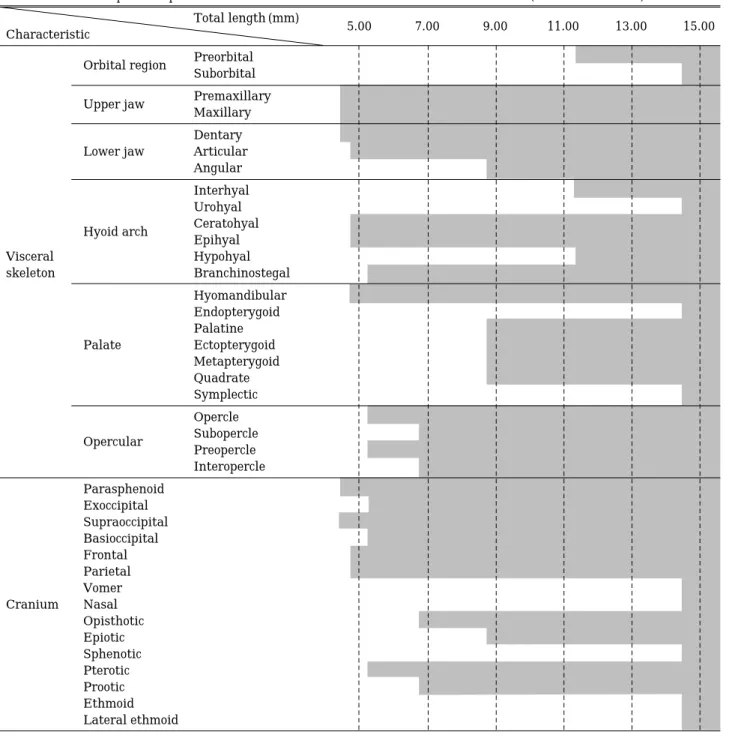
Table 1. The developmental process of visceral skeleton and cranium of Sebasticus tertius (Barsukov et Chen) Total length (mm)
Characteristic 5.00 7.00 9.00 11.00 13.00 15.00
Orbital region Preorbital Suborbital
Upper jaw Premaxillary Maxillary Dentary Lower jaw Articular
Angular Interhyal Urohyal Hyoid arch Ceratohyal
Epihyal Visceral Hypohyal
skeleton Branchinostegal
Hyomandibular Endopterygoid Palatine
Palate Ectopterygoid
Metapterygoid Quadrate Symplectic Opercle Opercular Subopercle
Preopercle Interopercle Parasphenoid
Exoccipital Supraoccipital Basioccipital Frontal Parietal Vomer Cranium Nasal
Opisthotic Epiotic Sphenotic Pterotic Prootic Ethmoid Lateral ethmoid
하면 骨, 骨, 骨이 최초로 骨 하여 산출 후39 일, 평균전장이14.21 mm일 때 모든 골격이 완성되었다. 쏨뱅이(김 등, 1997a, b)는 산출 후 5일, 평균 전장이 3.35 mm일 때 최초로 骨 가 시작되어 산출 후 39일, 평균 전장이 14.36 mm일 때 대부분의 骨 가 완성되었 고, 조피볼락(김과 한, 1991)은 산출 후 6~7일, 평균 전 장이 7.0 mm일 때 처음으로 骨 가 시작되어 산출 후 30~31일, 13.4~16.7 mm의 개체에서 骨 가 완성되었 다. 이 것으로 보아 붉은쏨뱅이의 骨 속도는 같은 어류인 쏨뱅이와 비슷하고, 볼락 어류인 조피볼락 보 다는 다소 느렸다.
붉은쏨뱅이는 骨의 骨, 骨, 骨과 肩 의 骨, 蓋 의 骨이 최초로 骨 하였는데 (Table 1), 이는 쏨뱅이(김 등, 1997a, b)와 동일하고, 조 피볼락(김과 한, 1991)과 볼락(김 등, 1993)은 새개부의 전새개골과 주새개골의 骨 가 함께 일어나 다소 차이 를 보였다. 하지만 이들 모두 턱을 구성하는 骨이 비교적 빠른 속도로 骨 하는데, 이것은 섭이와 영양 등 생존을 위한 적응으로 보인다(김 등, 1993).
붉은쏨뱅이의 새개부 骨 는 주새개골과 전새개골이 동시에 骨 되기 시작하여 하새개골과 간새개골이 骨 되었는데(Table 2), 이는 쏨뱅이(김 등, 1997a, b), 조피볼 락(김과 한, 1991), 볼락(김 등, 1993)과 유사하였다.
붉은쏨뱅이의 骨의 骨 (Table 2)는 쏨뱅이(김 등, 1997a, b), 조피볼락(김과 한, 1991), 볼락(김 등,
1993)과 동일하게 骨의 앞쪽에서 뒤쪽으로 骨 가
진행되었고, 骨의 骨 가 완료 되기전에
骨의 骨 가 시작되었지만, 조피볼락(김과 한, 1991)은 의 가 거의 骨 된 후에 骨이 骨
하여 붉은쏨뱅이와 차이를 나타내었다.
Mook (1997)에 의하면, 어류의 는 그들의 생활 방 식에 의해 骨 가 통제되며, 이러한 생활방식의 차이는 骨 되는 정도와 순서에 변화를 초래하여 자치어의 골 격발달에 다양한 변화를 줄 수 있다고 지적하였는데, 자 치어의 골격 연구는 자치어기의 동정 뿐 아니라, 성 어의 골격 이해 및 계통의 추정에 도움을 주므로 붉은 쏨뱅이 뿐만 아니라 양볼락科 어류들에 대한 체계적인 연구가 계속되어야 하겠다.
적 요
1997년 1월부터 전라남도 완도군 연안에서 연승어업 에 의해 어획된 붉은쏨뱅이를 육상수조에서 사육하던 중, 4월과 5월에 자연산출한 자치어를 사육하면서 관찰 한 자치어의 골격발달과정은 다음과 같다.
산출 후 8일의 후기자어는 전장이 4.23~4.60 mm (평 균 4.42 mm, n = 5)로 가장 먼저 肩 (shoulder girdle) 에 骨(clavicle)과 骨(jaw bones) 부분의 위턱에
骨(premaxillary), 骨(maxillary), 아래턱에 骨(dentary)이 骨 하기 시작하였으며, 蓋 (crani- um)의 배쪽면에 가늘고 긴 막대기 모양의 骨 (parasphenoid)이 骨 되었다
산출 후 15일의 후기자어는 전장이 4.81~5.51 mm (평균 5.23 mm, n = 5)로 蓋 에 骨(pterotic), 基
骨(basioccipital), 骨(exoccipital)이 骨 하기 시작하였고, 에 1개의 棘이 형성되었으며, 새개부(opercular)에 주새개골(opercle)과 1개의 棘을 가진 전새개골(preopercle)이 최초로 骨 하기 시작하였 Table 2. The developmental process of fin skeleton and vertebrae of Sebasticus tertius (Barsukov et Chen)
Total length (mm)
Characteristic 5.00 7.00 9.00 11.00 13.00 15.00
Clavicle Supraclavicle Postclavicle Fin Shoulder girdle
Coracoid
skeleton Actinost
Scapula
Pelvic girdle Pelvic girdle bone Vertebrae
Parapophysis Hypural bone 1, 2 Hypural bone 3, 4, 5 Vertebrae
Free interneural spine Urostyle
Pleural Epipleural
산출 후 27일의 후기자어는 전장이 7.94~9.85 mm (평균 8.81 mm, n = 5)로 전새개골에 5개의 棘이 형성되 었고, 骨은 머리쪽에서 꼬리쪽으로 骨 하여 15개 의 (centrum)가 骨 되었다.
산출 후 39일의 치어는 전장이 14.14~14.27 mm (평 균 14.21 mm, n = 5)로 모든 골격이 완성되었다.
인 용 문 헌
Barsukov, V.V. and L.C. Chen. 1978. Review of the sub- genus Sebastiscus (Sebastes, Scorpaenidae) with a de- scription of a new species. J. Ichthyol., 18(2) : 179~
193.
Fujita, S. 1958. On the egg development and larval stages of a viviparous Scorpanidae f ish, Sebastes oblongus Günther. Bull. Japan Soc. Sci. Fish., 24 : 475~479.
Kawamura, K. and K. Hosoya. 1991. A modif ied double staining technique for making a transparent fish~
skeletal specimen. Bull. Natl. Res. Inst., Aquaculture, 20 : 11~18.
Kendall, W. 1991. Systematics and identification of larvae and juveniles of the genus Sebastes. Env. Biol. Fish., 30 : 173~190.
Masuda, H., K. Amaoka, C. Araga, T. Uyeno and T.
Yoshino. 1984. The Fishes of the Japanese Archipelago.
Tokai University Press, pp. 437.
Mio, S. 1960. Studies on population biology of coastal fishes in Kyushu. I. Biology of Sebastes inermis Cuvier et Valenciennes. Rec. Oceanogr. Works Japan. 5(2) : 419~
436.
Mizue, K. 1958. Studies on a Scorpaenous f ish Sebasticus marmoratus Cuvier et Valenciennes - II. The seasonal cycle of mature testis and the spermatogenesis. Bull.
Fac. Fish. Nagasaki Univ., 6 : 27~38.
Mook, D. 1977. Larval and osteological development of sheephead, Archosagus probatocephalus (Pisces:
Sparidae). Copeia, 1 : 126~133.
Okiyama, M. 1988. An Atlas of the Early Stage Fishes in Japan. Tokai Univ. Press. pp. 1154.
Shinomiya, A. and O. Ezaki. 1991. Mating habits of the rockf ish, Sebastes inermis. Environment. Biol. Fish., 30 : 9~13.
Shiokawa, T. and H. Tsukahara. 1961. Studies on habits of
history of the purple rockfish, Sebastes pachycephalus pachycephalus. Rec. Oceanogr. Works Japan, 5 : 123~
127.
Shiokawa, T. 1962. Studies on habits of coastal fishes in the Amakusa Islands. Part II. Early life history of the purple rockfish, Sebastes pachycephalus pachycephalus.
Rec. Oceanogr. Works Japan, 6 : 103~111.
Takai, T. and T. Fukunaga. 1971. The life history of a ovoviviparous scorpaenoid fish, Sebastes longisponis (Matsubara). I. Eggs and larval stages. Bull. Simonoseki Univ. Fish., 20 : 25~29.
김광수∙임상구∙한경호∙오성현∙노병율. 1999. 붉은쏨뱅이, Sebasticus tertius (Barsukov et Chen)의 초기생활사 1.
난의 형태 및 산출 자치어의 성장에 따른 형태발달. 한국 양식학회지, 12(1) : 15~23.
김용억∙한경호. 1991. 조피볼락, Sebastes schlegeli의 초기생 활사. 한국어류학회지, 3 : 67~83.
김익수∙이완옥. 1993. 한국산 양볼락과 어류의 분류 및4 미 기록종. 한국동물학회지, 36 : 452~475.
김용억∙한경호. 1993. 볼락, Sebastes inermis의 초기생활사 에 관한 연구 1. 인위적 방법에 의한 수조내에서의 난발 생과정과 부화자어의 형태. 한국수산학회지, 26 : 458~
464.
김용억∙한경호∙변순규. 1993. 볼락, Sebastes inermis의 초 기생활사에 관한 연구 2. 산출 자치어의 외부형태 및 골 격발달. 한국수산학회지, 26 : 465~476.
김용억∙한경호∙강충배∙김진구∙변순규. 1997a. 쏨뱅이, Sebasticus mamoratus 1. 인위적인 방법에 의한 수조 내 에서의 난발생과정과 자어기의 형태. 한국어류학회지, 9(2) : 175~185.
김용억∙한경호∙강충배∙김진구∙변순규. 1997b. 쏨뱅이, Sebasticus mamoratus 2. 산출 자치어의 외부형태 및 골 격 발달. 한국어류학회지, 9(2) : 186~194.
한경호∙김용억∙김충만. 1996. 불볼락, Sebastes thompsoni 과 개볼락, Sebastes pachycephalus pachycephalus의 난 형태 및 자어의 형태발달. 한어지, 8(2) : 1~10.
한경호∙유동재∙변순규. 1999.한국산 붉은쏨뱅이(Sebasticus tertius)의 형태 및 골격. 여수대학교논문집, 14(2) : 537~
545.
한국동물분류학회. 1997. 한국동물명집(곤충제외). 도서출판 아카데미서적, 서울, pp. 243~281.
區 究 . 1995. シナ ∙ 鑑.
, 京, pp. 288.
Received : January 25, 2001 Accetped : March 17, 2001