서 론
어류의 종묘생산에 있어서 자치어 골격발달 연구는 사육 초 기에 골격 이상을 확인하고, 제거하는 데 필수적이며, 종의 동 정뿐만 아니라 성어의 골격특성에도 중요한 자료가 될 수 있 으므로 구체적이고 체계적인 연구가 필요하다(Koumoundou
ros et al., 1997a, b; Park et al., 2014). 또한 유어기의 형태발달
단계는 계통분류학적 기초형질 발현과 기능적인 발달경향 및 환경에 대한 선호도를 이해하는 데 도움을 주고 있어 이 분야 의 연구는 다양하다(Mook, 1977; Potthoff et al., 1988; Fuku
hara, 1992; Faustino and Power, 1999; Koumoundouros et al., 1999; Liu, 2001; Sfakianakis et al., 2004).
능성어(Epinephelus septemfasciatus)는 농어목(Perciformes) 바리과(Serranidae)에 속하는 어류로 바리과는 우리나라에서 12속 27종이 서식하고 있고, 남해안을 비롯한 제주도, 일본 및 중국에 분포하고 있다(Kim et al., 2005). 능성어에 관한 연구 로는 배란유도(Kim et al., 1997a), 유어기의 성장과 형태(Mi
—189 — http://www.fishkorea.or.kr
능성어 (Epinephelus septemfasciatus) 자치어의 골격발달
박종연·홍창기1·조재권·손맹현1·한경호2·박재민3,
*
국립수산과학원 양식관리과, 1남서해수산연구소 해역산업과,
2전남대학교 양식생물학전공, 3경상북도 토속어류산업화센터
Early Osteological Development of the Larvae and Juveniles in Sevenband Grouper, Epinephelus
septemfasciatus(Pisces: Serranidae)
by Jong Youn Park, Chang Gi Hong1, Jae Kwon Cho, Maeng Hyun Son1, Kyeong Ho Han2 and Jae Min Park3,* (Aquaculture Management Division, Aquaculture Research Institute, NFRDI, Busan 46083, Korea; 1Southwest Sea Fisheries Research Institute, NFRDI, Yeosu 59780, Korea; 2Chonnam National University, Department of Aqualife Science, Yeosu 59626, Korea; 3Gyeongsangbuk-do Native Fish Business Center, Uiseong 37366, Korea)ABSTRACT This research has been launched in order to observe the osteological development of sevenband grouper, Epinephelus septemfasciatus larvae and juveniles and will be used for the basic data of phylogenetically systematics research. We got some samples of larvae and juveniles from a adult fish which belongs to the sea cage in Geomun-do, Yeosu-si, Jeolla-namdo province, and we have watched them. The average of breeding water temperature was 21.5~24.5 °C (average 23.0±1.5
°C). In yolk-sac larvae just after hatching, ossification was not observed at all, and when the average total length reached 2.66 mm, the premaxillary and maxillary forming the upper jaw, dentary forming the lower jaw, and parasphenoid forming the skull base were ossified. In addition, the preopercle and opercle forming the opercular region began to be ossified. The caudal skeleton supporting the caudal fin is built with the caudal complex consisting of the malleus and incus. Until the average total length reached 2.59 mm, ossification did not occur at all and the urostyle bent at 45-degree angle. As to the ossification of the pterygiophore, when the average total length was 6.12 mm, the first interhemal spine began to be ossified in articulation with two spines of the anal fin, and the synapse also began to be ossified in articulation with two spines of the dorsal fin. When the average total length was 21.9 mm, individuals with completed ossification of the interhemal spines and synaptic clefts began to appear.
Key words: Epinephelus septemfasciatus, juvenile, larvae, osteological development
* Corresponding author: Jae Min Park Tel: 82548308831, Fax: 82548308809, Email: [email protected] ISSN: 1225-8598(Print), 2288-3371(online)
Accepted: September 23, 2015
yailara et al., 1989), 난 발생 및 자치어 형태발달(Kitajima et al., 1991), 번식기술개발(Lee and Go, 2003), 성전환 유도(Song et al., 2005a), 자치어 기형(Nagano et al., 2007) 및 초기생활사 (Park et al., 2014) 등이 수행되었다. 최근 능성어의 종묘생산 기술개발 연구가 활발히 진행되면서 초기 성장에 있어 골격형 태 이상과 기형발생이 문제가 되고 있다(Nagano et al., 2007).
이러한 골격발달 이상은 어미관리 부족과 선천적인 기형으로 인해 초기 자어의 생존에 큰 영향을 미치고 있는 것으로 보고 있다(NFRDI, 2014). 따라서 본 연구에서는 자치어의 골격발 달 과정을 관찰하여 골격에 대한 기초자료를 확보하고, 자치 어 시기의 종 동정을 위한 분류학적 연구자료로 이용하고자 한다.
재료 및 방법
1. 자치어 사육
능성어 자치어는 2012년 6월 전남 여수시 거문도에 위치한 해상가두리에서 사육하던 어미로부터 채란 및 인공수정하여 사육한 것을 대상으로 관찰하였다. 자치어는 25톤 사각수조 (800×800×40cm)에서 부화 후 35일까지 지수식으로 사육하 였고, 에어레이션으로 약하게 폭기시켜주었으며, 부화 후 35일
부터는 유수식으로 사육하였다. 사육 중 수온범위는 21.5~
24.5°C(평균 23.0±1.5°C)였고, 염분은 32.5~33.5psu(평균 33.0psu) 범위를 유지시켜 주었다. 먹이는 부화 후 3일부터 35 일까지 기수산 로티퍼(Branchionus rotundiformis)를 mL당 10~15개체 공급하였고, 35일부터 70일까지는 로티퍼와 알테 미아(Artemia nauplius)를 mL당 10~20개체씩 혼합 공급하였 으며, 55일 이후에는 초기배합사료(Love larva, Japan)를 공급 하였다.
2. 시료확보 및 골격염색
자치어는 부화 직후부터 매일 10마리씩 무작위로 채집하였 고, 5% 중성포르말린에 고정시킨 뒤 Walker and Kimmel(2007) 의 이중염색법에 의해 염색하였으며, KOH 0.1%와 Glycerol 50%에 보존하였다. 염색한 자치어는 실체현미경(Nikon JP SMZ800, Japan)을 이용하여 관찰하고 스케치하였다. 자치어의 골격 각 부위 명칭은 Kang et al.(2012)에 따랐다.
결 과
능성어의 자치어 골격발달은 두개골(cranium), 내장골(vis
ceral skeleton), 척추골(vertebrae), 견대골(pectoral girdle bone),
Fig. 1. Development of the caranium and visceral skeleton in sevenband grouper, Epinephelus septemfasciatus. A: 2.66mm in TL; B: 4.11mm in TL; C: 11.9mm in TL; D: 14.4mm in TL; E: 17.9mm in TL; F: 21.9mm in TL. al, alisphenoid; an, angular; ar, articular; bac, basioccipital; bas, basisphenoid; brs, branchiostegal rays; d, dentary; ecp, ectopterygoid; eh, epihyal; ep, epiotic; enp, endopterygoid; exo, exoccipital; fr, frontal; h, hypohyal; hm, hyomandibular; ih, interhyal; iop, interopercle; le, lateral ethmoid; lu, preorbital=lucrymal; me, mesethmoid; mp, metapterygoid;
mx, maxillary; na, nasal; op, opercle; opi, opisthotic; pa, parasphenoid; pal, palatine; par, parietal; pt, pterotic; pop, preopercle; pr, prootic; pmx, premaxillary; q, quadrate; soc, supraoccipital; sp, sphenotic; sop, subopercle; sor, suborbital; spm, supramaxillary; sy, sympletic. Scale bars=0.5 mm.
A B C
D E F
미골(caudal skeleton), 지느러미 및 담기골(pterygiophore)로 구분하여 관찰하였다.
1. 두부골격
두부골격은 두개골과 내장골로 구성되어 있고, 발육단계에 따른 골격발달 과정은 다음과 같다(Fig. 1, Table 1).
부화 직후의 난황을 가진 자어는 골화가 전혀 이루어지지
않았고, 평균 전장 2.66mm에 달하면 윗턱을 구성하는 전상악 골(premaxillary)과 주상악골(maxillary)이 골화되었고, 아래턱 을 구성하는 치골(dentary)이 골화하였으며, 두개골의 기저를 형성하는 부설골(parasphenoid)이 골화하였다. 새개부에는 전 새개골(preopercle)과 주새개골(opercle)의 골화가 시작되었다 (Fig. 1A).
평균 전장 4.11mm에 달하면 아래턱에 관절골(articular)과 각골(angular)이 골화되었고, 설궁부에는 상설골(epihyal)과 4 Table 1. Development process of the cranium and visceral skeleton in sevenband grouper, Epinephelus septemfasciatus
Elements Total length(mm)
2.66 2.72 4.11 4.21 6.12 6.31 11.9 14.4 17.9 19.3 21.9
Carnium Parasphenoid Supraoccipital Epiotic Alisphenoid Parietal Prootic Basioccipital Sphenotic Pterotic Frontal Lateral ethmoid Nasal
Exoccipital Opisthotic Mesethmoid Basisphenoid Orbital region
Suborbital Preorbital Hyoid arch Epihyal Branchiostegal Hypohyal Interhyal Palate
Metapterygoid Palatine Ectopterygoid Sympletic Endopterygoid Hyomandibular Quadrate Opercular Opercle Preopercle Interopercle Subopercle Jaw bone
Maxillary Premaxillary Dentary Supramaxillary Angular Articular
개의 새조골(branchiostegal rays)이 골화하였으며, 구개부에는 설악골(hyomandibular)이 골화하였다(Fig. 1B).
평균 전장 11.9mm에 달하면 두개골에는 눈의 위쪽에 액골 (frontal), 아래쪽 안와부에는 안전골(preorbital=lucrymal)이 골화하였고, 기저후두골(basioccipital)이 골화하였다. 설악골 위쪽에는 설이골(sphenotic)과 익이골(pterotic)이 골화하였고, 구개부에는 외익상골(ectopterygoid), 구개골(palatine) 방골 (quadrate) 및 접속골(sympletic)이 골화하였다. 새개부에는 간 새개골(interopercle)과 하새개골(subopercle)이 골화하였다 (Fig. 1C).
평균 전장 14.4mm에 달하면 두개골에는 비골(nasal), 측사 골(lateral ethmoid), 익설골(alisphenoid), 노정골(parietal), 전 이골(prootic), 상이골(epiotic) 및 상후두골(supraoccipital)이 골화하기 시작하였고, 구개부에는 내익상골(endopterygoid) 및 후익상골(metapterygoid)이 골화하였으며, 설궁부에는 하설골 (hypohyal) 및 간설골(interhyal)이 골화하였다(Fig. 1D).
평균 전장 17.9mm에 달하면 두개골에는 외후두골(exocci
pital), 후이골(opisthotic) 및 중사골(mesethmoid)이 골화하 였으며, 악골에는 상주상악골(supramaxillary)이 골화하였다 (Fig. 1E). 평균 전장 21.9mm에 달하면 두개골에는 기저설골 (basisphenoid), 안와부에는 안하골(suborbital)이 골화하면서 골격들의 골화가 완료되었다(Fig. 1F).
2. 미골
꼬리지느러미를 지지하는 미골(caudal skeleton)은 추골과
골편으로 이루어진 미골복합체로 구성된다(Fig. 2, Table 2).
평균 전장 2.59mm까지는 전혀 골화가 일어나지 않았고, 미부 봉상골(urostyle)이 45°로 휘어져 있었다(Fig. 2A).
평균 전장 4.11mm로 성장하면서 미부봉상골의 일부가 골 화하였고, 5개의 하미축골(hypural bone)이 골화하였다(Fig.
2B). 평균 전장 6.12mm에는 신경극(neural spine)과 혈관극 (hemal spine)이 골화하였고, 1개의 준하미축골(parhypural bone)이 골화하기 시작하였다(Fig. 2C).
평균 전장 11.9mm에는 첫 번째 상미축골(epural bone)과 미골판(caudal bony plate)의 골화가 시작되었다(Fig. 2D). 평 균 전장 14.4mm에는 두 번째 상미축골이 골화되었고, 미부 봉상골과 5번째 하미축골 사이에는 신경골(uroneural)이 골화 하였다(Fig. 2E). 평균 전장 21.9mm에는 제3번째 상미축골이 골화되어 이 시기에 모든 골편들의 골화가 완성되어 성어의 형태를 갖추게 되었다(Fig. 2F).
3. 견대골
가슴지느러미를 지지하는 견대골(pectoral girdle bone)은 수 개의 막골이 부속되며, 후측두골(posttemporal)에 의해 두개골 에 관절되었다(Fig. 3, Table 2). 평균 전장 2.59mm에서는 쇄골 (cleithrum)이 골화하기 시작하였고(Fig. 3A), 평균 전장 4.11 mm에서는 쇄골의 윗부분에 상쇄골(supracleithrum)이 골화하 여 후측두골과 관절되었고, 후쇄골(ventral post cleithrum)이 골화하였다(Fig. 3B). 평균 전장 6.12mm에서는 1개의 후쇄골 이 골화하기 시작하였고(Fig. 3C), 평균 전장 11.9mm에서는
Fig. 2. Development of the caudal skeleton in sevenband grouper, Epinephelus septemfasciatus. A: 2.59mm in TL; B: 4.11mm in TL; C: 6.12 mm in TL; D: 11.9mm in TL; E: 14.4mm in TL; F: 21.9mm in TL. cbp, caudal bony plate; ep l~3, epural l~3; hs, hemal spine; hy l~5, hypural l~5; ns, neural spine; ph, parhypural; uro, uroneural; us, urostyle. Scale bars=0.5mm.
A B C
D E F
us hy5 ns
hs ph
ep3 ep2 uro
cbp ep1
hy4 hy3 hy2 hy1
오훼골(coracoid), 견갑골(scapula), 사출골(actinost=radial) 이 골화하였고, 가슴지느러미 줄기가 분화하였다(Fig. 3D). 평 균 전장 14.4mm에서는 상측두골(supraposttemporal)이 골화
하였고, 견갑골은 사각형으로 오훼골은 망치 모양으로 골화가 진행되었다(Fig. 3E). 평균 전장 21.9mm에서는 상측두골의 끝이 뾰족해지면서 길어졌고, 후쇄골의 길이가 길어지면서 쇄 Table 2. Development process of the pectoral girdle bone, caudal skeleton in sevenband grouper, Epinephelus septemfasciatus
Elements Total length(mm)
2.66 2.72 4.11 4.21 6.12 6.31 11.9 14.4 17.9 19.3 21.9
Pectoral girdle Cleithrum Dosal post leithrum Ventral post leithrum Supracleithrum Posttemporal Supraposttemporal Coracoid Scapula Actinost(radial) Caudal skeleton
Urostyle
Hypural 1~5th
Parhypural
Epural 1st
2nd3nd Uroneural 4nd
Caudal bony plate
Fig. 3. Development of the pectoral girdle bone in sevenband grouper, Epinephelus septemfasciatus. A: 2.59mm in TL; B: 4.11mm in TL; C:
6.12mm in TL; D: 11.9mm in TL; E: 14.4mm in TL; F: 21.9mm in TL. cl, cleithrum; cr, coracoid; dpcl, dosal post cleithrum; pt, porttemporal; r, radial; sc, scapula; scl, supracleithrum; spt, supraposttemporal; vpcl, ventral post cleithrum. Scale bars=0.5mm.
A B C
D E F
cl
scl
vpcl
dpcl
spt
sc
cr
pt
r
골을 형성하는 모든 골격이 완성되었다(Fig. 3F).
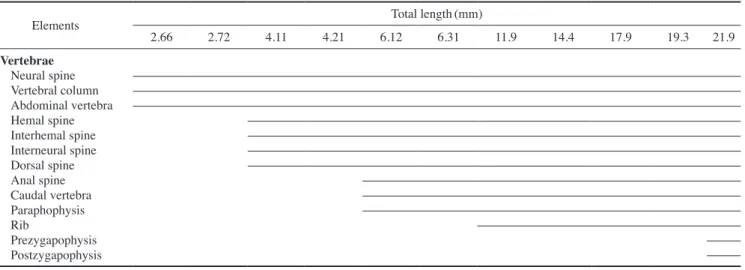
4. 척추골
몸의 중축을 이루는 척추골의 골화과정은 다음과 같았다 (Fig. 4, Table 3). 평균 전장 2.66mm에서는 복추골(abdominal vertebrae)에서 1~2번째 추체(centrum)의 골화가 신경극 (neural spine)과 함께 시작되었다(Fig. 4A). 이후 평균 전장 4.11mm로 성장하면서 복추골의 추체와 신경극이 모두 골화 되었고, 혈관극(hemal spine)과 신경극의 골화가 꼬리 쪽으로 진행되었다(Fig. 4B).
평균 전장 6.12mm에서는 7개의 복추골과 13개의 미추골 (caudal vertebrae)이 골화하였고, 두 번째 복추골에서부터 꼬 리쪽으로 5개의 측돌기(paraphophysis)가 형성되었다(Fig.
4C). 평균 전장 11.9mm에서는 8개의 복추골이 골화하였고, 6 개의 측돌기가 길게 형성되었으며, 늑골(rib)이 길게 형성되어 15개의 미추골이 골화하였다. 이 시기에는 신경극과 혈관극에 신경궁문(neural arch)과 혈도궁문(hemal arch)이 골화되면서 모든 추체의 골화가 완료되었다(Fig. 4D).
평균 전장 21.9mm에 달하면 미추골의 등 쪽에 전관절돌기 (prezygapophysis)와 후관절돌기(postzygapophysis)가 발달하 면서 모든 척추골을 형성하는 골격이 완성되었다(Fig. 4E).
5. 지느러미 및 담기골
각 지느러미와 등지느러미를 지지하는 신경간극과 뒷지느 러미를 지지하는 혈관간극 즉, 담기골(pterygiophore)의 골화 과정을 Fig. 4에 나타냈다. 지느러미 중 꼬리지느러미의 줄기 수는 부화 후 31일경인 평균 전장 11.9mm에 이미 정수에 달 하여 골화되었으며, 평균 전장 17.9mm에 달하면 꼬리지느러 미의 부속줄기의 골화가 완료되어 위쪽이 5~6개, 아래쪽이 7
~8개에 달하여 골화가 완료되었다. 등지느러미의 경우 두 번
Table 3. Development process of the vertebrae in sevenband grouper, Epinephelus septemfasciatus
Elements Total length(mm)
2.66 2.72 4.11 4.21 6.12 6.31 11.9 14.4 17.9 19.3 21.9
Vertebrae Neural spine Vertebral column Abdominal vertebra Hemal spine Interhemal spine Interneural spine Dorsal spine Anal spine Caudal vertebra Paraphophysis RibPrezygapophysis Postzygapophysis
Fig. 4. Development of the vertebrae, spinous and soft fin in seven
band grouper, Epinephelus septemfasciatus. A. 2.66mm in TL; B:
4.11mm in TL; C: 6.12mm in TL; D: 11.9mm in TL; E: 21.9mm in TL. as, anal spine; dp, dorsal spine; hs, hemal spine; ihs, interhemal spine; ins, interneural; ns, neural spine; pp, parapophysis; r, rib; v, vertebrae column. Scale bars=0.5mm.
A
B
C
D
E
째 등지느러미 가시의 극이 첫 번째보다 길어진 것이 평균 전 장 4.11mm에서 나타났고, 다시 극이 줄어들어 성어와 같은 완전한 형태를 갖춘 것은 평균 전장 21.9mm에서 나타났다.
배지느러미는 평균 전장 4.11mm에 최초로 1개의 줄기가 골 화되었고, 그중 첫 번째 배지느러미 가시의 극이 길어졌다. 평 균 전장 21.0mm에서는 첫 번째 가시의 극이 줄어들면서 성 어와 같은 형태를 갖추었다. 뒷지느러미는 꼬리지느러미와 같 은 평균 전장 11.9mm에서 골화가 진행됨과 동시에 그 수가 정수에 달하였다. 담기골의 골화는 평균 전장 6.12mm에서 첫 번째 혈관간극이 뒷지느러미 2개의 가시와 관절되어 가장 먼 저 골화가 시작되고, 신경간극 또한 등지느러미 2개의 가시와 관절되어 골화하기 시작하였으며, 혈관간극과 신경간극은 평 균 전장 21.9mm에 골화가 완료되는 개체가 출현하였다.
고 찰
일반적으로 경골어류는 두개골과 지느러미의 발달이 부화 이후에 관찰되는 것과 부화 이전에 골격 발달이 진행되는 것 으로 나누어진다. 주로 연어과 어류는 부화 이전에 골격이 발 달하는 것으로 알려져 있고, 농어목 어류는 부화 이후에 골격 이 발달되어 능성어의 골격발달은 후자에 속하였다(Matsuoka, 1985; Koumoundouros et al., 1997b; Faustion and Power, 1999;
Sfakianakis et al., 2004).
능성어는 최초 견대부의 쇄골과 두개골의 기저를 형성하는 부설골이 골화하였다. 점농어 Lateolabrax maculatus(Kang et al., 2012)는 쇄골이 가장 먼저 골화하였고, 이어서 두개골을 형성하는 전상악골, 치골 및 부설골의 골화가 진행되었다. 쏨 뱅이 Sebastiscus marmoratus(Kim et al., 1997b)와 붉은쏨뱅 이 Sebastiscus tertius(Han et al., 2001)는 쇄골과 악골 및 부설 골이 동시에 골화하였고, 황점볼락 Sebastes oblongus(Byun et al., 2012)은 쇄골이 먼저 골화한 뒤 전상악골 및 노정골이 골화하였다. 저울베도라치 Entomacrodus stellifer(Kim et al., 1992a)는 쇄골과 부설골이 먼저 골화된 다음 악골이 골화하였 고, 졸복 Takifugu pardalis(Han et al., 2005)은 쇄골과 부설골 및 악골이 동시에 골화하여 어종별로 차이를 나타내었다.
대부분의 어류들은 부화 후 가장 먼저 턱을 구성하는 악골이 골화하는 특징을 나타내는데 이것은 섭이와 호흡을 위해 우선 적으로 골화가 일어나는 부위이며(Vandewalle et al., 1997), 입의 개구와 섭이 기능에 sternohyoideus mescle을 지지하는 쇄골이 중요한 역할을 하기 때문이다(Wagemans and Vande
walle, 1999; Kang et al., 2012).
능성어는 가슴지느러미를 지지하는 골격 중 하나인 견갑골 에 한 개의 구멍이 형성되었는데 이러한 구멍은 농어목 어류의 전형적인 형질로 알려져 있다(Koumoundouros et al., 2001).
견갑골에 구멍이 형성되는 종으로는 참돔 Pagrus major(Matsu
oka, 1987), 황돔 Dentex dentex(Koumoundouros et al., 2001), 점농어(Kang et al., 2012), 자리돔류(Emery, 1973) 등의 농어목 어류를 비롯하여 쏨뱅이목 어류의 쏨뱅이(Kim et al., 1997b), 붉은쏨뱅이(Han et al., 2001), 조피볼락 Sebastes schlegeli(Kim and Han, 1991), 동갈치목의 전력날치 Cheilopogon heterurus doederleini(Dasilao and Yamaoka, 1998), 복어목 어류의 Bal
istes capriscus(Matsuura and Katsuragawa, 1984) 등 다른 분 류군에서도 형성되는 것으로 보아 농어목 어류에서 볼 수 있 는 전형적인 형질이 아닌 것으로 판단된다.
능성어의 새개부는 전새개골과 주새개골이 동시에 골화하 였고, 이후 간새개골과 하새개골이 골화하였는데 이는 쏨뱅이 목 어류의 쏨뱅이(Kim et al., 1997b), 붉은쏨뱅이(Han et al., 2001), 조피볼락(Kim and Han, 1991) 및 볼락(Kim et al., 1993) 등과 일치하였으며, 전력날치(Dasilao and Yamaoka, 1998), 참 돔(Matsuoka, 1987), 미끈망둑 Luciogobius guttatus(Kim et al., 1992b), 잿방어 Seriola dumerili(Liu, 2001) 등도 같은 경 향을 보여 골화 순서는 다른 어종과 특별한 차이를 보이지 않 는 것으로 생각된다.
능성어는 평균 전장 2.66mm일 때 척추골의 1~2번째 추체 가 골화하기 시작하여 점차 미추골 쪽으로 진행되었으며, 이러 한 경향은 꽁치 Cololabis saira(Fujita and Oozeki, 1994), 복섬 Takifugu niphobles(Fujita, 1992), 황점볼락(Byun et al., 2012), 쏨뱅이(Kim et al., 1997b), 붉은쏨뱅이(Han et al., 2001), 조피 볼락(Kim and Han, 1991) 및 점농어(Kang et al., 2012) 등에서 나타나 추체의 골화방향은 어종에 따른 차이가 없는 것으로 판단된다.
능성어는 추체가 모두 골화한 뒤 미부봉상골이 골화되었으 나 황점볼락(Byun et al., 2012), 복섬(Fujita, 1992), 꽁치(Fuji ta and Oozeki, 1994) 및 점농어(Kang et al., 2012) 등은 추체가 모두 골화되기 전에 미부봉상골의 골화가 시작되어 능성어와 차이를 보였다. 능성어는 추체가 골화되면서 신경극이 발달 하였고, 이어서 혈관극이 발달하였다. 꽁치(Fujita and Oozeki, 1994), 복섬(Fujita, 1992) 및 황점볼락(Byun et al., 2012)은 추 체가 완전히 골화되기 전에 신경극과 혈관극이 골화하여 차이 를 보였다.
농어목 어류의 지느러미 발달은 전방의 등지느러미 발달이 되기 이전에 후방의 등지느러미와 뒷지느러미가 먼저 발달 하기 시작하는 것이 일반적이다(Johnson, 1984; Faustino and Power, 1999). 능성어는 등지느러미와 뒷지느러미의 발달이 일반 농어목 어류와 같은 경향을 보였고, 담기골은 등지느러 미의 골화가 시작되면서 발달하였으며, 골화의 진행방향은 전 방에서 후방으로 진행되는 특징을 보였다. Park et al.(2014)의 보고에 따르면 능성어는 부화 후 9~12일 사이에 등지느러미 와 배지느러미의 극조가 형성되기 시작하는데 이 중 등지느러
미 두 번째 극조와 배지느러미 첫 번째 극조의 길이가 다른 극 조와 연조에 비해 길게 신장하며, 부화 후 50일경부터 다시 길 이가 짧아진다. 이러한 현상은 바리과 어류에 나타나는 특징으 로 붉바리 Epinephelus akaara(Fukuhara and Fushimi, 1988), 자바리 Epinephelus bruneus(Song et al., 2005b) 및 가시우럭 Liopropoma japonicum(Kim et al., 2004) 등에서 볼 수 있다.
능성어의 등지느러미는 전방에서 후방으로 순차적으로 골 화가 진행되었고, 점농어(Kang et al., 2012)는 전, 후방의 등지 느러미와 뒷지느러미가 동시에 골화하여, 능성어와 차이를 보 였으나 담기골은 전방에서 후방으로 발달이 진행되어 같은 경 향을 나타내었다. Diplodus puntazzo(Sfakianakis et al., 2005) 와 Diplodus sargus(Komoundouros et al., 2001)는 신경극과 혈관극의 골화가 전, 후방에서 거의 동시에 이루어졌고, Pagel
lus erythrinus(Sfakianakis et al., 2004)는 신경극의 후방에서 골화가 시작되어 이후 전, 후방에서 신경극의 골화가 동시에 이루어졌다. 참돔(Matsuoka, 1985)은 신경극과 혈관극의 골화 가 동시에 진행되나 신경극의 경우 전, 후방의 골화가 진행된 이후 중앙 쪽으로 골화가 진행되어 어종별로 다양한 골화과정 을 나타냈다.
경골어류의 꼬리지느러미를 지지하는 미골은 추골과 골편 으로 이루어진 미골복합체로 구성되어 있다. 능성어는 부화 직후부터 평균 전장 2.59mm까지는 골화가 일어나지 않았고, 이후에 추체가 모두 골화되기 전에, 미부봉상골(urostyle)이 45°로 휘어지기 시작하였다. 이와 같은 경향은 대부분의 경골 어류에서 나타났으며, 황점볼락(Byun et al., 2012), 붉은쏨뱅 이(Han et al., 2001), 쏨뱅이(Kim et al., 1997b) 및 볼락(Kim et al., 1993) 등의 경우 척추골의 미부 쪽 골화가 완전하지 않 았을 때 미부봉상골의 골화가 완료되었다. 다만 조피볼락(Kim and Han, 1991)의 경우 추체의 골화가 완료된 후에 미부봉상 골이 골화하기 시작하여 차이를 보였다. 하미축골은 미골을 형성하는 주요골격 중 하나로 능성어의 경우 외관상 3개의 하 미축골(하미축골 1+2,3,4+5)이 형성되었다. 황점볼락은 3개 의 하미축골(1+2,3+4,5)이 융합되었고 이와 같은 형태의 양 상을 보이는 어류는 볼락(Kim et al., 1993), 조피볼락(Kim and Han, 1991), Balistes capriscus(Matsuura and Katsuragawa, 1984) 등에서 알려져 있다. 방어류(Kohno, 1997; Liu, 2001)는 하미축골이 1+2,3+4+5가 서로 융합되었고, Hemilepidotus spp.(Kendall and Vinter, 1984)는 외관상 2개의 하미축골(준 하미축골+하미축골 1+2의 융합, 하미축골 3+4,5번째 하미 축골 소실)을 형성하는 종류도 있으며, 어종마다 다양한 융합 형태를 보여 하미축골의 융합현상은 계통학적 연구의 기초자 료로서 매우 중요하다고 판단된다. 이와 같이 어류의 해부학 적 정보는 유사종과의 식별이나 생태학 분야에서 유용한 자료 로 활용될 수 있으며, 향후 종묘생산에서 문제가 되고 있는 자 치어 시기의 기형발생과 원인을 파악하기 위해서는 먹이, 수온
및 수질환경 등 자치어 사육에 대한 구체적인 연구가 필요할 것으로 생각된다.
요 약
본 연구는 능성어 자치어의 골격발달을 관찰하여 분류학적 연구의 기초자료로 이용하고자 실시하였다. 자치어는 전남 여 수시 거문도에 위치한 해상가두리에서 사육하던 어미로부터 채란하여 사육한 것을 대상으로 관찰하였다. 자치어의 사육 수온범위는 21.5~24.5°C(평균 23.0±1.5°C)였다. 부화 직후 의 난황을 가진 자어는 골화가 전혀 이루어지지 않았고, 평균 전장 2.66mm에 달하면 윗턱을 구성하는 전상악골과 주상악 골이 골화되었고, 아래턱을 구성하는 치골이 골화하였으며, 두 개골의 기저를 형성하는 부설골이 골화하였다. 새개부를 형성 하는 전새개골과 주새개골의 골화가 시작되었다. 꼬리지느러 미를 지지하는 미골은 추골과 골편으로 이루어진 미골복합체 로 구성된다. 평균 전장 2.59mm까지는 전혀 골화가 일어나지 않았고, 미부봉상골이 45°로 휘어져 있다. 담기골의 골화는 평 균 전장 6.12mm에 첫 번째 혈관간극이 뒷지느러미 2개의 가 시와 관절되어 가장 먼저 골화가 시작되고, 신경간극 또한 등 지느러미 2개의 가시와 관절되어 골화하기 시작하였으며, 혈 관간극과 신경간극은 평균 전장 21.9mm에 골화가 완료되는 개체가 출현하였다.
사 사
이 연구는 국립수산과학원 시험연구사업비(RP2015AQ
074)의 지원에 의해 수행되었으며, 본 연구가 수행되기까지 많은 도움을 주신 청솔수산 윤낙진, 주일수산 박주일 사장님 께 감사의 말씀을 전합니다.
referenCes
Byun, S.G., C.B. Kang, J.G. Myoung, B.S. Cha, K.H. Han and C.G.
Jung. 2012. Early osteological development of the larvae and juveniles in Sebastes oblongus(Pisces: Scorpaenidae).
Kor. J. Ichthyol., 24: 6776.(in Korean)
Dasilao, Jr J.C. and K. Yamaoka. 1998. Osteological and functional development of the flyingfish, Cypselurus heterurus doeder
leini(Teleostei: Exocoetidae). Bull. Mar. Sci. Fish. Kochi Univ., 18: 1326.
Emery, A.R. 1973. Ecology and functional osteology damselfish (Pisces; Pomacentridae) at Alligator reef, Florida Keys.
Bull. Mar. Sci., 23: 649770.
Faustino, M. and D.M. Power. 1999. Development of the pectoral, pelvic, dorsal and anal fins in cultured sea bream. J. Fish.
Biol., 54: 10941110.
Fujita, K. 1992. Development of the caudal skeleton in the tetra
odontid fish, Takifugu niphobles. Japan J. Ichthyol., 38: 438
440.
Fujita, K. and Y. Oozeki. 1994. Development of the caudal skeleton in the saury, Cololabis saira. Japan J. Ichthyol., 41: 334
337.
Fukuhara, O. 1992. Study on the development of functional mor
phology and behavior of the larvae of eight commercially valuable teleost fishes. Contr. Fish Res. Jpn. Sea Block, 25:
1122.
Fukuhara, O. and T. Fushimi. 1988. Fin differentiation and squa
mation of artificially reared grouper, Epinephelus akaara.
Aquaculture, 69: 379386.
Han, K.H., J.K. Cho, S.H. Lee, S.Y. Hwang, S.M. Yoon, W.I. Seo and C.C. Kim. 2005. Osteological development of the larvae and juveniles of Takifugu pardalis(Teleostei: Tetraodonto
dae). Kor. J. Ichthyol., 17: 2935.(in Korean)
Han, K.H., S.K. Lim, K.S. Kim, C.W. Kim and D.J. Yoo. 2001.
Osteological development of the larvae and juveniles of Sebasticus tertius(Barsukov et Chen) in Korea. Kor. J. Ich
thyol., 13: 6368.(in Korean)
Johnson, G.D. 1984. Percoidei: development and relationship. In:
Moser H.G., W.J. Richards, D.M. Cohen, M.P. Fahay, A.W.
Kendall, S.L. Richardson(eds). Ontogeny and systematics of fishes. American Society of Ichthyologists and Herpetol
ogists, special publication no 1, Allen Lawrence KS, 464
498.
Kang, C.B., J.G. Myoung, Y.U. Kim and H.C. Kim. 2012. Early Os
teological development and squamation in the spotted sea bass Lateolabrax maculates(Pisces: Lateolabracidae). Kor.
J. Fish Aquat. Sci., 45: 271282.(in Korean)
Kendall, A.W. and B. Vinter. 1984. Development of Hexagrammids (Pisces: Scorpaeniformes) in the northeastern Pacific Ocean.
US Dep. Commer NOAA Tech. Rep. NMFS. 2: 44pp.
Kim, B.H., K.M. Kim, Y.D. Lee, C.B. Song and S. Rho. 1997a. Re
productive biology of the sevenband grouper, Epinephelus septemfasciatus Ⅰ. The effect of HCG on ovulation induction.
J. Aquaculture, 10: 5561.(in Korean)
Kim, I.S., Y. Choi, C.L. Lee, Y.J. Lee, B.J. Kim and J.H. Kim. 2005.
Illustrated book of Korean fishes. KyoHak Publishing, Seoul, pp. 276286.(in Korean)
Kim, S., J.M. Yoo and E.K. Lee. 2004. Description of a postflexion larva specimen of Liopropoma japonicum(Doderlein) off jeju island, Korea. Ocean and Polar Res., 26: 685688.(in Korean)
Kim, Y.U. and K.H. Han. 1991. The early life history of rockfish Sebastes schlegeli. Kor. J. Ichthyol., 3: 6783.(in Korean) Kim, Y.U., K.H. Han and C.B. Kang. 1992a. Morphology and skel
etal development of larvae and juveniles of Entomacrodus
stellifer lighti(Herre). Kor. J. Ichthyol., 4: 3143.(in Kore
Kim, Y.U., K.H. Han and S.K. Byun. 1993. The early life history of an) the rockfish, Sebastes inermis 2. Morphological and skele
tal development of larvae and juveniles. Bull. Korean Fish.
Soc., 26: 465476.(in Korean)
Kim, Y.U., K.H. Han, C.B. Kang and J.W. Ryu. 1992b. Early life history and spawning behavior of the gobiid fish, Luciogo- bius guttatus Gill. Kor. J. Ichthyol., 4: 113.(in Korean) Kim, Y.U., K.H. Han, C.B. Kang, J.K. Kim and S.K. Byun. 1997b.
The early life history of the rockfish, Sebastiscus marmor- atus 2. Morphology and skeletal development of larvae and juvenile. Kor. J. Ichthyol., 9: 186194.(in Korean)
Kitajima, C., M. Takaya, Y. Tsukashima and T. Arakawa. 1991. De
velopment of eggs, larvae and juvenile of the grouper, Epi- nephelus septemfasciatus, reared in the laboratory. Japan J.
Ichthyol., 38: 4755.
Kohno, H. 1997. Osteological development of the caudal skeleton in the carangid, Seriola lalandi. Ichthyol. Res., 44: 219221.
Koumoundouros, G., F. Gagliardi, P. Divanach, C. Boglione, S.
Cataudella and M. Kentouri. 1997a. Normal and abnormal osteological development of caudal fin in Sparus aurata L.
fry. Aquaculture, 149: 215226.
Koumoundouros, G., G. Oran, P. Divanach, S. Stefanakis and M.
Kentouri. 1997b. The opercular complex deformity in in
tensive gilthead sea bream(Spartus aurata L.) larviculture.
Moment of apparition and description. Aquaculture, 156:
165177.
Koumoundouros, G., P. Divanach and M. Kentouri. 1999. Osteolog
ical development of the vertebral column and of the caudal complex in Dentex dentex. J. Fish. Bio., 54: 424436.
Koumoundouros, G., P. Divanach and M. Kentouri. 2001. Osteo
logical development of Dentex dentex(Osteichthyes: Spar
idae): dorsal, anal, paired fins and squamation. Mar. Biol., 38: 399406.
Lee, Y.D. and K.M. Go. 2003. Aquatic industrialization by devel
opment of reproductive technology of sevenband grouper, Epinephelus septemfasciatus. Jeju Univ., pp. 186.(in Kore
an)
Liu, C.H. 2001. Early osteological development of the yellow tail Seriola dumerili(Pisces: Carangidae). Zool. Stud., 40: 289
Matsuoka, M. 1985. Osteological development in the red sea bream, 298.
Pagrus major. Japan. J. Ichthyol., 32: 3551.
Matsuoka, M. 1987. Development of the skeletal tissues and skel
etal muscles in the red sea bream. Bull. Seikai Red. Fish Res. Lab., 65: 114.
Matsuura, Y. and M. Katsuragawa. 1984. Osteological development of fins and their supports of larval grey triggerfish, Balistes capriscus. Japan J. Ichthyol., 31: 411421.
Miyailara, J., T. Arakawa and M. Takaya. 1989. Growth and mor
phology of young hybrids of Epinephelus akaara with E.
septemfasciatus and E. akaara with E. moara. Bull. NPIF,
15: 511.
Mook, D. 1977. Larval and osteological development of the sheeps
head, Archosargus probatocephalus(Pisces: Sparidae). Co
peia, 126133.
Nagano, N., A. Hozawa, W. Fujiki, T. Yamada, K. Miyaki, Y. Saka
kura and A. Hagiwara. 2007. Skeleton development and deformities in cultured larval and juvenile sevenband grou
per, Epinephelus septemfasciatus(Thunberg). Aqua. Res., 38: 121130.
NFRDI(National Fisheries Research & Development Institute).
2014. Technical development for the mass seedling produc
tion of sevenband grouper. Report of Nat. Fish. Res. & De
vel. Ins., pp. 6870.(in Korean)
Park, J.M., J.K. Cho, K.H. Han, N.R. Kim, H.K. Hwang, K.M. Kim, J.I. Myeong and M.H. Son. 2014. Early life history of the sevenband grouper, Epinephelus septemfasciatus from Ko
rea. Dev. Reprod., 18: 1323.(in Korean)
Potthoff, T., S. Kelley and L.A. Collins. 1988. Osteological devel
opment of the red snapper, Lutjanu scampechanus(Lutjani
dae). Bull. Mar. Sci., 43: 140.
Sfakianakis, D.G., C.K. Doxa, S. Kouttouki, G. Koumoundouros, E.
Maingot, P. Divanach and M. Kentouri. 2005. Osteological development of the vertebral column and of the fins in Di-
plodus puntazzo(Cetti, 1777). Auqa., 250: 3646.
Sfakianakis, D.G., G. Koumoundouros, P. Divanach and M. Ken
touri. 2004. Osteological development of the vertebral column and of the fins in Pagellus erythrinus(L. 1758).
Temperature effect on the developmental plasticity and morphoanatomical abnormalities. Auqa., 232: 407424.
Song, Y.B., H.J. Baek, H.B. Kim, K.J. Lee, S. Kiyoshi and Y.D. Lee.
2005a. Induced sex reversal of sevenband grouper, Epi- nephelus septemfasciatus by 17α-methyltestosterone. J.
Aquaculture, 18: 167172.(in Korean)
Song, Y.B., S.R. Oh, J.P. Seo, B.G. Ji, B.S. Lim, Y.D. Lee and H.B.
Kim. 2005b. Larval development and rearing of longtooth grouper Epinephelus bruneus in Jeju Island, Korea. J. World Aquacult. Soc., 36: 209216.
Vandewalle, P., I. Gluckmann, E. Baras, F. Huriaux and B. Focant.
1997. Postembryonic development of the cephalic region in Heterobranchus longifilis. J. Fish Bio., 50: 227253.
Wagemans, F. and P. Vandewalle. 1999. Development of the carti
laginous skull in Solea solea: trends on Pleuronectiforms.
Ann. Sci. Nat., 1: 3952.
Walker, M.B. and C.B. Kimmel. 2007. A twocolor acidfree car
tilage and bone stain for zebrafish larvae. Biotechnic and Histochemistry, 82: 2328.