서 론
망둑어과(Gobiidae) 어류는 전 세계 약 212속 1875종이 알 려져 있으며(Kim et al., 2005), 우리나라에는 약 39속 76종 이 보고되어 있다(NIBR, 2017). 두줄망둑(Tridentiger trigo
nocephalus)은 농어목(Perciformes) 망둑어과 검정망둑속 (Tridentiger)에 속하는 어류로 우리나라 전 연안, 일본, 중국 및 러시아 등에 분포하는 것으로 알려져 있다(Chyung, 1977;
Kim et al., 2005; Fishbase, 2018).
두줄망둑에 대한 연구로는 산란행동 및 초기생활사(Kim and Han, 1990), 치어의 내성(Kang et al., 2004), 샌디에고 만 서식 기록(Pondella and Chinn, 2005), 배체발달 및 적응 력(Feng et al., 2009), 흑해에 유입된 두줄망둑(Boltachev and Karpova, 2010), 자치어의 먹이생물(Kim and Yoon, 2016), 번 식특성(Hwang et al., 2018) 등이 있다. 또한 망둑어과 어류의 골격에 대한 연구로는 미끈날망둑, Chaenogobius laevis(Kim
and Han, 1989), 말뚝망둑어, Periophthalmus modestus(Lee, 1990), 미끈망둑, Luciogobius guttatus(Kim et al., 1992)에 한 해 이루어졌을 뿐이다.
자치어의 골격발달 특성은 종자생산 과정 중 사육초기 에 발생하는 골격의 이상을 탐지하고 제거하는데 필수적이 며(Koumoundouros et al., 1997a, b), 유어기의 계통분류학적 기초형질을 제공하고 있어 이 분야에 대한 연구가 활발하다 (Mook, 1977; Potthoff et al., 1987, 1988; Potthoff and Tellock, 1993; Liu, 2001; Sfakianakis et al., 2004; Coban et al., 2009).
두줄망둑은 민물두줄망둑(T. bifasciatus)과 외부 형태적으로 매우 유사하여 구분하기 어려우며, 두 종간의 차이점을 확인 할 수 있는 분류학적 연구 자료가 필요한 실정이다. 따라서 이 연구에서는 두줄망둑의 발육단계별 자치어 내부 골격발달 과 정을 관찰하고, 유연종인 같은 속간의 발달양상을 비교 고찰 하여 종별 외부형태가 유사한 망둑어과 어류의 분류학적 연구 기초자료로 이용하고자 한다.
—137 — http://www.fishkorea.or.kr
* Corresponding author: JaeMin Park Tel: 82548308821, Fax: 82548308809, Email: [email protected] ISSN: 1225-8598(Print), 2288-3371(Online)
Accepted: September 30, 2018
두줄망둑 (Tridentiger trigonocephalus) 자치어의 골격발달
한경호·이성훈·김두용1·서원일1·박재민2,*
전남대학교 해양기술학부, 1전라남도 해양수산기술원, 2경상북도 토속어류산업화센터
Osteological Development of the Larvae and Juvenile of Chameleon Goby, Tridentiger trigonoce
phalus by KyeongHo Han, SungHun Lee, DooYoung Kim1, WonIl Seo1 and JaeMin Park2,* (Marine Technology Undergraduate, Chonnam National University, Yeosu 59626, Republic of Korea; 1Jeollanam-Do Institute of Ocean and Fisheries Technology, Yeosu 59713, Republic of Korea; 2Gyeongsangbuk-Do Native Fish Business Center, Uiseong 37366, Republic of Korea)
ABSTRACT This study was carried out to observe the development of the autonomous skeletal development of the Tridentiger trigonocephalus. Seven days after hatching, the skeleton of the two line cranes began to ossify at premaxillary, maxilla and dentary when the average total length was 4.44 mm(n=5). At thirteen days after hatching, the frontal, parietal, and epiotic ossicles were observed in the cranium when the average total length was 5.62mm(n=5). At thirty-two days after hatching, actinost and post cleithrum were ossified at the shoulder when the average total length was 11.8mm (n=5). At forty days after hatching, the lateral ethmoid of the skull was ossified with an average total length of 13.3mm(n=5) and all skeletal development was completed.
Key words: Chameleon goby, juvenile, larvae, osteological, Tridentiger trigonocephalus
재료 및 방법
1. 채집 및 동정
연구에 사용된 시료는 2015년 5월 부산광역시 동백섬 인근 연안에서 암, 수 각각 10마리를 포획하여 연구실로 운반하였 다. 종 동정은 Kim et al.(2005)를 이용하였다.
2. 수정란 관리 및 자치어 사육
어미는 원형 유리수조(Ø50×30cm)에 수용하였고, 사육 중 수조 내부에 있는 작은 돌 아래에 산란된 수정란을 연구시료 로 이용하였다. 수정란은 산소공급을 위해 에어레이션을 해주 었고, 사육수온은 18.0~22.0℃(평균 20.0℃)를 유지하였다.
자치어 사육은 수정란과 환경조건이 동일하였고, 먹이생물은 부화 직후부터 치어기로 이행하는 기간 동안 기수산 로티퍼 (Brachionus rotundiformis), 알테미아(Artemia sp. nauplius) 부 화 유생을 순차적으로 공급하였다.
3. 골격염색
자치어는 부화 직후부터 치어기까지 매일 5마리씩 5% 중 성포르말린에 고정하였고, 골격은 Walker and Kimmel(2007) 의 방법에 따라 이중염색 하였다. 각 부위별로는 만능투영기 (Nikon JP V12B, Japan)와 실체현미경(Nikon NM40, Japan) 을 이용하여 두개골, 견대골, 척추골 및 미골을 중심으로 관찰 하였으며, 크기는 0.01mm까지 측정하였다. 각 부위별 명칭은 Kim and Han(1989) 및 Kimura(2010)에 따랐다.
결 과
1. 종 동정
채집한 어미는 몸길이가 비교적 짧고, 원통형으로 꼬리지느 러미 가장자리가 둥글며, 몸의 중앙에는 2개의 세로 줄무늬가 이어져 있었다. 머리 부분에서 눈은 위쪽으로 돌출되어 있었 고, 아가미 뚜껑 위에는 둥근 반점이 흩어져 있었다. 각 부위 별 지느러미 줄기 수는 등지느러미 극조 6개, 연조 11개였고, 뒷지느러미 극조 1개, 연조 11개였으며, 측선비늘 수는 50~60 개를 가져 두줄망둑으로 동정되었다.
2. 자치어 골격발달
두줄망둑의 자치어 골격발달 과정은 Figs. 1, 2와 Tables 1~3과 같았다.
골격의 골화가 시작되는 시기는 부화 후 7일째 중기자어
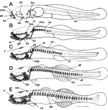
Fig. 1. Development stage of flextion and postflextion larvae of skel
eton in Tridentiger trigonocephalus. A: 7 days after hatching, 4.44 mm in total length (TL); B: 10 days after hatching, 5.02mm in TL;
C: 13 days after hatching, 5.62mm in TL; D: 16 days after hatching, 6.40mm in TL; E: 22 days after hatching, 7.57mm in TL. af: anal fin; an: angular; ar: articular; av: abdominal vertebrae; bo: basioccip
ital; br: branchiostegal rays; cl: clavicle; ch: ceratohyal; co: coracoid;
cv: caudal vertebrae; df: dorsal fin; dt: dentary; eh: epihyal; et: epi
otic; exo: exoccipital; f: frontal; gh; glossohyal; hh: hypohyal; hm:
hyomandibular; hs: hemal spine; iop: interopercle; mx: maxillary; n:
nasal; nc: notochord; ns: neural spine; op: opercle; pa: parapophysis;
pmx: premaxillary; ps: parasphenoid; pt: pterotic; sop: subopercle;
spc: supraoccipital. Scale bars=1.0mm.
Fig. 2. Development stage of postflextion larvae and juvenile of skeleton in Tridentiger trigonocephalus. A: 28 days after hatching, 9.32mm in TL; B: 32 days after hatching, 11.8mm in TL; C: 40 days after hatching, 13.3mm in TL. a: actinost; as: alisphenoid; ed: en
dopterygoid; em: lateral ethmoid; ep: epural bone; hy: hypural bone;
ins: interneural spine; ihs: interhemal spine; mt: metapterygoid; pat:
palatine; pcl: postcleithrum; pel: pelvic girdle; pf: prefrontal; ph:
parhypural bone; pro: prootic; r: rib; sca: scapula; uh: urohyal; uro:
urostyle. Scale bars=1.0mm.
A
B
C
D
E
A
B
C
전장 4.20~4.60mm(평균 4.44±0.28mm, n =5)로 섭이기 능 역할을 하는 위턱의 전상악골(premaxillary), 주상악골 (maxillary), 아래턱의 치골(dentary)이 골화하기 시작하였다.
또한 이 시기에 치골과 관절 되는 각골(angular)이 골화하기 시작하였으며, 두개골에는 부설골(parasphenoid)과 기저후두 골(basioccipital)이 최초로 골화하기 시작하였다. 설궁부에는 상설골(epihyal)이 골화하였고, 새개부에는 주새개골(opercle), 견대부에는 쇄골(clavicle)이 골화하였으며, 척추는 완성되지 않은 상태인 척색(notochord)으로 이루어져 있었다(Fig. 1A).
부화 후 10일째 중기자어는 전장 4.85~5.20mm(평균 5.02±0.24mm, n=5)로 구개부에 설악골(hyomandibular), 관 절골(articular)이 골화하였고, 설궁부에는 인설골(glossohyal), 각설골(ceratohyal), 새조골(branchiostegal rays)이 골화하기 시작하였으며, 두개골에는 외후두골(exoccipital)이 골화하였 다. 척추골에는 최초 10개의 복추골(abdominal verbrae)이 골 화하기 시작하였고, 척추의 골화는 머리 뒤쪽에서 꼬리 쪽 방 향으로 진행되었다(Fig. 1B).
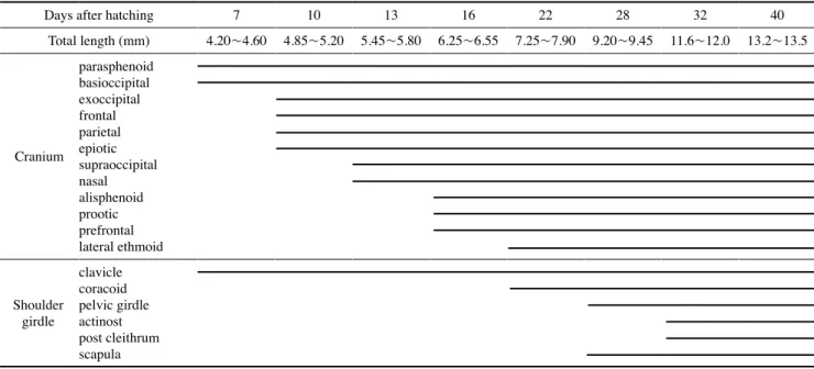
부화 후 13일째 후기자어는 전장 5.45~5.80mm(평균 Table 1. The development process of cranium and shoulder girdle of Tridentiger trigonocephalus
Days after hatching 7 10 13 16 22 28 32 40
Total length (mm) 4.20~4.60 4.85~5.20 5.45~5.80 6.25~6.55 7.25~7.90 9.20~9.45 11.6~12.0 13.2~13.5
Cranium
parasphenoid basioccipital exoccipital frontal parietal epiotic supraoccipital nasal alisphenoid prootic prefrontal lateral ethmoid
Shoulder girdle
clavicle coracoid pelvic girdle actinost post cleithrum scapula
Table 2. The development process of vertebrae and caudal skeleton of Tridentiger trigonocephalus
Days after hatching 7 10 13 16 22 28 32 40
Total length (mm) 4.20~4.60 4.85~5.20 5.45~5.80 6.25~6.55 7.25~7.90 9.20~9.45 11.6~12.0 13.2~13.5
Vertebrae
notochord
abdominal vertebrae neural spine caudal vertebrae hemal spine parapophysis ribinterneural spine interhemal spine
Caudal skeleton
urostyle hypural bone 4th5th 2nd3rd 1stparhypural bone epural bone 1st2nd 3rd
5.62±0.24mm, n =5)로 두개골에는 액골(frontal), 노정골 (parietal), 상이골(epiotic)이 골화하기 시작하였고, 새개부에는 하새개골(subopoercle)이 골화하기 시작하였다. 척추골에는 복추골 위쪽에 신경극(neural spine)이 골화하기 시작하였고, 5개의 미추골(caudal vertebrae)이 골화하기 시작하였다(Fig.
1C).
부화 후 16일째 후기자어는 전장 6.25~6.55mm(평균 6.40±0.21mm, n=5)로 설궁부에는 하설골(hypohyal), 새개 부에는 간새개골(interopercle)이 골화하기 시작하였다. 등지느 러미와 뒷지느러미에는 기조가 형성되기 시작하였고, 복추골 아래쪽에는 혈관극(hemal spine)이 골화하기 시작하였다(Fig.
1D).
부화 후 22일째 후기자어는 전장 7.25~7.90mm(평균 7.57
±0.45mm, n =5)로 두개골에는 상후두골(supraoccipital), 비골(nasal)이 골화하기 시작하였고, 견대부에는 오훼골 (coracoid)이 골화하였으며, 복추골의 아래쪽에는 측돌기(par
apophysis)가 골화하기 시작하였다(Fig. 1E).
부화 후 28일째 후기자어는 전장 9.20~9.45mm(평균 9.32±0.17mm, n =5)로 구개부에는 후익상골(metaptery
goid), 구개골(palatine)이 골화하였고, 배쪽에는 요대골(pelvic girdle)이 골화하기 시작하였다. 견대부에는 견갑골(scapula) 이 골화하기 시작하였고, 복추골의 아래쪽에는 늑골(rib) 이 형성되었으며, 등 쪽에는 지느러미를 지지하는 신경간극 (interneural spine)과 배 쪽에 혈관간극(interhemal spine)이 골
화하기 시작하였다. 미골부에는 미부봉상골(urostyle)이 골화 하기 시작하였고, 신경간극과 미부봉상골 사이에는 상미축골 (epural bone) 2개가 골화하기 시작하였다. 미부봉상골의 아래 쪽에는 2개의 하미축골(hypural bone)이 골화하면서 접합되었 다(Fig. 2A).
부화 후 32일째 치어는 전장 11.6~12.0mm(평균 11.8±
0.28mm, n =5)로 두개골에는 전액골(prefrontal), 익설골 (alisphenoid), 전이골(prootic)이 골화하였고, 구개부에는 내익 상골(endopterygoid), 설궁부에는 미설골(urohyal), 견대부에 는 사출골(actinost), 후쇄골(post cleithrum)이 골화하였다. 미 골부에는 하미축골 아래에 준하미축골(parhypural bone)이 골 화하였고, 2개의 하미축골이 증가하면서 접합되었다(Fig. 2B).
부화 후 40일째 치어는 전장 13.2~13.5mm(평균 13.3±
0.21mm, n=5)로 두개골에는 측사골(lateral ethmoid)이 골화 하였고, 미골부에는 1개의 하미축골과 상미축골이 추가로 골 화하면서 모든 골격의 골화가 완료되었다(Fig. 2C).
고 찰
두줄망둑은 부화 직후 골격의 발달이 관찰되지 않았으며, 성장함에 따라 부화 후 7일 평균전장 4.44mm일 때 섭이기 능 역할을 하는 전상악골, 주상악골, 치골이 먼저 골화하기 시 작하였다. 부화 후 10일째 평균전장 5.02mm일 때는 구개부 Table 3. The development process of visceral skeleton of Tridentiger trigonocephalus
Days after hatching 7 10 13 16 22 28 32 40
Total length (mm) 4.20~4.60 4.85~5.20 5.45~5.80 6.25~6.55 7.25~7.90 9.20~9.45 11.6~12.0 13.2~13.5
Visceral skeleton
Upper jaw premaxillarymaxillary Lower jawdentary
articular angular
Hyoid arch epihyal
branchiostegal rays ceratohyal glossohyal hypohyal urohyal
Palate
hyomandibular sympletic metapterygoid palatine endopterygoid ectopterygoid Opercular
preopercle opercle subopercle interopercle
의 설악골, 관절골 등 두개골의 발달에 비해 악골을 비롯한 구 개부, 설궁부가 우선적으로 골화하는 것이 관찰되었다. 두줄망 둑과 마찬가지로 부화 직후에도 골격발달이 진행되지 않은 종 이 있는 반면 난 내에서부터 골격발달이 진행되어 부화 직후 에는 섭이기능 역할이 대부분 갖추어진 상태에서 부화하는 종 이 있는 것으로 알려져 있다(Park et al., 2017). 같은 망둑어과 어류인 미끈망둑과 미끈날망둑은 부화 후 상당 시간이 지나서 섭이기능 역할을 하는 악골이 골화하기 시작하였고, 동사리과 어류인 얼록동사리, Odontobutis interrupta(Park et al., 2017) 와 연어과 어류인 연어, Oncorhynchus keta(Myoung and Kim, 1993)는 부화 직후 지느러미를 지지하는 신경간극, 혈관간극 등이 골화되어 있었고, 미골부의 미부봉상골이 45º로 휘어진 상태에서 지느러미 줄기가 발달되어 있었으며, 난황을 가지고 있었으나 섭이기능을 하는 골격의 골화가 진행되어 있어 두줄 망둑의 골격발달은 이들과 상이한 양상을 보였다.
입의 개구와 섭이기능의 발달은 쇄골의 역할과 관련이 있는 것으로 알려져 있으며(Wagemans and Vandewalle, 1999), 두줄 망둑의 견대골은 부화 후 7일째 발달하기 시작하여 쇄골, 오훼 골, 사출골 등의 순으로 골화하였고, 같은 망둑어과 어류인 미 끈망둑은 부화 후 11일째 평균전장 5.50mm일 때 쇄골, 오훼 골, 견갑골, 사출골 순으로 골화하였으며, 미끈날망둑은 부화 후 13일째 전장 6.20~6.35mm일 때 쇄골, 견갑골, 사출골 순 으로 골화하여 비교적 유사하게 발달하였다. 두줄망둑은 부화 후 40일째 평균전장 13.3mm일 때 견갑골에 1개의 체공이 형 성되는 것이 관찰되었고, 미끈망둑은 부화 후 47일째 평균전 장 13.4mm, 미끈날망둑은 부화 후 33일째 평균전장 12.5mm 일 때 관찰되었다. 두줄망둑은 미끈망둑과 발달시기가 유사하 였고, 미끈날망둑 보다는 다소 늦게 발달하여 차이를 보였다.
견갑골에 형성되는 체공은 농어목 어류에서 나타나는 분류학 적 형질로 알려져 있으나 이미 다양한 어류에서 관찰되어 일 반적인 형질로 보고 있다(Han et al., 2018).
두줄망둑은 부화 후 10일째 평균전장 5.02mm일 때 10개의 복추골이 머리에서 꼬리방향으로 발달하기 시작하였고, 미끈 망둑은 부화 후 16일째 평균전장 6.10mm일 때 복추골이 골 화하면서 동시에 신경극이 골화하였으며, 미끈날망둑은 부화 후 9~10일째 평균전장 6.00mm일 때 11~12개의 복추골과 3~4개의 미추골이 골화하기 시작하였다. 두줄망둑의 척추골 발달방향은 비교 종들과 동일하였고, 미끈망둑은 복추골의 골 화 이전에 신경극이 골화한다는 점에서 두줄망둑과 차이를 보 였다. 두줄망둑은 신경극이 먼저 발달하면서 혈관극이 골화하 기 시작하였고, 미끈망둑과 미끈날망둑의 발달양상과 유사하 였으나 척추골의 골화가 시작되기 전 신경극이 골화하는 미끈 망둑과 발달순서에서 다소 차이를 보였다.
농어목 어류의 지느러미 발달양상은 후방의 등지느러미, 뒷 지느러미가 먼저 발달하고, 전방의 등지느러미가 발달한다고
알려져 있다(Johnson, 1984; Faustino and Power, 1999). 두줄 망둑은 부화 후 16일째 평균전장 6.40mm일 때, 후방의 등지 느러미가 먼저 발달하였고, 뒷지느러미에 줄기가 4~5개 발달 하기 시작하여 일반적인 농어목 어류의 발달양상과 일치하였 다. 같은 망둑어과 어류인 모치망둑, Mugilogobius abei(Kim and Han, 1991)은 부화 후 12일째 평균전장 3.20mm일 때 후 방의 등지느러미와 뒷지느러미에 각각 3~4개가 발달하기 시 작하였으며, 미끈날망둑은 부화 후 17일째 평균전장 7.05mm 일 때 후방의 등지느러미 5개, 뒷지느러미 4개에서 줄기가 먼 저 발달하였다. 미끈망둑은 부화 후 16일째 전장 5.90~6.30 mm일 때 후방의 등지느러미 5개, 뒷지느러미 5개가 발달하기 시작하였고, 모치망둑(Kim and Han, 1991) 보다는 늦게 발달 하였으며, 미끈날망둑, 미끈망둑과 발달시기가 유사하였다. 등 지느러미는 후방에서 전방으로 발달하여 비교 종들과 동일한 양상을 보였다.
두줄망둑의 담기골은 추체의 발달이 시작된 이후에 이루어 졌고, 이러한 양상은 같은 망둑어과 어류인 미끈망둑, 미끈날 망둑에서도 나타났으며, 지느러미의 줄기 발달이 완성된 후 담기골의 형태가 완전한 모습을 갖추었다. 척추골과 지느러미 의 발달이 완료되었을 때 담기골이 발달하는 것은 어류의 유 영능력에 있어 추진력을 증가시키는 것과 관련 있는 것으로 보인다(Lee et al., 2001).
꼬리지느러미를 지지하는 미골부는 미부봉상골을 중심으로 상미축골, 하미축골, 준하미축골로 이루어져 있다. 두줄망둑은 부화 후 28일째 평균전장 9.32mm일 때 미부봉상골이 골화하 기 시작하였고, 미끈망둑은 부화 후 25일째 평균전장 7.60mm 일 때, 미끈날망둑은 부화 후 20일째 평균전장 7.25mm일 때 골화하기 시작하여 두줄망둑이 보다 늦게 발달하였다. 하미축 골의 융합은 부화 후 40일째 평균전장 13.3mm일 때 완료되 었고, 3개의 하미축골(1+2, 3+4, 5)을 형성하였다. 미끈날망 둑과 미끈망둑은 2개의 하미축골(1+2, 3+4+5)을 형성하여 차이를 보였다. 이와 같이 하미축골의 융합형태가 종에 따라 다르게 나타나는 것으로 보아 계통분류학적 연구의 기초자료 로 매우 중요하다고 사료된다. 자치어의 골격에 대한 연구는 종 동정과 성어의 골격특성을 이해하는데 매우 중요한 기초자 료로 이용될 것이고, 향후 망둑어과 어류의 분류 및 골격학적 특성을 규명하기 위해서는 많은 연구가 이루어져야 할 것으로 보인다.
요 약
이 연구의 목적은 우리나라 연안에 서식하는 두줄망둑의 자 치어 골격발달을 관찰하여 분류학적 연구자료로 이용하는 것 이다. 부화 후 7일째 평균전장 4.44±0.28mm(n=5) 일 때 섭
이기능을 하는 윗턱에는 전상악골, 주상악골이 골화하였고, 아 래턱에는 악골이 골화하기 시작하였다. 부화 후 13일째 평균 전장 5.62±0.24mm(n=5) 일 때 두개골의 액골, 노정골, 상 이골이 골화하였다. 부화 후 32일째 평균전장 11.8±0.28mm (n=5) 일 때는 견대부에 사출골, 후쇄골이 골화하였고, 부화 후 40일째 평균전장 13.3±0.21mm(n=5) 일 때 두개골의 측 사골이 골화하면서 모든 골격의 골화가 완료되었다.
REFERENCES
Boltachev, A.R. and E.P. Karpova. 2010. Naturalization of the Pacific Chameleon Goby Tridentiger trigonocephalus(Per
ciformes, Gobiidae) in the Black Sea(Crimea, Sevastopol Gulf). J. Ichthyol., 50: 188196.
Coban, D., C. Suzer, H.O. Kamaci, S. Saka and K. Firat. 2009.
Early osteological development of the fins in the hatch
eryreared red porgy, Pagrus pagrus(L. 1758). J. Appl.
Ichthyol., 25: 2632.
Faustino, M. and D.M. Power. 1999. Development of the pectoral, pelvic, dorsal and anal fins in cultured sea bream. J. Fish.
Biol., 54: 10941110.
Feng, G.P., P. Zhuang, L.Z. Zhang, J.Y. Liu, Y. Zhao, L.H. Chen and Q. Liang. 2009. Embryonic and prelarval development of Tridentiger trigonocephalus and adaptability to salinity.
Acta Hydrobio. Sin., 33: 176181.
Fishbase. <http://www.fishbase.org/summary/Tridentigertrigono cephalus.html>(on 01 September 2018).
Han, K.H., J.T. Park, S.H. Lee, D.S. Jin and J.M. Park. 2018. Oste
ological Development of the Larvae and Juveniles in Aucha Perch Coreoperca kawamebari(Perciformes: Centropomi
dae) in Korea. Kor. J. Fish. Aqua. Sci., 51: 170177.
Hwang, I.J., S.J. Choi and H.J. Baek. 2018. Changes in Repro
ductive Characteristics of Chameleon Goby Tridentiger trigonocephalus by Carbon Dioxide Exposure. Kor. J. Fish.
Aqua. Sci., 51: 5463.
Johnson, G.D. 1984. Percoidei: development and relationship. In:
Moser H.G., W.J. Richards, D.M. Cohen, M.P. Fahay, A.W.
Kendall, S.L. Richardson(eds). Ontogeny and systematics of fishes. American Society of Ichthyologists and Herpetol
ogists, special publication no 1, Allen Lawrence KS, 464
498.
Chyung, M.K. 1977. The fishes of Korea. Ilji-sa Publishing, Seoul, 727pp.(in Korean)
Kang, J.C., J.H. Jee, S.Y. Park, S.G. Kim and K.S. Park. 2004. Tol
erance of juvenile gobiidae, Tridentiger trigonocephalus exposed to various salinity. Kor. J. Environ. Biol., 22: 153
158.
Kim, I.S., Y. Choi, C.L. Lee, Y.J. Lee, B.J. Kim and J.H. Kim. 2005.
Illustrated book of Korean fishes. KyoHak Publishing, Seoul, p. 420443.(in Korean)
Kim, J.Y. and J.M. Yoon. 2016. Food Organisms of Juveniles of Tridentiger trigonocephalus from the Intertidal Zone in the Western Coast of Korea. J. Kor. Soc. Fish. Mar. Sci. Edu., 28: 180185.
Kim, Y.U. and K.H. Han. 1990. Early life history and spawning be
havior of the gobiid fish, Tridentiger trigonocephalus(Gill) reared in the laboratory. Kor. J. Ichthyol., 3: 110.(in Kore
an)
Kim, Y.U. and K.H. Han. 1991. Early life history and spawning behavior of the gobiid fish, Mugilogobius abei(Jordan et Snyder) reared in the laboratory. Kor. J. Ichthyol., 2: 5362.
Kimura, S.S. 2010. New atlas of fish anatomy. pp. 168-170.
Koumoundouros, G., F. Gagliardi, P. Divanach, C. Boglione, S.
Cataudella and M. Kentouri. 1997a. Normal and abnormal osteological development of caudal fin in Sparus aurata L.
fry. Aquaculture, 149: 215226.
Koumoundouros, G., G. Oran, P. Divanach, S. Stefanakis and M.
Kentouri. 1997b. The opercular complex deformity in in
tensive gilthead sea bream(Spartus aurata L.) larviculture.
Moment of apparition and description. Aquaculture, 156:
165177.
Kim, Y.U. and K.H. Han. 1989. 1. Egg development, larvae and juveniles of Chaenogobius laevis(Steindachner) early life history of the marine animals. Bull Kor. Fish. Soc., 22: 317
Kim, Y.U., K.H. Han, C.B. Kang and J.W. Ryu. 1992. Early life his331.
tory and spawning behavior of the gobiid fish, Luciogobius guttatus Gill. Kor. J. Ichthyol., 4: 113.
Lee, C.Y. 1990. Osteological Study of the mudhopper, Periopthal- mus cantonensis(Perciformes, Gobiidae) from Korea. Kor.
J. Zool., 33: 402410.
Liu, C.H. 2001. Early osteological development of the yellow tail Seriola dumerili(Pisces: Carangidae). Zool. Stud., 40: 289
298.
Mook, D. 1977. Larval and osteological development of the sheeps
head, Archosargus probatocephalus(Pisces: Sparidae).
Copeia, 1: 126133.
Myoung, J.G. and Y.U. Kim. 1993. Morphological study of Onco- rhynchus spp.(Pisces : Salmonidae) in Korea - Ⅰ Egg devel
opment and morphology of alevin, fry and smolt of chum salmon, Oncorhynchus keta. Kor. J. Ichthyol., 5: 5367.
NIBR(National Institute of Biological Resources). 2017. National list of species of Korea. National institute of biological resources, online at <http://kbr.go.kr>. accessed on 01 Sep
tember 2018.(in Korean)
Park, J.M., J.H. Han, S.M. Yun and K.H. Han. 2017. Early osteo
logical development of larvae and juveniles in the Korean spotted sleeper Odontobutis interrupta from Korea. Kor. J.
Fish. Aqua. Sci., 50: 396405.
Pondella, D.J. and Z.K.J. Chinn. 2005. Records of Chameleon Goby, Tridentiger trigonocephalus, in San Diego Bay, Cali
fornia. California Fish and Game, 91: 5759.
Potthoff, T. and J.A. Tellock. 1993. Osteological development of
the snook, Centropomus undecimalis(Teleostei, Centropo
midae). Bull Mar. Sci., 52: 669716.
Potthoff, T., S. Kelly, V. Saksena, M. Moe and F. Young. 1987. De
scription of larval and juvenile damselfish Microspathodon chrysurus, Pomacentridae, and their osteological develop
ment. Bull Mar. Sci., 40: 140.
Potthoff, T., S. Kelley and L.A. Collins. 1988. Osteological devel
opment of the red snapper, Lutjanu scampechanus(Lutjani
dae). Bull. Mar. Sci., 43: 140.
Sfakianakis, D.G., G. Koumoundouros, P. Divanach and M. Ken
touri. 2004. Osteological development of the vertebral column and of the fins in Pagellus erythrinus(L. 1758).
Temperature effect on the developmental plasticity and morphoanatomical abnormalities. Aqua., 232: 407424.
Walker, M.B. and C.B. Kimmel. 2007. A twocolor acidfree car
tilage and bone stain for zebrafish larvae. Biotechnic and Histochemistry, 82: 2328.
Wagemans, F. and P. Vandewalle. 1999. Development of the carti
laginous skull in Solea solea: trends on Pleuronectiforms.
Ann. Sci. Nat., 1: 3952.