170
Copyright © 2018 The Korean Society of Fisheries and Aquatic Science pISSN:0374-8111, eISSN:2287-8815
서 론
꺽저기
(Coreoperca kawamebari)
는농어목(Perciformes)
꺽 지과(Centropomidae)
꺽지속(Coreoperca)
에 속하는 어류로 우리나라에는꺽저기를비롯한꺽지(C. herzi) 2
종이분포한다(Kim et al., 2005).
서식지는물의흐름이느리고,
바닥에자갈 과돌이깔려있으며,
수초가있는곳을선호하며,
우리나라서·
남해를흐르는하천과동해남부로흘러드는하천에서식하고,
탐진강,
보성강및영산강등일부하천에서만서식하는것으 로알려져있다.
또한분포지역은우리나라뿐만아니라일본 교토,
후쿠야마,
큐슈지쿠젠상류및산요오의유하강상류에 도분포하는것으로알려져있다(Kim and Park, 2002; Kim et al., 2005).
그러나최근하천의댐과보건설,
하상정비등으로서식지가파괴되어그수가급격히줄어들고있는실정이다
.
따 라서환경부에서는개체수의급감을막기위해2012
년멸종위 기야생동식물II
급으로지정하여보호하고있다(NIBR, 2018).
꺽저기에대한연구로는산란습성및초기생활사
(Han et al., 2000),
분포및어류군집구조(Kim et al., 2013)
등이수행되었 으나골격에대한기초자료는부족한실정이며,
같은과에속하 는꺽지(Han et al., 2017)
및쏘가리(Siniperca scherzeri, My- oung et al., 2001)
등에대해서는자치어골격발달연구를통해 내부형태에대한분류학적차이점을확인할수있었다.
자치어골격발달에대한관심과지식은종자생산과정중사 육초기에발생하는골격의이상을탐지하고제거하는데필수 적이며
(Koumoundouros et al., 1997a, b),
유어기의계통분류 학적기초형질을제공하고있어이분야에대한연구가활발하한국산 꺽저기(Coreoperca kawamebari) 자치어의 골격발달
한경호·박준택 1 ·이성훈·진동수 2 ·박재민 3 *
전남대학교 해양기술학부, 1전라남도 해양수산과학관, 2경상남도청, 3경상북도 토속어류산업화센터
Osteological Development of the Larvae and Juveniles in Aucha Perch Coreoperca kawamebari (Perciformes: Centropomidae) in Korea
Kyeong Ho Han, Jun Taek Park
1
, Sung Hun Lee, Dong Soo Jin2
and Jae Min Park3
*Marine Technology Undergraduate, Chonnam National University, Yeosu 59626, Korea
1Maritime and Fisheries Science Museum, Yeosu 59771, Korea
2The Provincial Office of Gyeongsangnam-Do, Changwon 51154, Korea
3Gyeongsangbuk-Do Native Fish Business Center, Uiseong 37366, Korea
Samples were obtained from broodstork in May, 1998, while naturally fertilized embryos were maintained and the process of skeletal development was observed from larvae and juveniles. Prelarvae immediately after hatching showed an average total length of 5.38±0.41 mm (n=10), premaxillary and dentary were ossified, parasphenoid was ossified in the cranium, and centrum and caudal bone did not ossify. Prelarvae showed ossification with maxillary, articular and epihyal and branchiostegal rays of hyoid arch were ossified at 5 days after hatching with an average total length of 6.40±0.39 mm (n=10). The vertebrae began to ossify in the direction of the tail, and neural spine began to ossify above the ossified vertebra. Postlarvae showed ossification of lateral ethmoid, parietal, and caudal skeleton in the cranium when the average total length was 7.30±0.12 mm (n=10) in 8 days after hatching. At 22 days after hatch- ing, postlarvae ossified maxillary in the cranium, and ossified endopterygoid and ectopterygoid, etc. in the palate, when the average length of 11.1±1.27 mm (n=10). At 32 days after hatching, with the average length was 12.8±1.97 mm (n=10), caudal skeleton had one additional epural bone ossification, resulting in ossification of a total of 3 epural bone to complete ossification of all spicules.
Key words: Coreoperca kawamebari , Juveniles, Larvae , Osteological development
This is an Open Access article distributed under the terms of the Creative Commons Attribution Non-Commercial Licens (http://creativecommons.org/licenses/by-nc/3.0/) which permits unrestricted non-commercial use, distribution, and reproduction in any medium, provided the original work is properly cited.
https://doi.org/10.5657/KFAS.2018.0170 Korean J Fish Aquat Sci 51(2) 170-177, April 2018
Received 31 January 2018; Revised 5 March 2018; Accepted 6 March 2018
*Corresponding author: Tel: +82. 54. 830. 8833 Fax: +82. 54. 830. 8809
E-mail address: [email protected]
꺽저기 자치어의 골격발달
171
다
(Mook, 1977; Potthoff et al., 1987; Potthoff et al., 1988; Pot- thoff and Tellock, 1993; Liu, 2001; Sfakianakis et al., 2004;
Coban et al., 2009).
꺽저기는같은속어류인꺽지와생김새가유사하여체고
,
눈 의크기,
양안간격및측선비늘수에서차이를나타내육안으로 분류하기다소어려움이있을수있다(Kim et al., 2005).
따라 서이연구에서는꺽저기자치어의발달단계별골격발달과정 을관찰하여종보존을위한기초자료확보와더불어유사종과 의차이점을밝히고,
종동정을위한분류학적연구자료로제 공하고자한다.
재료 및 방법
시료확보 및 사육관리
실험에사용된꺽저기는멸종위기야생동식물
II
급으로지정 되기전인1998
년2
월부터6
월까지전남장흥군에위치한탐 진강지류하천에서반두를이용하여어미를채집후연구실로 운반하였고,
사육수조(90×45×30 cm)
에서관리하던중5
월 경어미(
전장8.0 cm, n=1)
로부터산란된수정란700-750
개(
평균730
개)
를확보하여사육하였다.
부화된자어는수온18.6- 22.0℃ (
평균20.3±2.40℃)
범위에서관리하였고,
먹이는난 황흡수완료후부터Artemia sp. nauplius
유생을mL
당2-3
개 체정도공급하였고,
치어기로이행하면서부터순차적으로배 합사료를공급하였다.
수질관리는저면여과기를이용해순환 여과식으로사육하였고,
사육수는1
주일에1
회50%
씩환수시 켜주었다.
골격염색
골격염색을위해자치어는부화직후부터치어기로이행하기 까지
4-5
일간격으로10
마리씩10%
중성포르말린에고정후 보관하였다.
보관된자치어샘플은2010
년전남대학교자원생 물실험실에서Walker and Kimmel (2007)
의이중염색방법을 사용하였으며,
염색된자치어는실체현미경(Nikon SMZ800,
Japan)
을이용하여 관찰및 스케치하였으며,
골격의각 부위명칭은
Kendall (1991), Okiyama (1988), Byun et al. (2012), Kang et al. (2012)
을인용하였다.
결 과
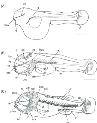
꺽저기자치어의골격발달과정은다음과같았다
(Fig. 1, 2;
Table 1-Table 3).
부화직후의전기자어는전장5.09-5.68 (
평 균5.38±0.41, n=10) mm
로입과항문이열려있었고,
난황을 가지고있었다.
두개골에는기저를형성하는부설골(parasphe-
noid)
이골화하였고,
섭이기능 역할을하는윗턱의전상악골(premaxillary),
아래턱에치골(dentary)
이골화하기시작하였 으며,
견대부에는쇄골이골화하였다.
척추와추체(centrum)
는 골화하지않았고,
척색(notochord)
으로이루어져있었으며,
꼬리끝의미부는
45°
로휘어져있었다.
꼬리지느러미에는10-12
개의줄기가형성되었다(Fig. 1A).
부화후
5
일째후기자어는전장6.12-6.68 (
평균6.40±0.39)
mm
로난황은대부분흡수가되었고,
먹이를섭취하는것이관찰되었다
.
윗턱에는주상악골(maxillary)
이골화하였고,
구개 부에는설악골(hyomandibular),
접속골(sympletic),
아래턱의 관절골(articular)
이 골화하였으며,
설궁부의 상설골(epihyal)
과4
개의새조골(branchiostegal rays)
이 골화하였다.
두개골 은 기저후두골(basioccipital)
과눈의위쪽에액골(frontal),
설 이골(sphenotic),
익이골(pterotic)
이골화하였고,
주상악골 위 쪽에는비골(nasal)
이골화하였다.
새개부에는주새개골(oper- cle)
이 골화하였고,
쇄골의아래쪽에는하후쇄골(ventral post
pa
pmx d
cl
fr sp pt bac
ar brs eh hm
pop
op vpcl mx
na sy
preo
epi al par
ch ih
pr soc
an le
sop scl
us ns
hs av
df
af cf (A)
(B)
(C)
sor
opi
iop
bas pal
ecp me
v
sc a
cr pos
pel
ph hy
1hy
2hy
3hy
4hy
5ep
1ep
2ep
3uro cdp ihs
ins
pp r mp
spt q
enp h smx
dpcl
vf (A)
(B)
(C)
exo
Fig. 1. Devleopment stage of prelarvae and postlarvae of skeleton in aucha perch Coreoperca kawamebari. A: Newly hatched larva, 5.38 mm in total length (TL); B: 5 days after hatching, 6.40 mm in TL; C: 8 days after hatching, 7.30 mm in TL. af, anal fin; al, ali- sphenoid; an, angular; ar, articular; av, abdominal vertebrae; bac, basioccipital; brs, branchiostegal rays; ch, ceratohyal; cl, clavicle;
cf, caudal fin; d, dentary; df, dorsal fin; eh, epihyal; epi, epiotic;
fr, frontal; hm, hyomandibular; hs, hemal spine; ih, interhyal; le, lateral ethmoid; mx, maxillary; na, nasal; ns, neural spine; op, opercle; pa, parasphenoid; par, parietal; pmx, premaxillary; pop, preopercle; pr, prootic; preo, preorbital=lucrymal; pt, pterotic; sp, sphenotic; scl, supracleithrum; soc, supraoccipital; sop, suboper- cle; sor, suborbital; sy, sympletic; us, urostyle; vpcl, ventral post cleithrum. Scale bars=1.0 mm.
한경호
ㆍ
박준택ㆍ
이성훈ㆍ
진동수ㆍ
박재민172
cleithrum)
이골화하였으며,
척추에는15
개의복추골(abdomi- nal vertebrae)
과위쪽에는신경극(neural spine)
이골화하기시 작하였다(Fig. 1B).
부화후
8
일째후기자어는전장7.22-7.39 (
평균7.30±0.12) mm
로두개골의전단부에는측사골(lateral ethmoid)
이골화하 였고,
후반부에익설골(alisphenoid),
노정골(parietal),
전이골(prootic),
상이골(epiotic),
상후두골(supraoccipital)
이골화하 였으며,
안와부에는안전골(preorbital=lucrymal),
안하골(sub- orbital)
이골화하였다.
설궁부에는각설골(ceratohyal),
간설골(interhyal)
이골화하였고,
구개부에는아래턱에각골(angular)
이골화하였으며,
새개부의전새개골(preopercle)
과주새개골 아래에하새개골(subopercle)
이골화하였다.
척추에는12
개의미추골이골화하였고
,
아래쪽에는혈관극(hemal spine)
이골화 하기시작하였으며,
꼬리끝에는미부봉상골(urostyle)
이골화 하였다.
견대부에는 쇄골상단부에 상쇄골(supracleithrum)
이 골화하였고,
각부위별지느러미줄기수는등지느러미(dorsal fin)
에 극조8
개,
연조10-11
개가 형성되었으며,
뒷지느러미(anal fin)
연조는7-10
개형성되었다(Fig. 1C).
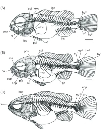
부화후
17
일째후기자어는전장8.72-9.42 (
평균9.07±0.49) mm
로 주상악골과치골에 작은이빨이여러 개 관찰되었고,
두개골에는 외후두골(exoccipital),
후이골(opisthotic)
이 골 화하였으며,
구개부에는 방골(quadrate),
설궁부에는 하설골(hypohyal)
이골화하였다.
새개부에는간새개골(interopercle)
이골화하였고,
주상악골의끝쪽상단부에는상주상악골(su- pramaxillary)
이골화하였다.
척추골은11+18
개,
총29
개로증 가하였고,
미골부에는미부봉상골아래쪽에준하미축골(par- hypural bone) 1
개와하미축골(hypural bone) 3
개가골화하였 고,
등지느러미와뒷지느러미를지지하는신경간극(interneural spine)
과혈관간극(interhemal spine)
이골화하였다.
견대부에 는상후쇄골(dorsal post cleithrum)
이골화하였고,
배지느러미(ventral fin)
는2
개의줄기가형성되었으며,
앞쪽에는지지할수 있는요대골(pelvic bone)
이골화하였다(Fig. 2A).
부화후
22
일째후기자어는전장10.2-12.0 (
평균11.1±1.27) mm
로두개골에는중사골(mesethmoid)
이골화하였고,
구개부 에는 내익상골(endopterygoid),
외익상골(ectopterygoid),
후 익상골(metapterygoid)
및구개골(palatine)
이골화하였다.
미 골부에는신경극과미부봉상골사이에2
개의상미축골(epural
bone)
과아래쪽에는하미축골2
개가추가로골화하였다.
견대부에는 상측두골
(supraposttemporal),
후측두골(posttempo- ral),
견갑골(scapula),
오훼골(coracoid)
및4
개의사출골(acti- nost)
이골화하였고,
복추골아래쪽에는측돌기(parapophysis)
와6
개의늑골(rib)
이골화하였다.
각부위별지느러미줄기수 는등지느러미극조12
개,
연조12-13
개,
뒷지느러미극조3
개,
연조8-10
개,
배지느러미극조1
개연조4-5
개로증가하였다(Fig. 2B).
부화후
32
일째치어는전장11.4-14.2 (
평균12.8±1.97) mm
로두개골에는기저설골(basisphenoid)
및서골(vomer)
이골화 하였고,
미골부에는1
개의상미축골이추가로골화하면서총3
개의상미축골이골화하였다.
상미축골과미부봉상골사이에 는미골판(caudal bony plate)
이골화하였고,
다섯번째하미축골위쪽에는신경골
(uroneural)
이골화하면서모든골편들의골화가완성되었다
(Fig. 2C).
고 찰
농어목어류는 두개골과 지느러미의 발달이 부화 이후에 관찰되나
(Matsuoka, 1985; Koumoundouros et al., 1997b, 2001a, 2001b; Faustion and Power, 1999; Sfakianakis et al.,
2004, 2005),
연어과어류는부화이전에두개골과지느러미의Fig. 2. Development stage of postlarvae and juveniles of skeleton in aucha perch Coreoperca kawamebari. A: 17 days after hatching, 9.07 mm in total length (TL); B: 22 days after hatching, 11.1 mm in TL; C: 32 days after hatching, 12.8 mm in TL. a, actinost; bas, basisphenoid; cdp, caudal bony plate; cr, coracoid; dpcl, dorsal post cleithrum; ecp, ectopterygoid; ep, epural bone; enp, endopter- ygoid; exo, exoccipital; h, hypohyal; hy, hypural bone; ihs, inter- hemal spine; ins, interneural spine; iop, interopercle; me, meseth- moid; mp, metapterygoid; opi, opisthotic; pal, palatine; pel, pelvic bone; ph, parhypural bone; pos, posttemporal; pp, parapophysis; q, quadrate; r, rib; sc, scapula; smx, supramaxillary; spt, suprapost- temporal; uro, uroneural; v, vomer; vf, ventral fin. Scale bars=1.0 mm.
pmx d
fr sp pt bac
ar brs eh hm
pop
op vpcl mx
na sy
preo
epi al par
ch ih
pr soc
an le
sop scl
us ns
hs av
df
af cf
(B)
(C)
sor
opi
iop
bas pal
ecp me
v
sc a
cr pos
pel
ph hy
1hy
2hy
3hy
4hy
5ep
1ep
2ep
3uro cdp ihs
ins
pp r mp
spt q
enp h smx
dpcl
vf (A)
(B)
(C)
exo
꺽저기 자치어의 골격발달
173
골격발달이시작되는것으로알려져
(Kendall et al., 1984),
본 종은농어목어류와유사한양상을나타내었다.
꺽저기는부화직후입의움직임과가슴의막지느러미로유 영이가능한것을관찰할수있었다
.
골격의발달과정을살펴본 결과윗턱의전상악골과아래턱의치골이최초로발달하면서 생존을위한섭이기능이먼저발달하는것으로보여지며,
유영 기능역할을하는견대부의쇄골과꼬리지느러미의줄기가발 달하는것이관찰되었다.
마찬가지로꺽저기의섭이,
유영기능 이우선적으로발달하는양상은같은과어류인꺽지(Han et al., 2017)
에서도관찰되었고,
다른과에속하는쏨뱅이Sebastiscus marmoratus (Kim et al., 1997)
와붉은쏨뱅이S. tertius (Han et al., 2001),
황점볼락Sebastes oblongus (Byun et al., 2012),
잿 방어Seriola dumerili (Liu, 2001),
볼락S. inermis (Kim et al., 1993)
및조피볼락S. schlegelii (Kim and Han, 1991),
복어목 어류인졸복Takifugu pardalis (Han et al., 2005),
망둑어과어 류인미끈날망둑Chaenogobius laevis (Kim and Han, 1989)
등에서도확인할수있었다.
턱을구성하는악골은섭이와호흡 에필요한중요한골격요소로서대부분의어류에서골화가우선적으로일어나는부위이며
(Vandewalle et al., 1997),
섭이기 능보다유영기능이우선적으로발달하는청베도라치과어류 저울베도라치Entomacrodus stellifer lighti (Kim et al., 1992),
점농어Lateolabrax maculatus (Kang et al., 2012),
능성어Epi- nephelus septemfasciatus (Park et al., 2015)
등어종별로다양 한골격발달양상을나타냈다.
입의개구와섭이기능과관련된골격이발달하면서쇄골이중 요한역할을하는것으로알려져있다
(Wagemans and Vande-
walle, 1999).
꺽저기는견대부골격중견갑골에둥근모양의체공
(foramen)
이1
개형성된다.
이러한현상은같은과어류인 꺽지(Han et al., 2017)
에서도관찰되었고,
참돔Pagrus major (Matsuoka, 1987),
황돔Dentex tumifrons (Koumoundouros et al., 2001b),
자리돔류(Emery, 1973)
에서도나타났으며,
주로 농어목어류에서나타나는전형적인형질로알려져있다고하 였으나점농어(Kang et al., 2012)
의경우견갑골에구멍이형 성된후오훼골에도체공이형성되었다사라지는특징을보여 다른농어목어류자치어의골격발달변화에대한자세한연구 가필요할것으로보인다(Koumoundouros et al., 2001b).
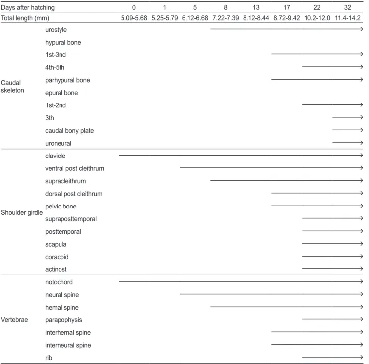
또 Table 1. The developmental process of cranium and orbital region of aucha perch Coreoperca kawamebariDays after hatching 0 1 5 8 13 17 22 32
Total length (mm) 5.09-5.68 5.25-5.79 6.12-6.68 7.22-7.39 8.12-8.44 8.72-9.42 10.2-12.0 11.4-14.2
Cranium
parasphenoid basioccipital nasal frontal sphenotic pterotic lateral ethmoid alisphenoid parietal prootic epiotic supraoccipital exoccipital opisthotic mesethmoid basisphenoid vomer Orbital region preorbital
suborbital
한쏨뱅이목어류인쏨뱅이
(Kim et al., 1997),
붉은쏨뱅이(Han et al., 2001),
조피볼락(Kim and Han, 1991),
동갈치목 어류 인전력날치Cheilopogon heterurus doederleini (Dasilao and Yamaoka, 1998),
복어목어류인Balistes capriscus (Matsuura and Katsuragawa, 1984)
등다른분류군에서도형성되는것으 로보아농어목어류뿐만아니라모든어류에서일반적으로형 성되는전형적인형질로보여진다.
꺽저기의척추골발달은두개골에서꼬리방향으로발달하였
고
,
척추골의골화가완료되기전미부봉상골이골화하기시작 하였다.
꺽저기의척추골수는27-30
개였고,
꺽지는30-34
개,
쏘가리는29
개로차이를보여분류학적형질로서유용하게활용될것으로판단된다
.
척추골발달은같은과어류인꺽지(Han
et al., 2017)
에서도유사하게나타났고,
두종은모두복추골이 먼저발달하였으며,
다음으로신경극이골화하였다.
그러나근 연종인쏘가리(Myoung et al., 2001)
는복추골의발달이전에 신경극이우선적으로골화하여차이를보였다.
Table 2. The developmental process of caudal skeleton, shoulder girdle and vertebrae of aucha perch Coreoperca kawamebari
Days after hatching 0 1 5 8 13 17 22 32
Total length (mm) 5.09-5.68 5.25-5.79 6.12-6.68 7.22-7.39 8.12-8.44 8.72-9.42 10.2-12.0 11.4-14.2
Caudal skeleton
urostyle hypural bone 1st-3nd 4th-5th
parhypural bone epural bone 1st-2nd 3th
caudal bony plate uroneural
Shoulder girdle clavicle
ventral post cleithrum supracleithrum dorsal post cleithrum pelvic bone
supraposttemporal posttemporal scapula coracoid actinost
Vertebrae
notochord neural spine hemal spine parapophysis interhemal spine interneural spine rib
꺽저기 자치어의 골격발달
175
지느러미의발달은전방의등지느러미가발달되기이전에후 방의등지느러미와뒷지느러미가먼저발달하기시작하는것이
농어목어류의일반적인발달양상으로알려져있다
(Johnson,
1984; Faustino and Power, 1999).
꺽저기는전방의등지느러 미와후방의등지느러미및뒷지느러미가순차적으로발달하였 고,
꺽지(Han et al., 2017)
는등지느러미가발달되기이전에후 방의등지느러미와뒷지느러미가발달하여전형적인농어목어류의지느러미발달양상을보였다
.
꺽저기를비롯한꺽지(Han
et al., 2017),
쏘가리(Myoung et al., 2001)
등은지느러미를지 지하는담기골이전방에서후방으로골화하였고,
모든담기골 은추체의골화및지느러미줄기발달이완성된후완료되었다.
이처럼척추골의골화와지느러미의줄기발달이완성된후에 담기골의골화가완료되는것은어류가유영하는데있어추진력을증가시키는것과관련이있는것으로보여진다
(Lee et al., 2001; Han et al., 2017).
꺽저기의미골부는부화직후미부봉상골의골화가진행되지 않았으나
45°
로휘어지기시작하였고,
부화후8
일째평균전 장7.30 mm
일때골화가완료되었다.
꺽지(Han et al., 2017)
의경우꺽저기와마찬가지로부화직후평균전장6.85 mm
일 때미부봉상골의골화는진행되지않았고,
부화후6
일째평균 전장9.00 mm
일때골화가완료되었다.
쏘가리(Myoung et al.,
2001)
는부화직후미부봉상골의골화가관찰되지않았고,
부화후
22
일째평균전장10.9 mm
일때미골부의골화가완료되 어근연종사이에서도시기와크기에따라미골부의다양한골 화형태를나타냈다.
경골어류의초기발육단계에서는체형과지느러미발달및이 Table 3. The developmental process of visceral skeleton of aucha perch Coreoperca kawamebari
Days after hatching 0 1 5 8 13 17 22 32
Total length (mm) 5.09-5.68 5.25-5.79 6.12-6.68 7.22-7.39 8.12-8.44 8.72-9.42 10.2-12.0 11.4-14.2
Visceral skeleton
Upper jaw
premaxillary maxillary supramaxillary Lower
jaw
dentary articular angular
Hyoid arch
epihyal
branchiostegal rays ceratohyal
interhyal hypohyal
Palate
hyomandibular sympletic angular quadrate endopterygoid ectopterygoid metapterygoid palatine
Opercular
preopercle opercle subopercle interopercle
빨의수증가
,
소화관발달등형태변화가후기자어시기에가장 많이일어나며(Myoung et al., 2001),
이시기에변화되는골격 발달의과정은종자생산시문제가되고있는육식성어류의공 식현상및기형발생등원인분석을하는데중요한기초자료가 될수있다.
또한근연종간의식별이나종동정등분류학및생 태학적정보로활용될수있어향우이분야의지속적인연구가 필요할것으로생각된다.
References
Byun SG, Kang CB, Myoung JG, Cha BS, Han KH and Jung CG. 2012. Early osteological development of the larvae and juveniles in Sebastes oblongus (Pisces: Scorpaenidae). Ko- rean J Ichthyol 24, 67-76.
Coban D, Suzer C, Kamaci HO, Saka S and Firat K. 2009. Early osteological development of the fins in the hatchery-reared red porgy, Pagrus pagrus (L. 1758). J Appl Ichthyol 25, 26- Dasilao Jr JC and Yamaoka K. 1998. Osteological and func-32.
tional development of the flyingfish, Cypselurus heterurus
doederleini (Teleostei: Exocoetidae). Bull Mar Sci Fish Ko-
chi Univ 18, 13-26.Emery AR. 1973. Ecology and functional osteology damselfish (Pisces; Pomacentridae) at Alligator reef, Florida Keys. Bull Mar Sci 23, 649-770.
Faustino M and Power DM. 1999. Development of the pectoral, pelvic, dorsal and anal fins in cultured sea bream. J Fish Biol 54, 1094-1110.
Han KH, Park JT, Kim BM, Oh SH, Lee SH and Jin DS. 2000.
Spawning behavior and early life history of aucha perch
Coreoperca kawamebari from Korea. Korean J Ichthyol 12,
129-136.Han KH, Cho JK, Lee SH, Hwang SY, Yoon SM, Seo WI and Kim CC. 2005. Osteological development of the larvae and juveniles of Takifugu pardalis (Teleostei: Tetraodontodae).
Korean J Ichthyol 17, 29-35.
Han KH, Park JT, Jin DS, Yoo DJ and Park JM. 2017. Osteolog- ical development of the larvae and juvenile in Coreoperca
herzi. Korean J Ichthyol 29, 32-40.
Han KH, Lim SK, Kim KS, Kim CW and Yoo DJ. 2001. Osteo- logical development of the larvae and juveniles of Sebastis-
cus tertius (Barsukov et Chen) in Korea. Korean J Ichthyol
13, 63-68.Johnson GD. 1984. Percoidei: development and relationship.
In: Moser HG, WJ Richards, DM Cohen, MP Fahay, AW Kendall, SL Richardson (eds). Ontogeny and systematics of fishes. Amer Soc of Ichthyol Herp, special publiction 1, Al- len Law KS 464-498.
Kang CB, Myoung JG, Kim YU and Kim HC. 2012. Early os- teological development and squamation in the spotted sea bass Lateolabrax maculates (Pisces: Lateolabracidae). Ko-
rean J Fish Aquat Sci 45, 271-282.
Kendall AW, Ahlstrom EH and Moser HG. 1984. Early life his- tory stages of fishes and their characters. In: HG Moser, WJ Richards, DM Cohen, MP Fahay, AW Kendall, SL Richard- son (eds). Ontogeny and systematics of fishes. Amer Soc of Ichthyol Herp, special publiction 1, Allen Law KS 11-12.
Kendall W. 1991. Systematics and identification of larvae and juveniles of the genus Sebastes. Env Biol Fish 30, 173-190.
Kim IS, Choi Y, Lee CL, Lee YJ, Kim BJ and Kim JH. 2005. Il- lustrated book of Korean fishes. Kyo Hak Publishing, Seoul, Korean,.
Kim IS and Park JY. 2002. Freshwater Fishes of Korea. Kyo Hak Publishing Co Ltd, Seoul, Korea.
Kim SH, Lee SH, Lee WO and Cho KH. 2013. Distribution of Coreoperca kawamebari and C. herzi and fish com- munity structure in relation to environmental differences in their sympatric area of the Boseong river, Korea. Ko- rean Soc Lim 46, 367-379. http://dx.doi.org/10.11614/
KSL.2013.46.3.367.
Kim YU and Han KH. 1989. Early life history of the marine animals 1. Egg development, larvae and juveniles of Chae-
nogobius laevis (Steindachner). Bull Korean Fish Soc 22,
317-331.Kim YU, Han KH and Kang CB. 1992. Morphology and skel- etal development of larvae and juveniles of Entomacrodus
stellifer lighti (Herre). Korean J Ichthyol 4, 31-43.
Kim YU, Han KH, Kang CB, Kim JK and Byun SK. 1997. The early life history of the rockfish, Sebastiscus marmoratus 2.
Morphology and skeletal development of larvae and juve- nile. Korean J Ichthyol 9, 186-194.
Kim YU, Han KH and Byun SK. 1993. The early life history of the rockfish, Sebastes inermis 2. Morphological and skeletal development of larvae and juveniles. Bull Korean Fish Soc 26, 465-476.
Kim YU and Han KH. 1991. The early life history of rockfish
Sebastes schlegeli. Korean J Ichthyol 3, 67-83.
Koumoundouros G, Sfakianakis DG, Maingot E, Divanach P and Kentouri M. 2001a. Osteological development of the vertebral column and of the fins in Diplodus sargus (Tele- ostei: Perciformes: Sparidae). Mar Biol 139, 853-862.
Koumoundouros G, Divanach P and Kentouri M. 2001b. Osteo- logical development of Dentex dentex (Osteichthyes: Spari- dae): dorsal, anal, paired fins and squamation. Mar Biol 38, 399-406.
Koumoundouros G, Gagliardi F, Divanach P, Boglione C, Cataudella S and Kentouri M. 1997a. Normal and abnormal osteological development of caudal fin in Sparus aurata L.
fry. Aquaculture 149, 215-226.
Koumoundouros G, Oran G, Divanach P, Stefanakis S and Kentouri M. 1997b. The opercular complex deformity in in- tensive gilthead sea bream (Spartus aurata L.) larviculture.
Moment of apparition and description. Aquaculture 156,
꺽저기 자치어의 골격발달
177
165-177.
Lee SJ, Kim YU and Han KH. 2001. Osteological development of larvae and juveniles of Hyporhampus sajori (Teleostei:
Hemiramphidae). Korean J Ichthyol 13, 173-180.
Liu CH. 2001. Early osteological development of the yellow tail
Seriola dumerili (Pisces: Carangidae). Zool Stud 40, 289-
Matsuoka M. 1985. Osteological development in the red sea 298.bream, Pagrus major. Japan J Ichthyol 32, 35-51.
Matsuoka M. 1987. Development of the skeletal tissues and skeletal muscles in the red sea bream. Bull Seikai Red Fish Res Lab 65, 1-14.
Matsuura Y and Katsuragawa M. 1984. Osteological develop- ment of fins and their supports of larval grey triggerfish,
Balistes capriscus. Japan J Ichthyol 31, 411-421.
Myoung JG, Mun JH, Kim JK, Park KD, Kang CB, Kim YU and Park JT. 2001. Osteological development of larvae and juveniles of Korean mandarine fish, Siniperca scherzeri (Perciformes: Centropomidae). Korean J Ichthyol 13, 129- Mook D. 1977. Larval and osteological development of the 135.
sheepshead, Archosargus probatocephalus (Pisces: Spari- dae). Copeia 1977, 126-133.
NIBR (national institute of biological resources). 2018. Endan- gered wild animals and plants. Retrieved from https://spe- cies.nibr.go.kr/home/mainHome.do?cont_link=009&subM enu=009002&contCd=009002&ktsn=120000058306 on Jan 26, 2018.
Okiyama M. 1988. An Atlas of the Early State Fishes in Japan.
Tokai University Press, Tokyo, Japan. 1157.
Park JY, Hong CG, Cho JK, Son MH, Han KH and Park JM.
2015. Early osteological development of the larvae and ju- veniles in sevenband grouper, Epinephelus septemfasciatus (Pisces: Serranidae). Korean J Ichthyol 27, 189-198.
Potthoff T, Kelly S, Saksena V, Moe M and Young F. 1987. De- scription of larval and juvenile damselfish Microspathodon
chrysurus, Pomacentridae, and their osteological develop-
ment. Bull Mar Sci 40, 1-40.Potthoff T, Kelley S and Collins LA. 1988. Osteological devel- opment of the red snapper, Lutjanu scampechanus (Lutjani- dae). Bull Mar Sci 43, 1-40.
Potthoff T and Tellock JA. 1993. Osteological development of the snook, Centropomus undecimalis (Teleostei, Centro- pomidae). Bull Mar Sci 52, 669-716.
Sfakianakis DG, Koumoundouros G, Divanach P and Kentouri M. 2004. Osteological development of the vertebral column and of the fins in Pagellus erythrinus (L. 1758). Tempera- ture effedt on the developmental plasticity and morphoana- tomical abnormalities. Auqaculture 232, 407-424.
Sfakianakis DG, Doxa CK, Kouttouki S, Koumoundouros G, Maingot E, Divanach P and Kentouri M. 2005. Osteologi- cal development of the vertebral column and of the fins in
Diplodus puntazzo (Cetti, 1777). Auqaculture 250, 36-46.
Vandewalle P, Gluckmann I, Baras E, Huriaux F and Focant B.
1997. Postembryonic development of the cephalic region in
Heterobranchus longifilis. J Fish Biol 50, 227-253.
Wagemans F and Vandewalle P. 1999. Development of the car- tilaginous skull in Solea solea: trends on Pleuronectiforms.
Ann Sci Nat 1, 39-52.
Walker MB and Kimmel CB. 2007. A two-color acid-free car- tilage and bone stain for zebrafish larvae. Biol Histochem 82, 23-28.