55
Open Access
Obesity and Preclinical Changes of Cardiac Geometry and Function
Joong Kyung Sung, MD1 and Jang-Young Kim, MD1,2
1Division of Cardiology, Department of Internal Medicine, Wonju Christian Hospital and
2Institute of Genomic Cohort, Yonsei University Wonju College of Medicine, Wonju, Korea
ABSTRACT
Overweight and obesity are rapidly increasing in prevalence due to adoption of the westernized life style in Korea.
Obesity is strongly associated with the development of cardiovascular risk factors such as diabetes, hypertension, and dyslipidemia. In addition, accumulating evidence suggests that obesity per se has a direct effect on cardiac func- tional and structural changes that may not be the result of atherosclerosis. In this review, we focus on the view that obesity can influence on the structural and functional changes of the heart, drawing evidence from human and animal studies. We also review influencing factors such as physical, neurohormonal, and metabolic altera- tions that are associated with changes of the heart in obesity. (Korean Circ J 2010;40:55-61)
KEY WORDS:Obesity; Left ventricular hypertrophy; Left ventricular dysfunction; Diastole.
Introduction
The prevalence of obesity is increasing due to the increasing adoption of westernized life styles in Korea.
Obesity is defined by body mass index (BMI), which is classified as underweight (<20 kg/m2), normal (20-24.9 kg/m2), overweight (25-29.9 kg/m2), and obese (≥30 kg/
m2). Korean National Health Examination and Nutri- tion Survey data in 2001 estimated that 3.0% of Korean adults were obese and 29.5% were overweight.1) The pre- valence of overweight Korean children and adolescents doubled from 5.4% in 1998 to 11.4% in 2001.2) Increas- ing degrees of obesity are closely correlated with the increasing rates of cardiovascular disease.3) Furthermore, a large body of evidence strongly supports the view that obesity itself is associated with preclinical structural and functional changes in the heart, which prelude heart failure.4) In this review, we discuss the changes of car- diac structure and function in obesity and focus on the
current evidence regarding the causative role of obesity in cardiac changes.
Structural Changes in the Heart in Obesity
Animal studies regarding structural changes of he- art in obesity
Two types of animal models are used in obesity stu- dies: genetic mutant models and high fat diet-induced obesity model. Genetic mutant mice models, such as ob/ob and db/db, present severe types of obesity. The ob/ob and db/db mice models become progressively obese, left ventricular (LV) hypertrophy develops, and LV mass increases. Interestingly, leptin infusion to these mice leads to decreased myocardial wall thickness.5) LV hypertrophy is a common cardiac phenotype in the res- ponse of genetic mutant mice to obesity. Use of high fat diet mice has demonstrated that increased non-es- terified fatty acid (NEFA) contents, which diminish the rate of glucose metabolism and increase oxygen con- sumption, result in reduced ability to recover from a workload.6) Furthermore, high fat diet mice also dis- play elevated blood pressure and impaired insulin re- ceptor activation.7) All of these factors contribute to the development of cardiac hypertrophy and LV dys- function in high fat diet mice.7)
Results from the two animal models indicate that
Correspondence: Jang-Young Kim, MD,Division of Cardiology, Department of Internal Medicine, Wonju Christian Hospital, Yonsei University Wonju College of Medicine, 162 Ilsan-dong, Wonju 220-701, Korea Tel: 82-33-741-0909, Fax: 82-33-741-1219
E-mail: [email protected]
○ cc This is an Open Access article distributed under the terms of the Creative Commons Attribution Non-Commercial License (http://creativecommons.
org/licenses/by-nc/3.0) which permits unrestricted non-commercial use, distribution, and reproduction in any medium, provided the original work is properly cited.
obesity may result in cardiac hypertrophy due to insulin resistance (IR), metabolism alteration such as leptin de- ficiency, or leptin resistance and elevated blood pressure.
Obesity and left ventricular hypertrophy in human
Obesity is an independent factor for LV hypertro- phy.8-11) Obesity itself may have hemodynamic effects that produce an increase in the total blood volume and cardiac output due to the high metabolic activity of excessive fat. In moderate to severe obesity, these increases may lead to LV dilation, increased LV wall st- ress, and compensatory (eccentric) LV hypertrophy.8)9) Despite this logical explanation, recent studies have shown that concentric LV hypertrophy is a predomi- nant form in obese subjects.10)11) Besides hemodynamic factors, hyperinsulinemia due to IR, which stimulates insulin like growth factor-1 (IGF-1) receptors, is also involved in the pathogenesis of LV hypertrophy.12) IGF-1 enhances anabolic effects on the myocardium, and fa- cilitates increased myocardial mass and concentric hy- pertrophy. Systolic hypertension is traditionally known as a major contributor to LV hypertrophy rather than obesity. However, in the Framingham heart study, the investigators found independent influences of BMI and blood pressure on LV mass index.13) Therefore, the effects of obesity and blood pressure were additive on LV changes. However, opinion is divided concerning which components are the stronger predictor of LV hy- pertrophy. Obesity was the strongest clinical predictor for LV hypertrophy in several studies,12)14) while another study showed that BMI and hypertension affect LV hy- pertrophy similarly.15)
In summary, obesity induces LV hypertrophy via he- modynamic changes caused by hyperinsulinemia, in- creased IGF-1 expression, and volume overload, which are similar to the effect caused by hypertension.
Obesity and cardiac adipocytes in human
Epicardial fat mass, as determined by echocardiogra- phic and magnetic resonance imaging studies, may re- flect intra-abdominal visceral fat. Therefore, epicardial fat mass could serve as a marker of visceral adiposity.16) The relationship between local adipose tissue in heart and cardiac geometry has recently been studied. Incre- ased epicardial fat mass and fatty infiltration of myo- cardium contributed to the increased cardiac mass.17) Al- though epicardial fat is normally accounted for about 20% of the total ventricular mass, total epicardial fat wei- ghts are significantly greater in hypertrophic heart.18)
The causal effect of epicardial adipose tissue on car- diac remodeling remains unsolved. However, epicar- dial adipose tissue serves as an endocrine organ, which releases monocyte chemotactic protein-1, interleukin-1β, interleukin-6 and tumor necrosis factor-alpha. These
adipokines may be important factors in cardiac remo- deling or hypertrophy in obese subjects.19)
Obesity and left atrial enlargement in human Left atrial (LA) size and LA volume is increased in the setting of obesity compared with non-obese indivi- duals.20) The possible mechanisms of increased LA size and volume in obesity are similar to those of LV hyper- trophy in obesity; increased BMI leads to volume over- load,21) which causes diastolic abnormality of LV filling defect. Diastolic dysfunction can contribute to LA en- largement or remodeling.22) In the Framingham heart study, obesity was found to be a strong risk factor for development of atrial fibrillation even after accounting for concomitant conditions such as hypertension, dia- betes mellitus, and myocardial infaction.23) In the cli- nical setting, dilated LA size in the absence of organic heart disease or atrial fibrillation is considered to be a risk factor for developing atrial fibrillation and long- term cardiovascular events.24)
Functional Changes of the Heart in Obesity
Changes of left ventricular systolic function The effects of obesity on LV systolic function are con- troversial. Several studies reported that LV systolic func- tion is normal or increased in obesity.8)9)25) However, some evidence from non-invasive or invasive techniques suggests that obesity causes a subclinical contractility abnormality.11)26)27) Obesity-induced myocardial dysfunc- tion can be explained by the derangement of myocar- dial metabolism. In animal models, IR in obesity cause alterations in myocardial fatty acid metabolism and ef- ficiency (cardiac work/myocardial oxygen consumption) that occur early in the cascade of events, leading to im- paired LV contractility.28) In human studies, obesity is a significant predictor of increased myocardial oxygen consumption and decreased efficiency, and IR is a ro- bust predictor of fatty acid uptake, utilization, and oxi- dation. These metabolic changes may play a role in the pathogenesis of decreased cardiac performance in obe- sity.29) Collectively, obesity might be a strong factor that can induce LV systolic dysfunction and eventually cause heart failure independent to coronary artery disease or other morbidities.
Changes of left ventricular diastolic function Echocardiography studies conducted with obese ani- mals and human have provided inconsistent results about E-wave velocity, deceleration time, A-wave velo- city.11)30) Prolongation of the isovolumic relaxation time may be the most consistent diastolic abnormality in obesity.31)32) In uncomplicated obese subjects, diastolic dysfunction is caused by hemodynamic and metabolic
factors. Hemodynamic changes cause diastolic dysfunc- tion in obese subjects through LV hypertrophy.33) Meta- bolic factors will be discussed later chapter. Other po- tential mediators include hormones and cytokines re- leased in association with obesity. Changes of adipo- kine concentration in serum, such as leptin and adipo- nectin, are observed in obese subjects, which may also be partly responsible for the LV diastolic dysfunction.
Studies with ob/ob mice (which are deficient in leptin) have demonstrated diastolic dysfunction by the changes of myocardial fatty acid and glucose metabolsim.28)34) Si- milarly, leptin resistance and hyperleptinemia are ob- served in obese subjects, which may lead to diastolic dysfunction. Circulating total and high-molecular-weight adiponectin are negatively correlated with LV wall thick- ness and diastolic dysfunction independent of age and metabolic factors.35) In the Otsuka Long-Evans Toku- shima Fatty rat model of pre-diabetes, pre-diabetic con- ditions cause an accumulation of myocardial collagen, leading to interstitial and perivascular fibrosis, which correlates with LV early diastolic dysfunction.36)
Collectively, the evidence indicates that the precli- nical diastolic dysfunction of obesity is related to hemo- dynamic alteration, various adipokines, and myocardial collagen accumulation.
Several Mechanisms that Influence the Structure and Function
of the Obese Heart
Hemodynamic changes
Obesity causes an increase in total blood volume and cardiac output.37) In addition, high blood pressure is commonly found in obesity, with an estimated preval- ence of up to 60%.38) These hemodynamic factors af- fect the cardiac geometry, especially LV hypertrophy.
Sleep apnea in obesity
Obstructive sleep apnea (OSA) is a very common abnormality in obese subjects. The Wisconsin Sleep Cohort Study demonstrated that the risk of develop- ment of hypertension in OSA patients is three times higher than non-OSA subjects.39) Likewise, OSA patients demonstrated not only day-time hypertension but night- time hypertension or non-dipper pattern.39) Repetitive episodes of airway obstruction cause hypoxemia and ch- anges of intra-thoracic pressure, which in turn causes sympathetic overactivity. Furthermore, chronic hypoxe- mia causes injury to myocytes and cardiac extracellular matrix. All of these factors-hypertension, sympathetic overactivity, hypoxemia-lead to ventricular remodeling.40) One study reported that 88% of OSA patients have LV hypertrophy and 64% of those have LA enlargement.41) The authors also demonstrated that the use of conti- nuous positive airway pressure for 6 months could re-
duce LV hypertrophy.41) All these findings suggest that the severity of nocturnal hypoxemia could be impor- tant in the development of LV hypertrophy in obese subjects with OSA.
Changes of cardiac metabolisms, mitochondrial dysfunction and oxidative stress
Energy sources of myocardial metabolism are differ- ent from diabetes compared to normal glucose tolerance subjects. In the setting of diabetes or IR, myocardium utilizes much more free-fatty acid and less glucose, which occurs even in the early course of obesity.42) Long- standing caloric excess or obesity activates peroxisome proliferator-activated receptor-α/peroxisome prolife- rator-activated receptor-γ coactivator signaling, which increases the expression of genes involved in fatty acid oxidation and fatty acid transporters such as FATP1 and CD36.43)44) In human, obesity is also associated with increased rates of fatty acid oxidation, increased myocardial oxygen consumption, and reduced cardiac performance proportionate to the degree of IR and obe- sity.29) Indeed, normalization of cardiac metabolism by overexpression of a human GLUT4 transgene in db/db mice reverses cardiac dysfunction, which suggests that altered myocardial metabolism contributes to contrac- tile dysfunction in this model.45) It has been demonst- rated that mitochondrial oxygen consumption rate and adenosine triphosphate (ATP) generation capacity are reduced in ob/ob mice, and that the protein levels of mitochondrial complexes I, III, V for the oxidative ph- osphorylation are significantly reduced in db/db mice.46) Because of mitochondrial response in obesity or dia- betes, the ratio of ATP generation and oxygen consump- tion is reduced (mitochondrial uncoupling), which is be- lieved to be a significant factor for declination of cardiac function.46) Mitochondria also represent a major source of superoxide production. In db/db mice, mitochondrial reactive oxygen species generation increases, which can damage myocardial cells.46)
In summary, cardiac metabolic changes, mitochondrial dysfunction, and oxidative stress can cause declination of cardiac function in obese subjects.
Insulin resistance
IR represents problems of insulin receptor, insulin signaling, and genetics. Among these problems, insu- lin signaling impairment could be the key factor for IR.
Impaired insulin-mediated activation of intracellular signaling has been described in ob/ob mice.42) In the animal models, obesity and IR can increase myocardial fatty acid uptake, which causes myocardial fatty acid oxidation and myocardial oxygen consumption. Persist- ence of this metabolic change causes an imbalance of fatty acid uptake and fatty acid oxidation, leading to accumulation of fatty acid intermediates and ceramide
production, which impairs myocardial function and increases apoptosis of myocardocytes.47)48) This pheno- menon has also been demonstrated in human.29) How- ever, in Zucker fatty rats treated with the insulin sen- sitizer thiazolinedione (TZD), myocardial glucose con- sumption was increased, fatty acid oxidation was dimi- nished, and myocardial injury was reduced.49) In ad- dition, hyperinsulinemia stimulates IGF-1 receptors, which is likely responsible in the pathogenesis of myo- cardial hypertrophy.12)
Neurohormonal activations
In general, obese subjects have activated sympathetic tone. This leads to concentric LV hypertrophy due to elevated afterload and increased cardiac contractility.
Additionally, catecholamine directly affects the myo- cardium without hemodynamic influence. The renin- angiotensin system also affects the heart via hemodyna- mic effects or direct signaling in obesity.50) Angioten- sinogen secretion and its messenger ribonucleic acid (mRNA) have been detected in fa/fa rat adipocytes and in human.51) Angiotensin is thought to be a con- tributor to the sympathoexcitation, therefore the ren- nin-angiotensin system could increase central sympa- thetic activation and have a role as an additive effect to blood pressure elevation and cardiac remodeling in obese subjects.52)
Changes of extracellular matrix and fibrosis The compositional changes of the extracellular ma- trix is an important contributor in cardiac remodel- ing.53) Serum levels of cardiac collagen synthesis have been significantly associated with IR in normotensive and non-diabetic obese subjects.54) In another study using a rabbit model of obesity, a high-fat diet caused fibrosis in coronary vessels as well as the accumulation of collagen in the cardiac interstitium.55)
Adipokines such as adiponectin or leptin play an important role in the extracellular matrix changes of the heart. Leptin is an adipokine that is produced in the obese gene (ob) located in adipocytes.56) In one st- udy, leptin increased matrix metalloproteinase-2 (MMP- 2) secretion and its mRNA expression, and attenuated collagen I mRNA synthesis and increased collagen III and IV, but did not change total collagen synthesis in human pediatric ventricular myocytes.57) In a diet-in- duced obesity murine model, elevated leptin level was detected after 20 weeks of a high fat diet, and this lep- tin level was correlated with reduced ventricular shor- tening and increased LV posterior wall thickness.58) In the coronary ligation model, procollagen I and III levels were elevated after 7 days postinfarction. Furthermore when the neutralizing leptin antibody was injected, en- hanced collagen was attenuated.59)
Adiponectin is considered a cardioprotective adipo-
kine. A recent study suggested that adiponectin level showed positive correlation with tissue inhibitor of metalloproteinase (TIMP), which is considered to exert an antifibrotic effect.60) The adiponetin levels are decreas- ed in the obese or IR subjects; the cardioprotective ef- fect of adiponectin, especially its antifibrotic effect, is di- minished in obese subjects.
In obese subjects, adipocytokines such as catechola- mine, rennin-angiotensin-aldosterone system, tumor ne- crosis factor-alpha, C-reactive protein, leptin, and adi- ponectin influence to MMP activity, TIMP expression, and collagen synthesis, which ultimately leads to car- diac remodeling.53)
Apoptosis of cardiomyocytes
The evidence of cardiac apoptosis in the genesis of heart failure has surged over the last decade. Apoptotic cardiomyocyte death has been proven from biopsies of dilated cardiomyopathy and end stage heart failure.61) Besides apoptotic cell death, activation of several pro- teases in the apoptotic cascade can cleave contractile proteins including actin, myosin, and troponins.62) Th- ere are numerous causes of apoptosis. Apoptosis appears to be ischemia or ischemia-reperfusion driven at the site of infarct and at sites remote from the ischemic area through neurohormonal effects. Myocardial stretch, wall stress, cytokines, and neurohormones such as nor- epinephrine and angiotensin II, which are commonly produced as a part of peripheral neurohormonal rear- rangements after an acute myocardial infarction, have been demonstrated to enhance apoptosis.63)64)
Studies for obesity associated cardiac apoptosis are lacking. Use of the Zucker fatty rat model has shown that cardiac contractility is reduced with increased car- diac cell apoptosis; however, when the rats were treated with TZD, cardiac cell apoptosis was decreased.48) An- other study involving a high fat diet induced IR rat model did not reveal a significant difference in apo- ptosis compared with control group, despite impaired cardiac function.65) Therefore, it is necessary to study the relationship between obesity, cardiac cell apoptosis, and impairment of cardiac function.
Therapeutic Implications
Weight reduction improves systolic blood pressure, IR, sleep apnea, and hyperlipidemia. Weight reduction also has beneficial effects on cardiac structure and func- tion such as reduction of LV diameter, LV wall thick- ness, LV mass, and LA dimension.66) However, removal of subcutaneous fat by liposuction does not produce a beneficial effect on metabolic changes, so it has little effect on the cardiovascular system.67) Pharmacologi- cal-mediated weight reduction is recommended for pa- tients in whom lifestyle modification has failed. Ori-
stat, which is a gastrointestinal lipase inhibitor, can reduce weight by about 3 kg and decreases progression to diabetes in high risk patients.68) Sibutramine, a mo- noamine reuptake inhibitor, can reduce weight by about 4-5 kg and improves serum lipid levels and insulin sen- sitivity,69) but it increases heart rate and blood pressure.
Rimonabant, an endocannabinoid receptor blocker, re- duces weight by 4-5 kg, decreases waist circumference, and improves serum high density lipoprotein choles- terol and triglyceride levels.70) However, there are no de- finitive data for the effect on cardiac structure and func- tion in use of these medications.71) Therefore, weight re- duction by diet and exercise is still the most important means to correct the functional and structural deteri- orations that occur in the heart in obese individuals.
Conclusions
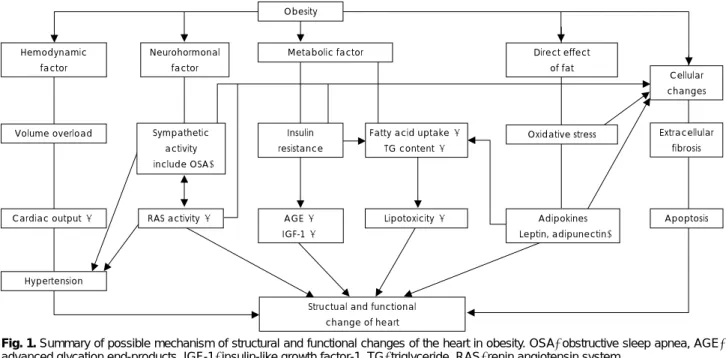
Obesity is a causative factor for development of pre- clinical changes of the heart. In obese subjects, initially volume overload develops, which leads to increased cardiac output and hypertension. Second, sympathetic and rennin-angiotensin-aldosterone system activity is increased, which causes hypertension. Third, hyperin- sulinemia (IR) increases advanced glycation end-pro- ducts and IGF-1 production and facilitates fatty acid uptake, which leads to lipotoxic cell damage. Fourth, vis- ceral adipose tissue affects oxidative stress and is relat- ed with decreased leptin level or leptin resistance and decreased adiponectin level; these changes affect cellu- lar changes and metabolic alteration. Fifth, cellular ch- anges are produced by neurohormonal and metabolic factors, oxidative stress, and adipokines, which leads to extracellular fibrosis and apoptosis. All these descrip-
tive factors eventually lead to structural and functional changes of the heart in obesity (Fig. 1). Therapeutically, life-style associated weight reduction may be more im- portant than any other molecular- or pathway-targeted therapy.
REFERENCES
1) Park HS, Park CY, Oh SW, Yoo HJ. Prevalence of obesity and metabolic syndrome in Korean adults. Obes Rev 2008;9:104-7.
2) Kim HM, Park J, Kim HS, Kim DH, Park SH. Obesity and cardiovascular risk factor in Korean children and adolescents aged 10-18 years from the Korean National Health and Nutri- tion Examination Survey, 1998 and 2001. Am J Epidemiol 2006;
164:787-93.
3) Hubert HB, Feinleib M, McNamara PM, Castelli WP. Obesity as an independent risk factor for cardiovascular disease: a 26-year follow-up of participants in the Framingham Heart Study. Circu- lation 1983;67:968-77.
4) Kenchaiah S, Evans JC, Levy D, et al. Obesity and the risk of heart failure. N Engl J Med 2002;347:305-13.
5) Barouch LA, Berkowitz DE, Harrison RW, O’Donnell CP, Hare JM. Disruption of leptin signaling contributes to cardiac hyper- trophy independently of body weight in mice. Circulation 2003;
108:754-9.
6) Korvald C, Elvenes OP, Myrmel T. Myocardial substrate meta- bolism influence left ventricular energetic in vivo. Am J Physiol Heart Circ Physiol 2000;278:H1345-51.
7) Ouwens DM, Boer C, Fodor M, et al. Cardiac dysfunction in- duced by high -fat diet is associated with altered myocardial in- sulin signaling in rats. Diabetologia 2005;48:1229-37.
8) Avelar E, Cloward TV, Walker JM, et al. Left ventricular hyper- trophy in severe obesity: interaction among blood pressure, noc- turnal hypoxemia, body mass. Hypertension 2007;49:34-9.
9) Wong CY, O’Moore-Sullivan T, Leano R, Byrne N, Beller E, Marwick TH. Alterations of the left ventricular myocardial ch- aracteristics associated with obesity. Circulation 2004;110:3081-7.
10) Iacobellis G, Ribaudo MC, Zappaterreno A, Iannucci CV, Di Mario U, Leonetti F. Adapted changes in left ventricular struc-
Cardiac output ↑ RAS activity ↑ Lipotoxicity ↑ Adipokines Apoptosis
(Leptin, adipunectin) AGE ↑
IGF-1 ↑
Hypertension Volume overload
Hemodynamic factor
Neurohormonal factor
Sympathetic activity (include OSA)
Insulin resistance
Metabolic factor
Fatty acid uptake ↑ TG content ↑
Direct effect of fat
Oxidative stress
Cellular changes
Extracellular fibrosis
Structual and functional change of heart Obesity
Fig. 1. Summary of possible mechanism of structural and functional changes of the heart in obesity. OSA: obstructive sleep apnea, AGE:
advanced glycation end-products, IGF-1: insulin-like growth factor-1, TG: triglyceride, RAS: renin angiotensin system.
ture and function in severe uncomplicated obesity. Obes Res 2004;
12:1616-21.
11) Peterson LR, Waggoner AD, Schechtman KB, et al. Alterations in left ventricular structure and function in young healthy obese women: assessment by echocardiography and tissue Doppler im- aging. J Am Coll Cardiol 2004;43:1399-404.
12) Sundstrom J, Lind L, Valind S, et al. Myocardial insulin-mediat- ed glucose uptake and left ventricular geometry. Blood Press 2001;10:27-32.
13) Lauer MS, Anderson KM, Levy D. Separate and joint influence of obesity and mild hypertension on left ventricular mass and geo- metry: the Framingham Heart Study. J Am Coll Cardiol 1992;19:
130-4.
14) Gottdiener JS, Reda DJ, Materson BJ, et al. Importance of obesity, race and age to the cardiac structure and functional ef- fect of hypertension. J Am Coll Cardiol 1994;24:1492-8.
15) Fox E, Taylor H, Andrew M, et al. Body mass index and blood pressure influence on left ventricular mass and geometry in Af- rican Americans: the Atherosclerotic Risk In Communities (ARIC) Study Hypertension 2004;44:55-60.
16) Iacobellis G, Leonetti F. Epicardial adipose tissue and insulin resistance in obese subjects. J Clin Endocardiol Metab 2005;90:
6300-2.
17) Iacobellis G, Ribaudo MC, Zappaterreno A, Iannucci CV, Leo- netti F. Relation between epicardial adipose tissue and left ven- tricular mass. Am J Cardiol 2004;94:1084 -7.
18) Corradi D, Maestri R, Callegari S, et al. The ventricular epicar- dial fat is related to the myocardial mass in normal, ischemic and hypertrophic hearts. Cardiovasc Pathol 2004;13:313-6.
19) Malavazos AE, Ermetici F, Coman C, Corsi MM, Morricone L, Ambrosi B. Influence of epicardial adipose tissue and adipocy- tokine levels on cardiac abnormalities in visceral obesity. Int J Cardiol 2007;121:132-4.
20) Ayer JG, Almafragy HS, Patel AA, Hellyer RL, Celermajer DS.
Body mass index is an independent determinant of left atrial size.
Heart Lung Circ 2008;17:19-24.
21) Di Bello V, Santini F, Di Cori A, et al. Obesity cardiomyopathy:
is it a reality? An ultrasonic tissue characterization study. J Am Soc Echocardiogr 2006;19:1063-71.
22) Pritchett AM, Mahoney DW, Jacobsen SJ, Rodeheffer RJ, Karon BL, Redfield MM. Diastolic dysfunction and left atrial volume:
a population-based study. J Am Coll Cardiol 2005;45:87-92.
23) Wang TJ, Parise H, Levy D, et al. Obesity and the risk of new- onset atrial fibrillation. JAMA 2004;292:2471-7.
24) Tsang TS, Barnes ME, Miyasaka Y, et al. Obesity as a risk factor for the pregression of paroxysmal to permanent atrial fibrillation:
a longitudinal cohort study of 21 years. Eur Heart J 2008;29:
2227-33.
25) Messerli FH, Ventura HO, Reisin E, et al. Borderline hyperten- sion and obesity: two prehypertensive states with elevated car- diac output. Circulation 1982;66:55-60.
26) Aurigemma GP, Silver KH, Priest MA, Gaasch WH. Geometric changes allow normal ejection fraction despite depressed myo- cardial myocardial shortening in hypertensive left ventricular hypertrophy. J Am Coll Cardiol 1995;26:195-202.
27) Garavaglia GE, Messerli FH, Nunez BD, Schmieder RE, Gros- sman E. Myocardial contractility and left ventricular function in obese patients with essential hypertension. Am J Cardiol 1988;
62:594-7.
28) Aasum E, Hafstad AD, Severson DL, Larsen TS. Age-dependent changes in metabolism, contractile function, and ischemic sensi- tivity in hearts from db/db mice. Diabetes 2003;52:434-41.
29) Peterson LR, Herrero P, Schechtman KB, et al. Effect of obesity and insulin resistance on myocardial substrate metabolism and
efficiency in young women. Circulation 2004;109:2191-6.
30) Pascual M, Pascual DA, Soria F, et al. Effect of isolated obesity on systolic and diastolic left ventricular function. Heart 2003;
89:1152-6.
31) Berkalp B, Cesur V, Corapcioglu D, Erol C, Baskal N. Obesity and left ventricular diastolic dysfunction. Int J Cardiol 1995;52:
23-6.
32) Morricone L, Malavazos AE, Coman C, Donati C, Hassen T, Caviezel F. Echocardiographic abnormalities in normotensive obese patients: relationship with visceral fat. Obes Res 2002;10:
489-98.
33) Chakko S, Mayor M, Allison MD, Kessler KM, Materson BJ, Myerburg RJ. Abnormal left ventricular diastolic filling in ec- centric left ventricular hypertrophy of obesity. Am J Cardiol 1991;
68:95-8.
34) Christoffersen C, Bollano E, Lindegaard ML, et al. Cardiac lipid accumulation associated with diastolic dysfunction in obese mice. Endocrinology 2003;144:3483-90.
35) Kozakova M, Muscelli E, Flyvbjerg A, et al. Adiponectin and left ventricular structure and function in healthy adults. J Clin Endocrinol Metab 2008;93:2811-8.
36) Mizushige K, Yao L, Noma T, et al. Alteration in left ventricular diastolic filling and accumulation of myocardial collagen at in- sulin-resistant prediabetic stage of a type II diabetic rat model.
Circulation 2000;101:899-907.
37) Messerli FH, Christie B, DeCarvalho JGR, et al. Obesity and es- sential hypertension; hemodynamics, intravascular volume, so- dium excretion, and plasma renin activity. Arch Intern Med 1981;
141:81-5.
38) Must A, Spadano J, Coakley EH, Field AE, Colditz G, Dietz WH.
The disease burden associated with overweight and obesity. JA- MA 1999;282:1523-9.
39) Peppard PE, Young T, Palta M, Skatrud J. Prospective study of the association between sleep-disordered breathing and hyper- tension. N Engl J Med 2000;342:1378-84.
40) Quan SF, Gersh BJ. Cardiovascular consequences of sleep- disordered breathing: past, present and future: report of a work- shop from the National Center on Sleep Disorders Research and the National Heart, Lung, Blood Institute. Circulation 2004;109:
951-7.
41) Cloward TV, Walker JM, Farney RJ, Anderson JL. Left ventri- cular hypertrophy is a common echocardiographic abnormality in severe obstructive sleep apnea and reverses with nasal con- tinuous positive airway pressure. Chest 2003;124:594-601.
42) Buchanan J, Mazumder PK, Hu P, et al. Reduced cardiac effici- ency and altered substrate metabolism precedes the onset of hy- perglycemia and contractile dysfunction in two mouse models of insulin resistance and obesity. Endocrinology 2005;146:5341-9.
43) Mazumder PK, O’Neill BT, Roberts MW, et al. Impaired cardiac efficiency and increased fatty acid oxidation in insulin-resistant ob/ob mouse hearts. Diabetes 2004;53:2366-74.
44) Coort SL, Hasselbaink DM, Koonen DP, et al. Enhanced sarco- lemmal FAT/CD 36 content and triacylglycerol storage in cardiac myocytes from obese zucker rats. Diabetes 2004;53:1655-63.
45) Semeniuk LM, Kryski AJ, Severson DL. Echocardiographic assessment of cardiac function in diabetic db/db and transgenic db/db-hGLUT4 mice. Am J Physiol Heart Circ Physiol 2002;283:
H976-82.
46) Boudina S, Sena S, Theobald H, et al. Mitochondrial energetics in the heart in obesity related diabetes: direct evidence for in- creased uncoupled respiration and activation of uncoupling pro- teins. Diabetes 2007;56:2457-66.
47) Unger RH. Lipotoxic disease. Annu Rev Med 2002;53:319-36.
48) Zhou YT, Grayburn P, Karim A, et al. Lipotoxic heart disease in
obese rats: implications for human obesity. Proc Natl Acad Sci U S A 2000;97:1784-9.
49) Sidell RJ, Cole MA, Draper NJ, Desrois M, Buckingham RE, Clarke K. Thiazolidinedione treatment normalizes insulin resist- ance and ischemic injury in the Zucker fatty rat heart. Diabetes 2002;51:1110-7.
50) Ruano M, Silvestre V, Castro R, et al. Morbid obesity, hyperten- sive disease and the rennin-angiotensin-aldosterone axis. Obes Surg 2005;15:670-6.
51) Hainault I, Nebout G, Turban S, Ardouin B, Ferre P, Quiqnard- Boulange A. Adipose tissue-specific increase in angiotensinogen expression and secretion in the obese (fa/fa) Zucker rat. Am J Physiol Endocrinol Metab 2002;282:E59-66.
52) Davy KP, Hall JE. Obesity and hypertension: two epidemics or one? Am J Physiol Regul Integr Comp Physiol 2004;286:R803-13.
53) Miner EC, Miller WL. A look between the cardiomyocytes: the extracellular matrix in heart failure. Mayo Clin Proc 2006;81:
71-6.
54) Quilliot D, Alla F, Bohme P, et al. Myocardial collagen turnover in normotensive obese patients: relation to insulin resistance. Int J Obes 2005;29:1321-8.
55) Carroll JF, Tyagi SC. Extracellular matrix remodeling in the he- art of the homocysteinemic obese rabbit. Am J Hypertens 2005;
18:692-8.
56) Halaas JL, Gajiwala KS, Maffei M, et al. Weight-reducing effects of the plasma protein encoded by the obese gene. Science 1995;
269:543-6.
57) Madani S, De Girolamo S, Munoz DM, Li RK, Sweeney G.
Direct effects of leptin on size and extracellular matrix compon- ents of human pediatric ventricular myocytes. Cardiovasc Res 2006;69:716-25.
58) Park S, Cho YR, Kim HJ, et al. Unraveling the temporal pattern of diet-induced insulin resistance in individual organs and car- diac dysfunction in C57BL/6 mice. Diabetes 2005;54:3530-40.
59) Purdham D, Zou MX, Rajapurohitam V, Karmazyn M. Rat heart is a site of leptin production and action. Am J Physiol Heart Circ Physiol 2004;287:H2877-84.
60) Kumada M, Kihara S, Ouchi N, et al. Adiponectin specifically increased TIMP-1 through interleukin-10 expression in human macrophages. Circulation 2004;109:2046-9.
61) Olivetti G, Abbi R, Quaini F, et al. Apoptosis in the failing hu- man heart. N Engl J Med 1997;336:1131-41.
62) Communal C, Sumandea M, de Tombe P, Narula J, Solaro RJ, Hajjar RJ. Functional consequences of caspase activation in cardiac myocytes. Proc Natl Acad Sci U S A 2002;99:6252-6.
63) Cigola E, Kajstura J, Li B, Meggs LG, Anversa P. Angiotensin II activates programmed myocyte cell death in vitro. Exp Cell Res 1997;231:363-71.
64) Communal C, Singh K, Sawyer DB, Colucci WS. Opposing ef- fects of beta 1- and beta2- adrenergic receptors on cardiac myo- cyte apoptosis: role of a pertussis-toxin sensitive G protein. Cir- culation 1999;100:2210-2.
65) Relling DP, Esberg LB, Fang CX, et al. High-fat diet-induced juvenile obesity leads to cardiomyocyte dysfunction and upregu- lation of Foxo3a transcription factor independent of lipotoxicity and apoptosis. J Hypertens 2006;24:549-61.
66) Alpert MA, Lambert CR, Panayiotou H, et al. Relation of dur- ation of morbid obesity to left ventricular mass, systolic function, and diastolic filling, and effect of weight loss. Am J Cardiol 1995;
76:1194-7.
67) Klein S, Fontana L, Young VL, et al. Absence of an effect of lipo- suction on insulin action and risk factors for coronary heart disease. N Engl J Med 2004;350:2549-57.
68) Torgerson JS, Hauptman J, Boldrin MN, Sjostrom L. Xenical in the prevention of diabetes in obese subjects (XENDOS) study.
Diabetes Care 2004;27:155-61.
69) Wadden TA, Berkowitz RI, Womble LG, et al. Randomised trial of lifestyle modification and pharmacotherapy for obesity. N Engl J Med 2005;353:2111-20.
70) Curioni C, Andre C. Rimonabant for overweight or obesity. Coch- rane Database Syst Rev 2006;4:CD006162.
71) Padwal RS, Majumdar SR. Drug treatments for obesity: oristat, sibutramine, and rimonabant. Lancet 2007;369:71-7.