Non-invasive Myocardial Strain Imaging to Evaluate Graft Failure in Cardiac Xenotransplantation
9
0
0
전체 글
(2) J Korean Soc TransplantㆍMarch 2017ㆍVolume 31ㆍIssue 1. in the xenograft: hyperacute rejection has been controlled. MATERIALS AND METHODS. with 1,3-galactosyltransferase gene-knockout (GT-KO) pigs expressing some human complement regulatory protein,. 1. Experimental animals. and other acute humoral or vascular rejection has been. From August 2013 to July 2015, we prospectively per-. overcome with various immunosuppressive regimens(1-3).. formed a total of six cardiac heterotopic xenotransplan-. Until now, heterotopic abdominal cardiac xenotransplantation. tations. The experimental animal model has been described. has been a standard model to define genetic modification. previously(9). In brief, as a donor, we used homozygous. or immune suppression regimens which ultimately prolong. GT-KO pigs (n=6, blood type A, 5 to 7 kg; Animal Biote-. graft contraction and perfusion without supporting mon-. chnology Division, National Institute of Animal Science,. key’s circulation. To detect the cardiac graft rejection, a. Suwon, Korea), as a recipient, we used cynomolgus monkeys. catheter-derived right ventricular endomyocardial biopsy. (Macaca fascicularis, n=6, blood type A, 4 to 7 kg; Orient. has been considered the gold-standard procedure. However,. Genia Inc., Seongnam, Korea). The protocols were approved. in the heterotopic small animal model, an invasive open bi-. by the Orient Genia Institutional Animal Care and Use. opsy on left ventricular (LV) seems inevitable, which sub-. Committee (IACUC No. ORIENT-IACUC-11104).. sequently can result in an increase of cardiac enzymes, poor incision wound healing, or infection. As xenograft survival. 2. Heterotopic cardiac xenotransplantation. increases, to detect early xenograft rejection during the pro-. Two cardiovascular specialty surgeons (J.S. Kim, H.K.. longed period, non-invasive surveillance tools to avoid com-. Chee) performed heterotopic abdominal xenotransplantation. plicated serial invasive open biopsies seem essential to in-. as described previously(9). The donor pig ascending aorta. crease survival.. root was anastomosed to the recipient monkey’s abdominal. Echocardiography is a well-established, widely used non-in-. aorta, and the pig main pulmonary artery to the monkey’s. vasive diagnostic tool to assess LV function. The conven-. inferior vena cava. The pig coronary arteries are perfused. tional LV ejection fraction implies changes in the LV cavity. from the abdominal aorta, the coronary venous blood enter-. dimensions or volumes regardless of LV wall mechanics—. ing to the right heart via the coronary sinus, and then eject-. the ejection fraction or stroke volume itself would not be. ed into the inferior cava via the pulmonary trunk(10).. so important in this heterotopic non-life-supporting xenotransplantation model. Tissue Doppler or myocardial speck-. 3. Immunosuppressive regimen. le-tracking strain imaging is a sensitive echocardiographic. For recipients’ induction therapy, we used rabbit an-. module to record tissue mechanics within the myocar-. ti-thymocyte globulin (5 mg/kg/day for 4 days; Genzyme,. dium(4). Clinical and experimental studies with two-dimen-. Cambridge, MA, USA), rituximab (10 mg/kg/day for 2. sional (2D) strain ultrasound imaging have been demon-. days; Roche, Basel, Switzerland), cobra venom factor (0.05. strated a significant relationship between strain indexes and. mg/kg/day for 5 days), and anti-CD154 (20 mg/kg/day ×7;. acute cellular rejection in posttransplant allograft surveil-. 5C8, provided by the NIH Nonhuman Primate Reagent. lance(5-7), but this is still under discussion(8).. Resource). For maintenance therapy, we applied FK 506. In this study, we investigate the feasibility of ultrasound. (by mouth at 4 mg/kg/day), mycophenolate mofetil (by mouth. myocardial speckle-tracking for heterotopic cardiac xeno-. at 100 mg/kg/day), and methylprednisolone (intravenous at. grafts in monkeys from GT-KO miniature pigs, and whether. 2 mg/kg/day for 14 days, at 1 mg/kg/day for the next 7. myocardial strain would better reflect cardiac xenograft. days, and at 0.5 mg/kg/day thereafter).. failure than routine M-mode or 2D LV ejection fractions. 4. Histopathologic analysis For each the cardiac xenograft, we performed an open surgical biopsy immediately after heterotopic heart transplantation (postoperative day [POD] 0), and after expiry as. 26.
(3) CS (%). to the International Society for Heart and Lung Transplantation guidelines with C4d immunostaining(11,12). Cause of. Before expiry. 5. Echocardiography and myocardial strain analysis We monitored the cardiac xenograft function via non-invasive echocardiography using the ultrasound LOGIQ plat-. Wall (mm). form (GE Healthcare, Waukesha, WI, USA), with a linear. M-EF (%). well as laboratory findings.. 2D-EF (%). RS (%). death or complications were determined from autopsy as. transducer (9 or 11 L) on immediate POD (POD 0), every. cumferential peak systolic strain, using TomTec software (TomTec Imaging systems GmbH, Image-Arena platform, Fulda, Germany). Radial strain represents the percentage of radial thickening which is presented as positive values; circumferential strain, the percentage of circumferential shortening, as negative ones(15). The M-mode or 2D ejection fraction or myocardial strain was analyzed by an investigator blinded to the biopsy results. 6. Statistical analysis We performed statistical analysis using IBM SPSS ver. 22. 6.80 25.57 29.33 0.85 3.82 2.33 13.75 26.68 30.67 2.25 7.96 3.78. ESD (mm) EDD (mm) CS (%). −10.790 −0.057 −1.492 −2.191 −6.201 −1.419 4.809 0.166 1.832 3.236 5.408 1.100 78 35 32 60 45 45 83 41 34 70 58 48 Concentric thickening. axis view of the LV at mid-level, on radial as well as cir-. 4.80 5.21 4.92 6.24 5.07 6.11. 0.6(14). We analyzed myocardial strain from the 2D short. 3.98 4.46 19.22 5.10 13.04 13.63. +posterior wall dimension)3−end-diastolic dimension]}+. 7.50 5.36 22.58 7.84 18.21 17.52. [(end-diastolic dimension+interventricular septal dimension. 4 9 9 43 38 9. from M-mode dimensions of LV mass (g)=0.8×{1.04. 1 2 6 3 4 5. wall-to-cavity dimension ratio). LV mass was calculated. Eccentric dilatation. tric thickening (increased LV wall thickening and. RS (%). (increased LV mass and end-diastolic dimension) or concen-. 2D-EF (%). diac xenograft geometric changes as eccentric dilatation. M-EF (%). formula and 2D Simpson’s method(13). We defined the car-. Wall (mm). were calculated from M-mode dimensions or the Teichholz. ESD (mm). chamber views. LV dimensions and the ejection fraction. Immediate postoperative. 2D short axis views of LV at mid-level, and 2D apical four. Survival LV No. (day) EDD remodeling (mm). X). Routine echocardiographic images include M-mode and. Table 1. Six cases of xenotransplantation: recipient survival and cardiac xenograft functional parameters. 3 to 7 days thereafter, and immediately before expiry (POD. Abbreviations: LV, left ventricular; EDD, LV end-diastolic cavity dimension; ESD, LV end-systolic cavity dimension; Wall, LV wall thickness; M-EF, M-mode LV ejection fraction by Teichholz’s method; 2D-EF, two-dimensional LV ejection fraction by Simpson’s method; RS, peak systolic radial strain; CS, peak systolic circumferential strain; ISHLT, International Society of Heart and Lung Transplantation; AMR, antibody mediated rejection.. body mediated rejection (AMR) was also assessed according. Bleeding Bleeding Dysfunction AMR Sepsis Sepsis. and Lung Transplantation Guidelines 2004(11). Acute anti-. −0.010 −0.046 −0.038 −0.455 −1.610 −0.510. evaluated according to the International Society for Heart. 0.010 0.126 0.116 0.928 0.050 0.424. Complication. nostics, Hamilton, CA, USA). Cell-mediated rejection was. 80 10 10 85 80 70. tioned, and stained with hematoxylin and eosin (Dako diag-. 85 10 10 94 87 75. formalin. The sections were embedded in paraffin, sec-. 4.12 3.62 4.67 14.71 11.10 8.61. ISHLT grade /AMR. an autopsy. The specimens were fixed in 10% buffered. 0R/0 0R/0 0R/0 0R/1 0R/0 0R/0. Hyun Suk Yang, et al: Strain Imaging in Cardiac Xenotransplantation. 27.
(4) J Korean Soc TransplantㆍMarch 2017ㆍVolume 31ㆍIssue 1. (IBM Co., Armonk, NY, USA). The differences between. 3); for the others, the causes of death were related to com-. the paired continuous values were analyzed by Wilcoxon. plications of bleeding, sepsis, or sudden death with a severe. signed-rank test. A P<0.05 (two-sided) was considered as. graft dysfunction. The xenograft survival was more than. statistically significant. Percent changes are expressed sub-. one month was in two cases (cases 3 and 4): one died with. traction of the value POD X from POD 0 divided POD 0.. suspicion of AMR after 43 days (Fig. 1), and the other. Percent change >50% of considered significant suppression. passed out with sepsis after 38 days (Fig. 2). The AMR case. of myocardial function.. revealed significantly depressed radial or circumferential strain at POD 43 compared with POD 0 (percent changes. RESULTS. −71% or −79%, respectively), but not in M-mode or with 2D LV ejection fractions (percent change 35% or 42%, re-. Routine echocardiographic imaging with myocardial. spectively).. speckle tracking analysis was feasible in all six consecutive. The changes of echocardiographic parameters depicted in. cases. Data are summarized in Table 1. Among the six cases,. Table 1 and Fig. 3. Changes were significant for radial as. there was only one xenograft-rejection-related death (case. well as circumferential strain (P=0.028), but not in conven-. Fig. 1. A donor heart (41.6 g) from a transgenic pig ( 1,3-galactosyltransferase gene-knockout, blood type A, 4-week-old, 6.7 kg) was transplanted into a cynomolgus monkey (blood type A, 5.5-year-old, 6.7 kg). Case number 3, pictured here, survived 43 days. (A, B) Hematoxylin and eosin staining of donor left ventricular walls at necropsy shows severe congestion, hemorrhages, myocyte necrosis, conspicuous endothelial cell changes, intravascular mononuclear cells (×400). No cellular rejection is present (International Society of Heart and Lung Transplantation [ISHLT] acute cellular rejection grade 0R). (C) Immunoglobulin G (IgG) staining of the donor left ventricular myocardium shows a positive stain in the necrotic area (×400). (D) Diffuse, multifocal capillary C4d staining with strong intensity of the corresponding specimen confirmed antibody-mediated rejection (ISHLT antibody mediated rejection) (×400).. 28.
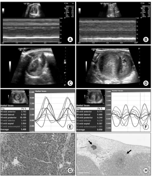
(5) Hyun Suk Yang, et al: Strain Imaging in Cardiac Xenotransplantation. Fig. 2. A donor heart (37.5 g) from a transgenic pig ( 1,3-galactosyltransferase gene-knockout, blood type A, 6.4 kg) expressing human complement regulatory protein CD46 was transplanted into a cynomolgus monkey (blood type A, 5.3 kg). Case number 4, pictured here, survived 38 days. An M-mode tracing echocardiogram of the mid-level of the left ventricular (LV) short axis view compared with (A) immediate postoperative and (B) just before expiry. Two-dimensional echocardiograms of the mid-level LV short axis view at the end-systolic phase, (C) different from at the immediate postoperative, (D) just before expiry note the markedly thickened LV walls with a very small LV cavity. (E) Radial strain of the corresponding short-axis view measures 5.408% at immediate postoperative (F) decreasing to 0.050% just before expiry. (G) Hematoxylin and eosin staining of the cardiac xenograft LV walls immediately postoperative from an open biopsy shows a relatively normal myocardium (×400). (H) At autopsy (postoperative day 38), multifocal bacterial colonies with microabscesses (arrows) confirmed the sepsis-involved myocardium (×100).. tional M-mode or 2D ejection fraction (P=0.600, P=0.340,. cases, the suppression of LV function was significant only. respectively) (Fig. 3). Compared with POD 0, POD X showed. in strain values (3/3), not in M-mode or with 2D ejection. eccentric dilatation in cases 1, 2, and 6; concentric thicken-. fractions (0/3).. ing in cases 3, 4, and 5. Among the eccentric dilatation cases, a significant LV suppression (>50% of percent change). DISCUSSION. was demonstrated in 2/3 (M-mode or 2D; radial strain or circumferential strain). Among the concentric thickening. This is the first heterotopic cardiac xenograft strain anal-. 29.
(6) J Korean Soc TransplantㆍMarch 2017ㆍVolume 31ㆍIssue 1. Fig. 3. In a total of six cases, changes of echocardiographic parameters of the left ventricular (LV) dimension or function between immediate postoperative (postoperative day [POD] 0) and just before expiry (POD X) are compared using a Wilcoxon signed-rank test. Radial and circumferential strain show significant changes (P=0.028). Blue lines with round points represent LV eccentric dilatation; red dots with triangular points represents LV concentric thickening. (A) LV end-diastolic cavity dimension (EDD), (B) LV end-systolic cavity dimension (ESD), (C) interventricular septum (IVS), (D) LV posterior wall dimension (PWD), (E) M-mode LV ejection fraction by Teichholz’s method (M-mode EF), (F) two-dimensional LV ejection fraction by Simpson’s method (2D-EF), (G) radial strain, and (H) circumferential strain.. ysis, to our knowledge, that has evaluated the xenograft. the surgical design or experimental animals. Heterotopic ab-. failure. It was feasible to use strain analysis in all consec-. dominal cardiac xenograft survival can be monitored by the. utive six cases. Progressive decrease of cardiac xenograft. abdominal palpation score(16), which represents the super-. function was significant in radial or circumferential strain. ficial ventricle, mostly on the right ventricular free walls.. (P=0.028). Especially in cases of concentric remodeling of. With the advent of the internet, video surveillance of recipi-. the LV (cases 3, 4, and 5), a small slit-like LV cavity gave. ent activities or graft telemetric signals also has been avail-. a higher M-mode or 2D ejection fraction; however, the con-. able(17,18). Immunologic monitoring or biomarkers such as. centric thickened walls revealed significantly declined radial. circulating organ-specific microRNAs have been implica-. or circumferential strain (percent changes >50%), which. ted(19). Compared with indirect measurement of xenograft. emphasized the incremental diagnostic role of myocardial. rejection, direct non-invasive visualization of graft con-. strains with an advantage of evaluating myocardial mechanics.. traction and understanding of myocardial mechanics seems. For the detection of cardiac xenograft rejection, several. promising for detecting early xenograft dysfunction or re-. diagnostic tools have been applied in xenotransplantation,. jection, and this study demonstrated the feasibility of my-. from manual palpations, blood sampling, and echocardiog-. ocardial strain monitoring in pig to monkey heterotopic car-. raphy, to the standard endomyocardial biopsy depending on. diac xenograft.. 30.
(7) Hyun Suk Yang, et al: Strain Imaging in Cardiac Xenotransplantation. Clinically, in patients with orthotopic heart allotrans-. multifocal microabscess related with sepsis involved myocar-. plantation, 2D strain parameters have a demonstrated diag-. dium in case 4 (Fig. 2). Abicht et al.(21) suggested that the. nostic role, and the European Association of Cardiovascular. LV wall hypertrophy was preclinical suspicion of humoral. Imaging mandates measurement after heart transplanta-. rejection or signs of thrombotic microangiopathy. In this. tion(20). Mingo-Santos et al.(7) suggested that LV longi-. small-number animal study with only one case of obvious. tudinal strain measurement to exclude acute cellular re-. AMR, it was difficult to confirm strain as early diagnosing. jection can reduce repeated endomyocardial biopsy. Indeed,. xenograft rejection. But we could speculate that radial or. real-world retrospective analysis used negative results to dif-. circumferential myocardial strain suggested a diagnostic role. ferentiate biopsy-proven cellular rejection during the first. in early xenograft dysfunction regardless of specific causes. year after orthotopic heart transplantation(8). In an ex-. such as sepsis, ischemia or rejection. At the time of sacrifice,. perimental model, radial strain seemed useful in early. a failing LV with a low contractile or perfusion status might. non-invasive detection of transplant rejection in a hetero-. be depicted in lower strain indexes. As this study demon-. topic rat cardiac transplantation model(5,6). Therefore, in. strated, strain imaging better diagnosed LV contractile dys-. this experimental animal study, we adopted the radial and. function than conventional M-mode or 2D ejection fractions. circumferential strain from short-axis view of LV. Differing. in cases with progressive concentric remodeling. In concen-. from the previous reports regarding the role of strain for. tric LV wall thickening, tissue Doppler derived indexes such. allograft rejection(5-8) which were focused on the de-. as strain were able to depict subtle changes of LV wall me-. tection of “rejection” rather than “graft dysfunction,” this. chanics or injury, at a time when conventional indices of. paper concentrated on xenograft dysfunction.. LV ejection fractions remained normal or super normal due. In this heterotopic xenograft study, the LV remodeling. to small, slit-like LV cavity dimensions.. progressed into two distinct patterns: eccentric dilatation. There are several limitations. First, the small number of. (n=3) versus concentric thickening (n=3). The eccentric di-. cardiac xenografts for the 2 years of the study period led. latation cases tended to end with poor survival (less than. to it containing only one antibody-mediated rejection.. 10 days). Among them, the cases with severe LV dysfunc-. Therefore more cases are needed to determine the diagnostic. tion (ejection fraction 10%, cases 2 and 6) had morphology. role of strain for xenograft rejection. Second, the funda-. similar to dilated cardiomyopathy; both M-mode and 2D. mental limitation of myocardial strain analysis in cardiac. ejection fractions well-appreciated the LV failure. In case. xenografts is lack of a normal reference value of strain in. 1, however, the conventional ejection fractions were within. miniature pigs. Therefore the authors demonstrate the per-. the normal range even at the time of sacrifice, but strain. cent changes instead comparisons with a normal value.. values were significantly decreased with the early detection. Third, the higher heart rate, relative to humans, makes it. of myocardial dysfunction. The percent change of strain in. more difficult to capture the proper peak systolic strain val-. case 2 was less than 50% (−24% or −19%, respectively),. ues, which difficulty could be overcome with better hard-. because the immediate postoperative radial or circum-. ware or software with higher temporal resolution. Finally,. ferential strain was quite low with a suspicion of myocardial. along with the concentric thickening of LV walls, the. stunning, and before expiry was also low with severe LV. end-systolic LV cavity size becomes closer to zero, limiting. dysfunction—the low absolute values of strain suggest my-. the application of Teichholz’s or Simpson’s formula, sug-. ocardial contractile dysfunction. The concentric thickening. gesting that another method is needed to evaluate LV func-. cases tended to show better survival (cases 3 or 4, 43 or. tion with thickened wall mechanics, such as measuring my-. 38 days, respectively) with remarkable progressive concen-. ocardial strain.. tric LV thickening. Histopathology of the thickened walls was noticeable with a substantial interstitial edema, hemor-. CONCLUSION. rhages and myocyte necrosis related with a C4d stain positive AMR in case 3 (Fig. 1), or mild interstitial edema, and. Non-invasive myocardial strain analysis in experimental. 31.
(8) J Korean Soc TransplantㆍMarch 2017ㆍVolume 31ㆍIssue 1. cardiac xenografts was feasible. Cardiac xenograft failure. al. Outcomes of alpha 1,3-GT-knockout porcine heart. appeared as two types: a dilated pattern with decreased ejec-. transplants into a preclinical nonhuman primate model.. tion fraction or a myocardial-thickening pattern with preserved ejection fraction. Radial and circumferential strains were significantly decreased in both types of xenograft failure irrespective of LV ejection fraction.. Transplant Proc 2013;45:3085-91. 10) Mohiuddin MM, Reichart B, Byrne GW, McGregor CG. Current status of pig heart xenotransplantation. Int J Surg 2015;23(Pt B):234-9. 11) Stewart S, Winters GL, Fishbein MC, Tazelaar HD, Kobashigawa J, Abrams J, et al. Revision of the 1990 working for-. REFERENCES. mulation for the standardization of nomenclature in the diagnosis of heart rejection. J Heart Lung Transplant. 1) Kuwaki K, Tseng YL, Dor FJ, Shimizu A, Houser SL,. 2005;24:1710-20.. Sanderson TM, et al. Heart transplantation in baboons using. 12) Behr TM, Feucht HE, Richter K, Reiter C, Spes CH, Pongratz. alpha1,3-galactosyltransferase gene-knockout pigs as do-. D, et al. Detection of humoral rejection in human cardiac. nors: initial experience. Nat Med 2005;11:29-31.. allografts by assessing the capillary deposition of comple-. 2) Mohiuddin MM, Corcoran PC, Singh AK, Azimzadeh A, Hoyt RF Jr, Thomas ML, et al. B-cell depletion extends the survival of GTKO.hCD46Tg pig heart xenografts in baboons for up to 8 months. Am J Transplant 2012;12:763-71.. ment fragment C4d in endomyocardial biopsies. J Heart Lung Transplant 1999;18:904-12. 13) Lang RM, Badano LP, Mor-Avi V, Afilalo J, Armstrong A, Ernande L, et al. Recommendations for cardiac chamber. 3) Mohiuddin MM, Singh AK, Corcoran PC, Hoyt RF, Thomas. quantification by echocardiography in adults: an update. ML 3rd, Ayares D, et al. Genetically engineered pigs and. from the American Society of Echocardiography and the. target-specific immunomodulation provide significant graft. European Association of Cardiovascular Imaging. J Am Soc. survival and hope for clinical cardiac xenotransplantation. J Thorac Cardiovasc Surg 2014;148:1106-13.. Echocardiogr 2015;28:1-39.e14. 14) Devereux RB, Alonso DR, Lutas EM, Gottlieb GJ, Campo. 4) Ferferieva V, Van den Bergh A, Claus P, Jasaityte R, La. E, Sachs I, et al. Echocardiographic assessment of left ven-. Gerche A, Rademakers F, et al. Assessment of strain and. tricular hypertrophy: comparison to necropsy findings. Am. strain rate by two-dimensional speckle tracking in mice:. J Cardiol 1986;57:450-8.. comparison with tissue Doppler echocardiography and. 15) Cameli M, Mondillo S, Solari M, Righini FM, Andrei V,. conductance catheter measurements. Eur Heart J Cardi-. Contaldi C, et al. Echocardiographic assessment of left ven-. ovasc Imaging 2013;14:765-73.. tricular systolic function: from ejection fraction to torsion.. 5) Shi J, Pan C, Shu X, Sun M, Yang Z, Zhu S, et al. The. Heart Fail Rev 2016;21:77-94.. role of speckle tracking imaging in the noninvasive de-. 16) Abbott CP, Dewitt CW, Creech O Jr. The transplanted rat. tection of acute rejection after heterotopic cardiac trans-. heart: histologic and electrocardiographic changes. Trans-. plantation in rats. Acta Cardiol 2011;66:779-85.. plantation 1965;3:432-45.. 6) Pieper GM, Shah A, Harmann L, Cooley BC, Ionova IA,. 17) Chen RH, Kadner A, Adams DH. Monitoring pig-to-primate. Migrino RQ. Speckle-tracking 2-dimensional strain echo-. cardiac xenografts with live Internet images of recipients. cardiography: a new noninvasive imaging tool to evaluate. and xenograft telemetric signals: histologic and immuno-. acute rejection in cardiac transplantation. J Heart Lung. histochemical correlations. J Heart Lung Transplant. Transplant 2010;29:1039-46.. 2000;19:591-7.. 7) Mingo-Santos S, Monivas-Palomero V, Garcia-Lunar I,. 18) Horvath KA, Corcoran PC, Singh AK, Hoyt RF, Carrier C,. Mitroi CD, Goirigolzarri-Artaza J, Rivero B, et al. Usefulness. Thomas ML 3rd, et al. Left ventricular pressure measure-. of two-dimensional strain parameters to diagnose acute. ment by telemetry is an effective means to evaluate trans-. rejection after heart transplantation. J Am Soc Echocardiogr. planted heart function in experimental heterotopic cardiac. 2015;28:1149-56.. xenotransplantation. Transplant Proc 2010;42:2152-5.. 8) Ambardekar AV, Alluri N, Patel AC, Lindenfeld J, Dorosz. 19) Zhou M, Hara H, Dai Y, Mou L, Cooper DK, Wu C, et. JL. Myocardial strain and strain rate from speckle-tracking. al. Circulating organ-specific microRNAs serve as bio-. echocardiography are unable to differentiate asymptomatic. markers in organ-specific diseases: implications for organ. biopsy-proven cellular rejection in the first year after car-. allo- and xeno-transplantation. Int J Mol Sci 2016;17:E1232.. diac transplantation. J Am Soc Echocardiogr 2015;28:478-85.. 20) Badano LP, Miglioranza MH, Edvardsen T, Colafranceschi. 9) Kim H, Chee HK, Yang J, Hwang S, Han KH, Kang J, et. AS, Muraru D, Bacal F, et al. European Association of. 32.
(9) Hyun Suk Yang, et al: Strain Imaging in Cardiac Xenotransplantation. Cardiovascular Imaging/Cardiovascular Imaging Department. 21) Abicht JM, Mayr T, Reichart B, Buchholz S, Werner F,. of the Brazilian Society of Cardiology recommendations. Lutzmann I, et al. Pre-clinical heterotopic intrathoracic. for the use of cardiac imaging to assess and follow patients. heart xenotransplantation: a possibly useful clinical tech-. after heart transplantation. Eur Heart J Cardiovasc Imaging. nique. Xenotransplantation 2015;22:427-42.. 2015;16:919-48.. 33.
(10)
수치

![Fig. 3. In a total of six cases, changes of echocardiographic parameters of the left ventricular (LV) dimension or function between immediate postoperative (postoperative day [POD] 0) and just before expiry (POD X) are compared using a Wilcoxon signed-ran](https://thumb-ap.123doks.com/thumbv2/123dokinfo/5443496.651225/6.918.84.810.156.635/echocardiographic-parameters-ventricular-dimension-immediate-postoperative-postoperative-wilcoxon.webp)
관련 문서
