519
ORIGINAL ARTICLE DOI 10.4070 / kcj.2009.39.12.519
Print ISSN 1738-5520 / On-line ISSN 1738-5555 Copyright ⓒ 2009 The Korean Society of Cardiology
Open Access
Relationship Between Serum Biochemical Markers of Myocardial
Fibrosis and Diastolic Function at Rest and With Exercise
in Hypertrophic Cardiomyopathy
Chi Young Shim, MD1, Jong-Won Ha, MD1, Eui-Young Choi, MD1, Hyun-Jin Lee, RN1,
Sun-Ha Moon, RN1, Jin-Mi Kim, RN1, Se-Joong Rim, MD1 and Namsik Chung, MD1,2
1Division of Cardiology, Cardiovascular Center and 2Research Institute, Yonsei University College of Medicine, Seoul, Korea
ABSTRACT
Background and Objectives:Recent studies have demonstrated the usefulness of biochemical markers of collagen turnover as markers of myocardial fibrosis in various diseases. In this study, we hypothesized that increased col-lagen markers in patients with hypertrophic cardiomyopathy (HCM) were correlated with diastolic function at rest and diastolic functional reserve during exercise. Subjects and Methods: Thirty-six patients with HCM and 21 controls with normal left ventricular thickness were studied. Mitral septal annular velocities and mitral in-flow velocities were measured at rest and during graded supine bicycle exercise (25 W, 3-minute increments) for the assessment of diastolic function at rest and during exercise. By radioimmunoassay, a byproduct of collagen III synthesis (PIIINP) and peptides resulting from collagen I synthesis (PINP) and degradation (ICTP) were measured. The patients with HCM were divided into two groups according to the median value of the PINP/ICTP ratio in the group. Results: At rest, the mitral annular early diastolic velocity (E’) was lower and the E/E’ was higher in the patients with HCM with high a PINP/ICTP ratio compared with patients with HCM with a low PINP/ICTP ra-tio and controls (p<0.001, p=0.012). With exercise, the Doppler parameters were increased in all groups, but there was no significant difference in the change in E’ and E/E’ during exercise according to collagen turnover markers. Conclusion: A higher PINP/ICTP ratio was associated with resting diastolic dysfunction in patients with HCM; however, there was no relationship with augmented diastolic dysfunction during exercise. We suggest that the type I collagen synthesis-to-degradation ratio is a useful marker of resting diastolic function in patients with HCM. (Korean Circ J 2009;39:519-524)
KEY WORDS:Cardiomyopathy hypertrophic; Collagen; Myocardial contraction, diastole; Exercise.
Introduction
The pathophysiology of hypertrophic cardiomyopathy (HCM) is complex, with structural, hemodynamic, and arrhythmic processes contributing to the symptoms and natural history in this disorder. However, the most
im-portant pathophysiologic features of HCM is diastolic dysfunction and a decrease in left ventricular compliance. Thus, HCM is regarded as the most representative dis-ease of left ventricular diastolic dysfunction.1)2) The
his-topathologic findings of HCM are myocardial hypertro-phy, and interstitial fibrosis and disarray. Previous studies based on autopsies have shown that the degree of myo-cardial fibrosis is severe and varies as compared with my-ocardium obtained from normotensive or hypertensive patients.3) In recent years, several studies have been
con-ducted to quantify the myocardial fibrosis and have re-ported that serum biochemical markers, which are syn-thesized by the production and degradation of matrix collagen, are elevated in subjects with hypertensive myo-cardial hypertrophy and HCM relating to the myomyo-cardial fibrosis.4-6) It has also been suggested that serum
bioche-mical markers are associated with diastolic dysfunction.7-10)
The most common symptom of HCM is shortness of
Received: January 9, 2009 Revision Received: May 6, 2009 Accepted: May 19, 2009
Correspondence: Jong-Won Ha, MD,Division of Cardiology, Cardiovascular Center, Yonsei University College of Medicine, 250 Seongsan-ro, Seodae-mun-gu, Seoul 120-752, Korea
Tel: 82-2-2228-8448, Fax: 82-2-2227-7943 E-mail: [email protected]
○cc This is an Open Access article distributed under the terms of the Creative Commons Attribution Non-Commercial License (http://creativecommons. org/licenses/by-nc/3.0) which permits unrestricted non-commercial use, distribution, and reproduction in any medium, provided the original work is properly cited.
520·Collagen Markers and Hypertrophic Cardiomyopathy
breath and it is usually aggravated by exercise.1) Diastolic
dysfunction seems to be the most important determinant of exercise capacity in patients with HCM. In HCM, the limited capability of increasing left ventricular end-di-astolic volume, especially during exercise at high heart rates, implies an inadequate increase in stroke volume.11)
Such a lack of increase in stroke volume is accompanied by a leftward shift of the left ventricular diastolic pres-sure-volume relation during exercise.11)12) Specifically,
bas-ed on a few studies which were conductbas-ed using exercise Doppler echocardiography, it has been demonstrated that left ventricular diastolic reserve, systolic reserve, and ex-ercise tolerance are decreased in patients with HCM.13)14)
However, there are many differences in exercise capacity and symptoms of heart failure, although there is a simi-lar extent of myocardial hypertrophy or the left ventric-ular dimension in patients with HCM. Thus, we can hy-pothesize that the degree of myocardial fibrosis might be associated with the left ventricular reserve and exercise tolerance. To prove our hypothesis, we sought to assess diastolic function not only at rest, but also with exercise by using exercise Doppler echocardiography in patients with HCM. To quantify the amount of myocardial fibro-sis, serum biochemical markers which are involved in collagen metabolism were analyzed. We then examined the relationship between serum biochemical markers of myocardial fibrosis and diastolic function at rest and with exercise in patients with HCM.
Subjects and Methods
SubjectsThe study population was comprised of 36 patients with HCM (20 males; mean age, 57±10 years; 31 pa-tients with non-obstructive HCM) and 21 age- and gen-der-matched control subjects with normal left ventric-ular thickness. The subjects with left ventricventric-ular systolic dysfunction (left ventricular ejection fraction <55%), significant valvular disease, arrhythmia, coronary artery disease, or renal insufficiency (serum creatinine >1.4 mg/mL) were excluded. Each subject provided informed, written consent to the protocol that had been approved by our Institutional Review Board.
The ratio between procollagen type I N-terminal pep-tide (PINP) and collagen type I pyridinoline cross-linked C-terminal telopeptide (ICTP), PINP/ICTP, which is known as the degree of synthesis and degradation of type I collagen, was 9.1±7.8 in the control group and 10.9± 6.5 in the HCM group. Patients with HCM (n=36) were divided into the following two groups based on the me-dian value of the PINP/ICTP: the lower group (PINP/ ICTP < median value; n=18; 10 males) and the higher group (PINP/ICTP≥median value; n=18; 10 males) for analysis. A comparison was made between the 3 groups (the control group, the HCM group with a lower PINP/
ICTP, and the HCM group with a higher PINP/ICTP). Two-dimensional and exercise doppler echocardi-ography (diastolic stress echocardiechocardi-ography)
Standard 2-dimensional measurements (left ventric-ular diastolic and systolic dimensions, ventricventric-ular sep-tum and posterior wall thickness, left atrial volume, and left ventricular outflow tract) were obtained with the sub-jects in the left decubitus position. The left ventricular ejection fraction was calculated by the modified method of Quinones et al.15) After obtaining the rest images from
the standard parasternal and apical views, a multistage supine bicycle exercise testing was performed with a var-iable load bicycle ergometer (Medical Positioning Inc., Kansas City, MO, USA). Subjects pedaled at constant speed beginning at a workload of 25 W with an incre-ment of 25 W every 3 minutes. Echocardiography was performed using an ultrasound system (System 7; GE Vingmed, Horten, Norway) with a 2.5-MHz transducer during rest, each stage of exercise, and recovery in the se-quence described as follows. From the apical window, a 1-2-mm pulsed Doppler sample volume was placed at the mitral valve tip, and mitral flow velocities from 5-10 cardiac cycles were recorded. The mitral inflow velocities were traced and the following variables were obtained: peak velocity of early (E) and late (A) filling, and decel-eration time of the E wave velocity. Tricuspid regurgitant jet velocity was also obtained to estimate pulmonary ar-tery systolic pressure using continuous wave Doppler, if measurable. Mitral annular velocity was measured by Doppler tissue imaging using the pulsed wave Doppler mode. The filter was set to exclude high-frequency sig-nals, and the Nyquist limit was adjusted to a range of 15-20 cm/s. Gain and sample volume were minimized to allow for a clear tissue signal with minimal background noise. E’ and systolic (S’) velocities of the mitral annulus were measured from the apical 4-chamber view with a 2-5-mm sample volume placed at the septal corner of the mitral annulus. Stroke volume (SV) was measured from the left ventricular outflow tract diameter and the pulse wave Doppler signal, as previously described.16) To
pro-vide a continuous variable that might estimate Ed, the E/E’ ratio was used as an estimation of mean left atrial pressure. Operant Ed was estimated as E/E’ divided by the volume SV of filling during diastole, based on a pre-vious study.17) These measurements were performed at
baseline, at each stage of exercise, and recovery in the same sequence. All data were stored digitally and meas-urements were made at the completion of each study. Serologic markers of collagen metabolism
To assess collagen markers, we measured serum levels of peptides released during collagen synthesis and degra-dation. Peripheral venous blood samples were centrifug-ed at 4℃. Aliquots of serum were immcentrifug-ediately storcentrifug-ed at
Chi Young Shim, et al.·521
-70℃ until the assay.
The measurement of serum PINP, procollagen type III amino terminal peptide (PIIIINP), and ICTP was per-formed using a commercially available radioimmunoas-say (Farmos Diagnostica, Oulu, Finland), all of which were indicators reflecting collagen synthesis and degradation. The accuracy of this method has been reported to have an inter- and intra-assay variation <10%.
Statistical analysis
All the results were expressed as the mean±standard deviation. To compare the categorical variables, a chi-square test was used. For comparison of the continuous variables, analysis of variance (ANOVA) was performed. Serum biochemical markers indicating myocardial fibro-sis did not follow a normal distribution, which were con-verted to logarithms for statistical analysis. Statistical sig-nificance was defined as <.05. Statistical analysis was per-formed using Statistical Package for Social Science (SPSS) 13.0 for Windows (SPSS, Inc., Chicago, IL, USA).
Results
Clinical characteristics of the study subjects The baseline characteristics of clinical variables and
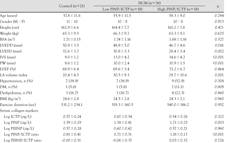
serum collagen markers are shown in Table 1. The age and gender distribution was not different between the three groups. The left ventricular diastolic and end-systolic dimensions were significantly lower in the HCM group, especially in the HCM group with a higher PINP/ ICTP ratio. The myocardial thickness of the interventri-cular septum and posterior wall were increased in the HCM group. The HCM group had a significantly larger left ventricular volume index, but there were no differ-ences in the left ventricular systolic function. There were no significant differences in the duration of exercise be-tween the three groups. Of the serum biochemical mark-ers indicating myocardial fibrosis, the log PINP and log PINP/ICTP were significantly higher in the HCM group. Furthermore, the patients with HCM with a higher NP/ICTP ratio had significantly higher levels of log PI-NP and log PIPI-NP/ICTP due to the study design. Hemodynamic variables at rest and with exercise
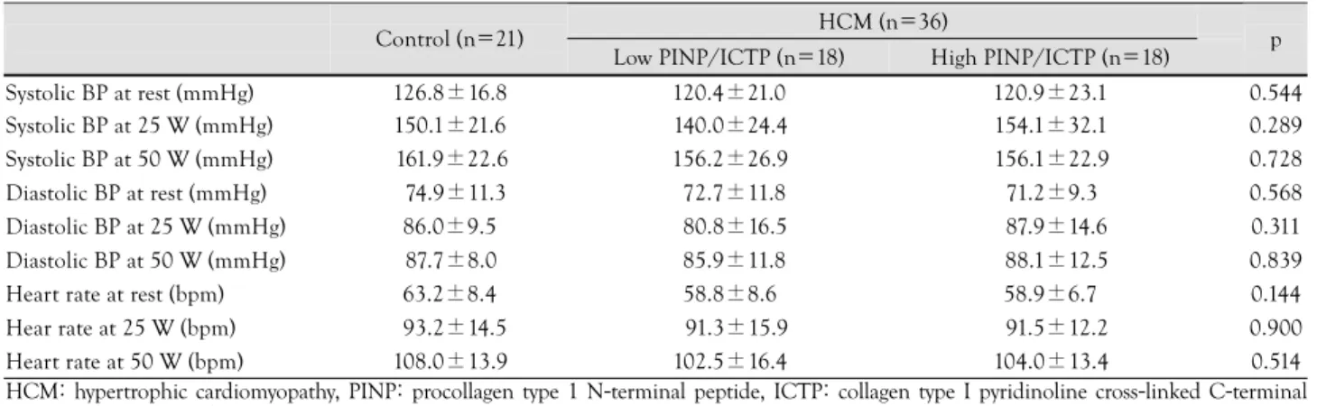
The changes in systolic blood pressure, diastolic blood pressure, and heart rate during exercise are shown in Ta-ble 2. In the three groups, the blood pressure and heart rate were increased gradually during exercise; however, there were no significant differences between the three groups in hemodynamic variables at rest and with exercise.
Table 1. Baseline characteristics of control and HCM patients
HCM (n=36) Control (n=21)
Low PINP/ICTP (n=18) High PINP/ICTP (n=18) p Age (years) 57.8±11.6 55.9±11.5 58.3±9.0 <0.784 Gender (M : F) 11 : 10 10 : 8 10 : 8 <0.973 Height (cm) 162.9±6.6 164.4±7.7 161.2±7.8 <0.471 Weight (kg) 65.3±9.5 66.3±9.7 63.3±9.1 <0.625 BSA (m2) 1.71±0.15 1.74±1.16 1.68±1.16 <0.527 LVEDD (mm) 50.9±3.5 48.8±5.0 46.7±4.6 <0.016 LVESD (mm) 32.6±3.7 30.8±3.3 28.4±3.4 <0.002 IVS (mm) 9.0±1.2 13.0±4.2 14.6±4.2 <0.001 PW (mm) 8.6±1.2 10.0±2.4 10.9±1.5 <0.001 LVEF (%) 68.9±6.4 69.6±3.4 71.2±6.7 <0.464 LA volume index 20.4±4.5 30.5±9.3 29.7±10.6 <0.001 Hypertension, n (%) 7 (38.9) 7 (38.9) 9 (52.9) <0.308 DM, n (%) 1 (5.6)0 1 (5.6)0 2 (11.1). <0.808 Dyslipidemia, n (%) 3 (16.7) 3 (16.7) 4 (22.3) <0.860 BMI (kg/m2) 24.6±2.8 24.5±2.6 24.3±2.1 <0.960
Exercise duration (sec) 531.2±234.1 519.3±160.5 540.0±186.2 <0.952 Serum collagen markers
Log ICTP (μg/L) 0.57±0.24 0.67±0.34 0.54±0.26 <0.322 Log PINP (μg/L) 1.39±0.29 1.38±0.41 1.71±0.25 <0.003 Log PIIINP (μg/L) 0.57±0.28 0.60±0.42 0.57±0.21 <0.960 Log PINP/ICTP ratio 0.81±0.41 0.71±0.31 1.18±0.13 <0.001 Log PIIINP/ICTP ratio -0.00±0.31 -0.06±0.37 0.03±0.32 <0.726 BSA: body surface area, LVEDD: left ventricular end-diastolic dimension, LVESD: left ventricular end-systolic dimension, IVS: interventricular septum, PW: posterior wall, LVEF: left ventricular ejection fraction, LV: left ventricular, LA: left atrium, DM: diabetes mellitus, BMI: body mass index, ICTP: collagen type I pyridinoline cross-linked C-terminal telopeptide, PINP: procollagen type 1 N-terminal peptide, PIIINP: procollagen type III amino terminal peptide, HCM: hypertrophic cardiomyopathy
522·Collagen Markers and Hypertrophic Cardiomyopathy
Left ventricular diastolic function at rest
As shown in Table 3, Doppler echocardiographic pa-rameters were compared at rest and each stage of exercise between the three groups. At rest, the E’ velocity was
sig-nificantly lower in the patients with HCM as compared with the control subjects. Interestingly, the HCM group with a higher PINP/ICTP revealed lower E’ velocities, a higher E/E’ ratio, and lower S’ velocities than the HCM
Table 3. Comparison of Doppler echo variables at rest and during exercise in the study groups
HCM (n=36) Control (n=21)
Low PINP/ICTP (n=18) High PINP/ICTP (n=18) p E (cm/sec) Rest 62.9±13.1 57.9±13.1 67.4±24.0 <0.268 25 W 95.5±19.6 83.9±17.1 92.3±24.7 <0.232 50 W 110.8±19.2 95.8±21.5 101.8±24.3 <0.163 A (cm/sec) Rest 68.2±17.1 54.4±12.5 62.8±15.0 <0.022 25 W 90.1±17.1 75.1±18.0 83.1±19.7 <0.054 50 W 102.3±18.8 87.6±14.0 92.9±22.8 <0.102
E’ (cm/sec) Rest 6.0±1.5 4.8±1.2 4.3±1.1 <0.001
25 W 7.8±1.8 6.1±1.7 5.7±1.3 <0.001 50 W 8.7±2.5 6.3±1.4 6.4±1.6 <0.002 E/E’ Rest 10.9±2.5 12.8±4.5 16.3±8.2 <0.012 25 W 12.8±2.9 14.6±4.8 17.0±7.0 <0.058 50 W 13.5±4.1 15.3±3.1 17.0±7.0 <0.177 SV (mL) Rest 60.6±14.2 66.8±18.9 66.4±13.9 <0.497 25 W 66.7±18.4 70.7±18.4 68.3±18.0 <0.853 50 W 68.1±12.2 71.3±21.5 67.4±13.2 <0.840 Ed Rest 0.19±0.04 0.18±0.03 0.22±0.05 <0.112 25 W 0.20±0.08 0.18±0.07 0.24±0.05 <0.278 50 W 0.20±0.06 0.21±0.06 0.22±0.05 <0.774 S’ (cm/sec) Rest 6.8±0.9 6.1±1.2 5.8±0.8 <0.011 25 W 7.8±1.1 6.8±1.2 6.3±1.3 <0.003 50 W 8.7±1.6 7.5±1.5 7.1±1.3 <0.005 Peak 9.5±2.3 8.0±1.8 8.4±3.3 <0.179
Change in E’ Rest to 25 W 1.8±1.3 1.2±1.5 1.3±1.3 <0.461 Rest to 50 W 2.9±2.5 1.5±1.2 2.1±1.4 <0.141 Change in E/E’ Rest to 25 W 1.5±2.7 1.4±5.0 1.5±3.3 <0.990 Rest to 50 W 1.8±2.4 1.3±4.5 0.1±4.7 <0.488 Change in S’ Rest to 25 W 1.0±1.3 0.7±1.3 0.5±1.2 <0.515 Rest to 50 W 2.0±1.8 1.2±1.7 1.2±1.1 <0.300 Rest to Peak 2.8±2.6 1.8±1.8 2.5±3.0 <0.522 E: early diastolic mitral inflow velocity, A: late diastolic mitral inflow velocity, E’: early diastolic mitral annular velocity, SV: stroke volume, Ed: dias-tolic elastance, S’: sysdias-tolic mitral annular velocity, HCM: hypertrophic cardiomyopathy, PINP: procollagen type 1 N-terminal peptide, ICTP: colla-gen type I pyridinoline cross-linked C-terminal telopeptide
Table 2. Hemodynamic response to exercise
HCM (n=36) Control (n=21)
Low PINP/ICTP (n=18) High PINP/ICTP (n=18) p Systolic BP at rest (mmHg) 126.8±16.8 120.4±21.0 120.9±23.1 0.544 Systolic BP at 25 W (mmHg) 150.1±21.6 140.0±24.4 154.1±32.1 0.289 Systolic BP at 50 W (mmHg) 161.9±22.6 156.2±26.9 156.1±22.9 0.728 Diastolic BP at rest (mmHg) 74.9±11.3 72.7±11.8 71.2±9.3 0.568 Diastolic BP at 25 W (mmHg) 86.0±9.5 80.8±16.5 87.9±14.6 0.311 Diastolic BP at 50 W (mmHg) 87.7±8.0 85.9±11.8 88.1±12.5 0.839 Heart rate at rest (bpm) 63.2±8.4 58.8±8.6 58.9±6.7 0.144 Hear rate at 25 W (bpm) 93.2±14.5 91.3±15.9 91.5±12.2 0.900 Heart rate at 50 W (bpm) 108.0±13.9 102.5±16.4 104.0±13.4 0.514 HCM: hypertrophic cardiomyopathy, PINP: procollagen type 1 N-terminal peptide, ICTP: collagen type I pyridinoline cross-linked C-terminal telopeptide, BP: blood pressure, bpm: beats per minute
Chi Young Shim, et al.·523
group with lower PINP/ICTP. From these results, we know that the left ventricular longitudinal diastolic and systolic function at rest were significantly impaired in pa-tients with HCM who had a higher PINP/ICTP ratio. Left ventricular diastolic function with exercise
At each stage of exercise, the E’ velocity, E/E’ ratio, and S’ velocity were significantly lower in the HCM group than the control group, and the values were ele-vated to a similar extent during exercise. Thus, the mag-nitude of changes in E’ velocity, E/E’, and S’ velocity from rest to each stage of exercise was not different be-tween the three groups. Therefore, the presence of HCM or the higher PINT/ICTP ratio were not associated with a significant decrease in diastolic functional reserve dur-ing exercise, even though there was significant left ven-tricular diastolic dysfunction at rest.
Discussion
The principle findings of this study are as follows: 1) in patients with HCM, the concentration of serum bio-chemical markers indicating myocardial fibrosis was ele-vated, and the left ventricular diastolic function at rest was significantly impaired; and 2) although the serum biochemical markers indicating myocardial fibrosis was elevated in patients with HCM, it remained unclear whe-ther the left ventricular diastolic functional reserve was more impaired during exercise.
In patients with HCM, the left ventricular diastolic dysfunction is the most important feature and it has been known as a major determinant of a patient’s symptoms.18)
The left ventricular diastolic function is determined by the degree of myocardial hypertrophy and interstitial fibrosis. In the normal heart, the intersitial tissue is com-posed of types I and III collagen. Type I collagen was abundantly present at a ratio of 70 : 30 and it is charac-terized by a higher degree of rigidity.19) In patients with
HCM, due to the presence of myocardial fibrosis, col-lagen is increased by approximately 20% of the total volume of the myocardium.20) According to this study,
type 3 collagen did not show a significant difference in the HCM groups as compared with the control group. Type I collagen and the indicator for the turnover of type I collagen, PINP/CITP in the HCM groups, was high-er than the control group. Especially, the patients with HCM with a higher PINP/CITP ratio had a larger sep-tal thickness, decreased diastolic and systolic longitudi-nal parameters, and increased E over E’, indicating a higher left ventricular filling pressure in this group. The differences in these parameters were consistent at rest and at each stage of exercise.
A microscopic examination based on the endomyo-cardial biopsy has been considered as the most accurate, standard test for the diagnosis of myocardial fibrosis. But,
this diagnostic method requires an invasive procedure and there are also limitations that a partial tissue sample of endomyocardium which is obtained from the right ventricle cannot accurately reflect the heterogeneity of myocardial fibrosis in the left ventricle. The type I colla-gen synthesis-to-degradation ratio, a turnover marker of type I collagen, could estimate the degree of myocardial fibrosis simply with non-invasive serologic tests. Lom-bardi et al.6) demonstrated a higher collagen turnover in
36 patients with HCM compared with 14 age- and gen-der-matched controls. In the study, the left ventricular diastolic dysfunction was assessed by the pulse-wave Dop-pler parameters of mitral inflow and pulmonic vein flow. In the present study, we also assessed the left ventricular longitudinal diastolic function by tissue Doppler imag-ing. From the tissue velocities on the mitral septal annu-lus, the E’ and S’ velocities could be obtained, then the E over E’ ratio calculated, indicating the left ventricular filling pressure.
The aim of this study was to prove the hypothesis that a higher level of serologic markers indicating collagen turnover would show a reduced diastolic functional re-serve during exercise within the HCM group. However, although the patients with HCM with a higher collagen turnover had significant diastolic dysfunction at rest, there were no significant differences in the degree of changes in diastolic parameters during exercise as compared with the patients with HCM with a lower collagen turnover. Therefore, we conclude that a higher collagen turnover ratio in patients with HCM is not associated with left ventricular diastolic functional reserve in this study pop-ulation.
The limitations of the current study were as follows: 1) In patients with HCM with a higher collagen turnover marker, there were significantly lower E’ and S’ velo-cities from the resting status. Because of these resting dif-ferences in the diastolic and systolic tissue parameters, it might be inappropriate to confirm the difference in dia-stolic function reserve during exercise. To prove the dif-ferent changes in diastolic function according to exercise, it is preferable that the subjects have similar profiles of E’ velocity, S’ velocity, and E over E’ at rest. 2) This study was limited by the small number of enrolled subjects. 3) This study did not consider the effects of cardiovascular medications which could affect the degree of myocardial fibrosis.
In conclusion, the non-invasive serologic test of type I collagen turnover (the PINP/ICTP ratio) was associ-ated with resting diastolic dysfunction in patients with HCM. However, there was no relationship with augment-ed diastolic dysfunction during exercise. Therefore, we suggest that the type I collagen synthesis-to-degradation ratio will be a useful marker of resting diastolic function in patients with HCM. Further studies are warranted to examine the diastolic functional reserve with exercise in
524·Collagen Markers and Hypertrophic Cardiomyopathy
patients with HCM with relatively well-preserved dia-stolic function at rest.
Acknowledgments
This work was financially supported by the grant of The Korean Society of Circulation in 2003.
REFERENCES
1) Maron BJ. Hypertrophic cardiomyopathy. Lancet
1997;350;127-33.
2) Jeong JW. Hypertrophic cardiomyopathy. Korean Circ J 2002;
32:7-14.
3) Tanaka M, Fujiwara H, Onodera T, Wu DJ, Hamashima Y, Kawai C. Quantitative analysis of myocardial fibrosis in normals,
hy-pertensive hearts, and hypertrophic cardiomyopathy. Br Heart J 1986;55:575-81.
4) Lopez B, Gonzalez A, Varo N, Laviades C, Querejeta R, Diez J.
Biochemical assessment of myocardial fibrosis in hypertensive heart disease. Hypertension 2001;38:1222-6.
5) Diez J, Laviades C, Mayor G, Gil MJ, Monreal I. Increased serum
concentrations of procollagen peptides in essential hypertension: relation to cardiac alterations. Circulation 1995;91:1450-6.
6) Lombardi R, Betocchi S, Losi MA, et al. Myocardial collagen
turnover in hypertrophic cardiomyopathy. Circulation 2003;108: 1455-60.
7) Fassbach M, Schwartzkopff B. Elevated serum markers for
col-lagen synthesis in patients with hypertrophic cardiomyopathy and diastolic dysfunction. Z Kardiol 2005;94:328-35.
8) Martos R, Baugh J, Ledwidge M, et al. Diastolic heart failure:
evidence of increased myocardial collagen turnover linked to di-astolic dysfunction. Circulation 2007;115:888-95.
9) Diez J, Laviades C, Monreal I, Gil MJ, Panizo A, Pardo J. Toward
the biochemical assessment of myocardial fibrosis in hypertensive patients. Am J Cardiol 1995;76:14D-7D.
10) Ihm SH, Youn HJ, Kim SR, et al. Relation between serum
car-boxy-terminal propeptide of type 1 procollagen (PIP), a marker
of myocardial fibrosis, and left ventricular diastolic function in patients with early type 2 diabetes mellitus. Korean Circ J 2005; 35:500-6.
11) Briguori C, Betocchi S, Romano M, et al. Exercise capacity in
hy-pertrophic cardiomyopathy depends on left ventricular diastolic function. Am J Cardiol 1999;84:309-15.
12) Lele SS, Thomson HL, Seo H, Belenkie I, McKenna WJ, Fren-neaux MP. Exercise capacity in hypertrophic cardiomyopathy:
role of stroke volume limitation, heart rate, and diastolic filling characteristics. Circulation 1995;92:2886-94.
13) Ha JW, Ahn JA, Kim JM, et al. Abnormal longitudinal
myocar-dial functional reserve assessed by exercise tissue Doppler echo-cardiography in patients with hypertrophic cardiomyopathy. J Am Soc Echocardiogr 2006;19:1314-9.
14) Choi EY, Ha JW, Rim SJ, et al. Incremental value of left
ven-tricular diastolic function reserve index for predicting exercise capacity in patients with hypertrophic cardiomyopathy. J Am Soc Echocardiogr 2008;21:487-92.
15) Quinones MA, Waggoner AD, Reduto LA, et al. A new, simplified
and accurate method for determining ejection fraction with two-dimentional echocardiography. Circulation 1981;64:744-53.
16) Oh JK, Seward JB, Tajik AJ. The Echo Manual. 2nd ed.
Philadel-phia: Lippincott Williams & Wilkins; 1999.
17) Ha JW, Lee CH, Park S, et al. Gender-related difference in left
ventricular diastolic elastance during exercise in patients with diabetes mellitus. Circ J 2008;72:1443-8.
18) Maron BJ, Bonow RO, Cannon RO 3rd, Leon MB, Epstein SE.
Hypertrophic cardiomyopathy: interrelations of clinical manifes-tations, pathophysiology, and therapy. N Engl J Med 1987;316: 780-9.
19) Burlew BS, Weber KT. Cardiac fibrosis as a cause of diastolic
dysfunction. Herz 2002;27:92-8.
20) Shirani J, Pick R, Roberts WC, Maron BJ. Morphology and
sig-nificance of the left ventricular collagen network in young pa-tients with hypertrophic cardiomyopathy and sudden cardiac death. J Am Coll Cardiol 2000;35:36-44.