CASE REPORT
초고령 환자에서 위의 위장관 기질 종양 수술 후 나타난 반복적인 문합부 재발
김혜진, 박재용, 김범진, 김재규, 김희성1, 박중민2
중앙대학교 의과대학 내과학교실, 병리학교실1, 외과학교실2
Gastric Gastrointestinal Stromal Tumor with Repeated Recurrence at the Anastomosis Site in a Very Elderly Patient
Hye-Jin Kim, Jae Yong Park, Beom Jin Kim, Jae Gyu Kim, Hee Sung Kim1 and Joong-Min Park2
Department of Internal Medicine, Pathology1 and Surgery2, Chung-Ang University College of Medicine, Seoul, Korea
Although high-risk gastrointestinal stromal tumors (GISTs) frequently recur, even after a complete resection and imatinib therapy, local recurrence at the suture line after complete resection is rare. The present case was an 88-year-old woman who was initially diagnosed with high-risk GIST without a distant metastasis. She underwent a complete surgical resection of the lesion and re- ceived adjuvant imatinib therapy for 18 months, which was discontinued due to severe drug-induced anemia. During the fol- low-up, an endoscopic examination performed 40 months after the initial surgery revealed local recurrence at the anastomosis site. Although a complete surgical resection was performed, repeated local recurrence was detected 18 months later, which pro- gressed rapidly to metastatic disease. This paper reports a case of a completely resected gastric GIST with repeated local re- currence, despite the complete surgical resections and adjuvant imatinib therapy. (Korean J Gastroenterol 2020;76:206-210) Key Words: Gastrointestinal stromal tumors; Surgery; Imatinib mesylate; Recurrence
Received May 25, 2020. Revised July 30, 2020. Accepted August 16, 2020.
CC This is an open access article distributed under the terms of the Creative Commons Attribution Non-Commercial License (http://creativecommons.org/licenses/
by-nc/4.0) which permits unrestricted non-commercial use, distribution, and reproduction in any medium, provided the original work is properly cited.
Copyright © 2020. Korean Society of Gastroenterology.
교신저자: 박재용, 서울시 동작구 흑석로 84, 중앙대학교 의과대학 내과학교실 소화기내과
Correspondence to: Jae Yong Park, Division of Gastroenterology, Department of Internal Medicine, Chung-Ang University College of Medicine, 84 Heukseok-ro, Dongjak-gu, Seoul 06974, Korea. Tel: +82-2-6299-3161, Fax: +82-2-6299-2064, E-mail: [email protected], ORCID: https://orcid.org/0000-0001-6114-8920 Financial support: None. Conflict of interest: None.
INTRODUCTION
A gastrointestinal stromal tumor (GIST) is the most common mesenchymal tumor of the gastrointestinal tract, accounting for 1-3% of all gastrointestinal (GI) malignancies.1 The present- ing symptoms of GIST include anemia, abdominal pain, and gastrointestinal bleeding. Most patients, however, are asympto- matic and are diagnosed incidentally during an endoscopic examination. Surgery for the complete removal of the tumor is the gold standard of therapy for a resectable GIST and the only chance of cure. On the other hand, recurrence after
surgery is common, particularly in high-risk GISTs. Approximately 40-50% of patients have disease recurrence in this subgroup.2 The two main prognostic factors are the mitotic activity and the tumor size. More than five mitoses per 50 high-power fields (HPFs) and a size greater than 10 cm are the factors associated with poor outcomes after surgery.2 Adjuvant im- atinib is the key treatment for postoperative high-risk patients with GISTs to decrease the incidence of relapse.3 Nevertheless, imatinib cannot completely prevent the recurrence of GIST in all cases, and side effects, such as hematologic abnormal- ities, can hinder the maintenance of imatinib therapy. This
A B
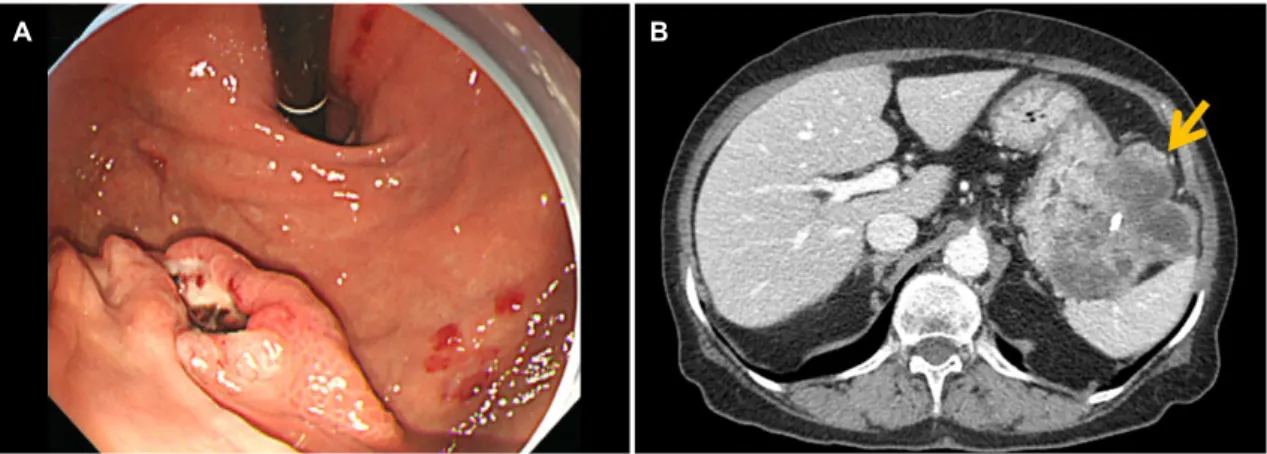
Fig. 1. Endoscopic images of the gastric lesion and abdominal computed tomography findings at the initial presentation. (A) A 2-cm sized, irregular shaped ulcerofungating mass on the greater curvature of the high body of the stomach. (B) An exophytic, heterogeneous enhancing mass with necrosis and central calcification, located adjacent to the gastric high body, closely abutting the splenic hilum (arrow).
AA BB CC
D
D EE
Fig. 2. Histologic features of the gastric gastrointestinal stromal tumor at the first surgery. (A) Histopathological evaluation reveals blandly spindle cells with faintly eosinophilic cytoplasm in a syncytial pattern. Elongated nuclei with inconspicuous nucleoli are noted (H&E, ×400).
(B) Diffuse positive cytoplasmic staining for CD117 (Immunohistochemical staining, ×20). (C) Diffuse positive staining for CD34 (Immunohistochemical staining, ×20). (D) Focal positive cytoplasmic staining for DOG-1 (Immunohistochemical staining, ×20). (E) Ki-67 proliferation index is less than 5% (Immunohistochemical staining, ×20).
paper reports a case of gastric GIST with repeated local recur- rences, even after a complete resection and adjuvant imatinib therapy, which eventually progressed to metastatic disease.
CASE REPORT
An 88-year-old woman was admitted to Chung-Ang University Hospital due to melena. She had a prior medical history of hypertension and chronic kidney disease. The pe-
ripheral blood cell counts showed anemia with a hemoglobin level of 3.8 g/dL. Esophagogastroduodenoscopy (EGD) was performed to determine the source of suspected upper GI bleeding. The endoscopic examination revealed a 2-cm sized, irregular-shaped ulcerofungating mass on the greater curva- ture, high body of the stomach (Fig. 1A). An endoscopic biopsy was performed to obtain a pathologic diagnosis of GIST.
Abdominal CT revealed an 8.9 cm sized exophytic growing, heterogenous enhancing mass adjacent to the gastric high
A B
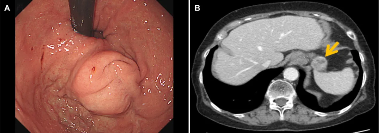
Fig. 3. Endoscopic images of the recurred gastric lesion and abdominal computed tomography findings at the first recurrence. (A) A 2-cm sized spherical shaped subepithelial mass lying on the anastomosis site. (B) An exophytic, heterogeneously enhancing mass at the anastomosis site of the stomach, measuring 2.6 cm, without signs of extragastric metastasis (arrow).
A B
Fig. 4. Histologic features of a gastric gastrointestinal stromal tumor at the second surgery. (A) Histopathological evaluation revealed spindle cell proliferation with frequent mitoses up to 159/50 high-power fields (H&E, ×40) (B) Ki-67 proliferation index is 30% (Immunohistochemical staining, ×20).
body (Fig. 1B). There was no evidence of a distant metastasis.
She had undergone a laparoscopic wedge resection of the stomach for the gastric GIST. A pathology evaluation con- firmed that the lesion was GIST with a high risk of progressive disease. The tumor size was 9.0×6.0×5.3 cm, and the mitotic index was a 16/50 HPF. The histologic type was the mixed spindle and epithelioid type (Fig. 2A). Immunohistochemistry confirmed GIST with diffuse positive staining for CD117, CD34, and DOG1, with a low Ki-67 proliferation index (Fig.
2B-E). There was no involvement of the tumor at the resection margin on the microscopic evaluation. After surgery, she re- ceived an adjuvant imatinib therapy at an initial dose of 400 mg/day to prevent a recurrence of the disease. On the other hand, recurrent anemia occurred during treatment, and the dose of imatinib was reduced. Despite the dose reduction,
however, the severe anemia persisted, and the hemoglobin level decreased to 4.6 g/dL. Imatinib therapy was dis- continued after 18 months of maintenance owing to severe drug intolerance. She had undergone EGD 1 year after the surgery and abdominal CT every 6 months, which showed no signs of tumor recurrence. She underwent a follow-up EGD 40 months after surgery, which revealed a 2-cm-sized round-shaped mass at the anastomosis site (Fig. 3A).
Subsequent abdominal CT was performed as local recurrence was suspected (Fig. 3B). A laparoscopic wedge resection of the gastric mass was performed because there was no sign of distant metastasis. The histology was a spindle cell type GIST with high risk, showing a tumor size of 2.6×2.3×2.0 cm (Fig. 4A). The mitotic index was more than 10/50 HPF, with a high Ki-67 proliferation index of 30% (Fig. 4B). The resection
A B
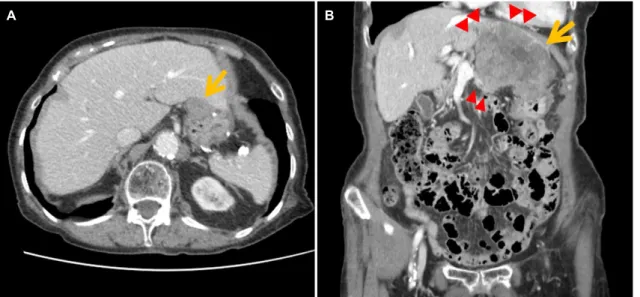
Fig. 5. Follow-up abdominal computed tomography findings after the second surgery. (A) A newly appeared 2.6-cm sized mass was found at the gastric anastomosis site (arrow), 1.5 years after the second surgery. (B) After 6 months of observation, the recurrent gastric lesion increased in size (2.6→9.4 cm, arrow) with invasion to the adjacent liver, pancreas, diaphragm, and pericardial fat (arrowheads).
margin was free from tumor involvement. After 2 months of adjuvant imatinib therapy at the lower dose (300 mg/day), severe anemia occurred again, which led to discontinuation of the drug. Eighteen months after the second surgery, a new- ly appeared 2.6 cm mass was detected on abdominal CT, and subsequent EGD revealed a mass located at the anasto- mosis line (Fig. 5A). Considering her very old age and poor performance status, she was followed up without treatment.
Six months later, an abdominal CT scan showed progressive disease with invasion to the adjacent organs and multiple intra-abdominal metastases (Fig. 5B). She is now on inter- mittent imatinib therapy with a reduced dose (200 mg/day) due to her old age and the drug intolerance, while carefully monitoring the hemoglobin count. Follow-up CT taken after 2 months of medication revealed a partial response of the recurred disease, and she is currently being treated in an outpatient clinic setting.
DISCUSSION
After complete surgical resection of a localized GIST, ap- proximately half of the patients showed tumor recurrence, and the 5-year disease-specific survival was below 60% in the pre-imatinib era.4 The risk of recurrence varies widely accord- ing to the characteristics of the tumor. A high mitotic index and large tumor size are almost uniformly considered factors associated with poor outcomes.2,3,5 Other factors, such as
non-gastric primary tumor location, exon 9 mutation, or “wild type” kit, also appear to be deleterious prognostic factors.
Intraperitoneal rupture or bleeding while manipulating the tu- mor during the surgery is another factor associated with a high risk of postoperative recurrence.2
Imatinib has shown its efficacy in treating metastatic and unresectable GISTs, and can also improve the recurrence-free survival after a surgical resection in patients with high-risk GIST. Currently, adjuvant imatinib is a standard therapy in pa- tients who have undergone surgery for GIST with a high risk of recurrence.6 On the other hand, the optimal duration of imatinib therapy after a surgical resection is still controversial.
Some studies have shown that adjuvant imatinib for more than 3 years, even 5 years, might decrease tumor recurrence in high-risk GIST and that most recurrences occur after the discontinuation of imatinib.4,5
Two main factors may hinder long-term treatment with imatinib.4 One is the side effects associated with imatinib, and another is the acquired drug resistance. The most com- mon toxic effects of imatinib at 400 mg/day include nausea, vomiting, edema, skin rash, and bone marrow toxicity.
Although the drug-related side effects of imatinib are usually mild and resolve with dosage reduction or discontinuation, relatively severe toxicity can occur.7 Previous studies have shown that all patients treated with imatinib experienced one or more adverse events of any grade, and 21×43% experi- enced at least one Grade 3 or 4 adverse event at a dose
of 400 mg/day.4,7,8 These might ultimately alter the efficacy of the imatinib treatment. In the present case, the patient had received adjuvant imatinib therapy only for 18 months.
During the maintenance of imatinib, the patient suffered re- current anemia. The anemia was not resolved completely after a transfusion and supplementation of oral iron. Owing to the severe hematologic adverse effects, she could not continue the imatinib therapy for a sufficient duration. In one report, grade 3-4 hematologic toxicity developed in approximately 20% of elderly patients who underwent imatinib therapy for chronic myelogenous leukemia (CML), which appeared to be related to myelosuppression.9 Nevertheless, imatinib therapy is generally regarded as a safe treatment and is still recom- mended in elderly patients with CML. Considering that myelo- suppression is less common in patients with GIST on imatinib than in patients with CML, a similar treatment strategy can be applied for GIST in elderly patients.7 Close monitoring for toxicity, followed by a temporary dose reduction and treatment continuation can be considered for the maintenance of re- mission in this patient population.10
In conclusion, this paper reported a case of a completely resected gastric GIST with repeated local recurrence in a very elderly patient. The hematologic side effects of imatinib were controlled with dose reduction and conservative manage- ment, and the patient is still alive for 6 years after the initial diagnosis.
REFERENCES
1. Sorour MA, Kassem MI, Ghazal Ael-H, El-Riwini MT, Abu Nasr A.
Gastrointestinal stromal tumors (GIST) related emergencies. Int J Surg 2014;12:269-280.
2. Deshaies I, Cherenfant J, Gusani NJ, et al. Gastrointestinal stro- mal tumor (GIST) recurrence following surgery: review of the clin- ical utility of imatinib treatment. Ther Clin Risk Manag 2010;
6:453-458.
3. Zhao R, Wang Y, Huang Y, et al. Adjuvant imatinib for patients with high-risk gastrointestinal stromal tumors: a retrospective cohort study. Sci Rep 2017;7:16834.
4. Raut CP, Espat NJ, Maki RG, et al. Efficacy and tolerability of 5-year adjuvant imatinib treatment for patients with resected in- termediate- or high-risk primary gastrointestinal stromal tumor:
the PERSIST-5 clinical trial. JAMA Oncol 2018;4:e184060.
5. Joensuu H, Eriksson M, Sundby Hall K, et al. One vs three years of adjuvant imatinib for operable gastrointestinal stromal tumor:
a randomized trial. JAMA 2012;307:1265-1272.
6. Judson I, Bulusu R, Seddon B, Dangoor A, Wong N, Mudan S. UK clinical practice guidelines for the management of gastro- intestinal stromal tumours (GIST). Clin Sarcoma Res 2017;7:6.
7. Demetri GD, von Mehren M, Blanke CD, et al. Efficacy and safety of imatinib mesylate in advanced gastrointestinal stromal tumors. N Engl J Med 2002;347:472-480.
8. Blanke CD, Rankin C, Demetri GD, et al. Phase III randomized, in- tergroup trial assessing imatinib mesylate at two dose levels in patients with unresectable or metastatic gastrointestinal stro- mal tumors expressing the kit receptor tyrosine kinase: S0033.
J Clin Oncol 2008;26:626-632.
9. Latagliata R, Ferrero D, Iurlo A, et al. Imatinib in very elderly pa- tients with chronic myeloid leukemia in chronic phase: a retro- spective study. Drugs Aging 2013;30:629-637.
10. Ogata K, Kimura A, Nakazawa N, et al. Long-term imatinib treat- ment for patients with unresectable or recurrent gastrointestinal stromal tumors. Digestion 2018;97:20-25.