Extra-gastrointestinal stromal tumor of the pancreas: report of a case
Hyung Jun Kwon
Department of Surgery, Kyungpook National University Chilgok Hospital, Kyungpook National University School of Medicine, Daegu, Korea
Gastrointestinal tumors (GISTs) of the pancreas are extremely rare with limited individual case reports and small num- ber of case series. Herein, we report a case of pancreatic extragastrointestinal stromal tumor (EGIST) along with liter- ature review. A 64-year-old female patient was referred to us for treatment of an abdominal mass detected by ultrasono- graphic examination. The tumor was located in the periamullary region. Under a preoperative diagnosis of a duodenal GIST, we performed a pylorus preserving pancreatoduodenectomy for this lesion. Laboratory examination results were within normal ranges. On pathologic gross examination, the tumor measured at 7 cm in its greatest dimension almost entirely involved the pancreatic head. Its cut surface was rubbery and white. It was surrounded by a thin pseudocapsule and well demarcated. Histopathological examination of the specimen showed a cellular lesion with compressed pancre- atic tissue at peripheral. Mitotic count was 5 per 50 high-power fields. Immunohistochemically, neoplastic cells were positive for antibodies against C-KIT (CD117), CD 34, and vimentin. However, smooth-muscle actin reactions with antibodies against S-100 or desmin were negative. Based on above findings, the tumor was finally diagnosed as GISTs originating from the pancreas. The patient has been followed up postoperatively for 72 months. There is no evidence of recurrence. Here we report this case of pancreatic EGIST presenting as a solid neoplasm along with literature review of cases previously described. Our review on pancreatic EGISTs is limited and insufficient to make a conclusion regard- ing its clinical features. Those manifested large masses tended to have an aggressive biological and clinical behavior.
Thus, pancreatic EGISTs need to be carefully differentiated. Adequate surgical intervention is necessary for pancreatic EGISTs. (Ann Hepatobiliary Pancreat Surg 2017;21:237-242)
Key Words: Diagnosis; Gastrointestinal stromal tumor; Pancreas; Pancreatectomy; Pancreatic neoplasm
Received: October 1, 2017; Accepted: October 23, 2017 Corresponding author: Hyung Jun Kwon
Department of Surgery, Kyungpook National University Chilgok Hospital, 807 Hoguk-ro, Buk-gu, Daegu 41404, Korea Tel: +82-53-200-2166, Fax: +82-53-200-2027, E-mail: kwonhj95@naver.com
Copyright Ⓒ 2017 by The Korean Association of Hepato-Biliary-Pancreatic Surgery
This is an Open Access article distributed under the terms of the Creative Commons Attribution Non-Commercial License (http://creativecommons.org/
licenses/by-nc/4.0) which permits unrestricted non-commercial use, distribution, and reproduction in any medium, provided the original work is properly cited.
Annals of Hepato-Biliary-Pancreatic Surgery ∙ pISSN: 2508-5778ㆍeISSN: 2508-5859
INTRODUCTION
GISTs are the most common primary mesenchymal tu- mors originated from the gastrointestinal tract. These tu- mors occur mainly in the stomach (50-60%), small bowel (20-30%), large bowel (10%), and esophagus (5%).1 In rare cases, these tumors are found as primary tumors out- side the GI tract, such as the omentum, mesentery, retro- peritoneum, and gallbladder.2,3 These tumors have been designated as “extra-gastrointestinal stromal tumors”
(EGISTs). EGISTs arising in the pancreas are extremely rare with limited individual case reports and small num- bers of case series. Herein, we report a case of a pancre- atic extra-gastrointestinal stromal tumor in a 64-year-old female patient along with a review of literature.
CASE
A 64-year-old woman was referred to our department for treatment of an abdominal mass incidentally detected by ultrasonographic examination during health screening in August 2010. She had no clinical symptom related to this tumor. She had no remarkable history. Physical ex- amination revealed no specific findings. Laboratory data were within normal limits. Tumor markers were also with- in normal limits. Abdominal computed tomography (CT) showed a well enhanced mass measuring 6.5×6.5 cm with central low dense lesion (Fig. 1A and B). Endoscopic ul- trasonography (EUS) revealed a well-defined homogenous hypoechoic mass like lesion without posterior acoustic shadowing at the pancreatic head. The main pancreatic duct was mildly dilated (Fig. 1C). EUS guided biopsy was
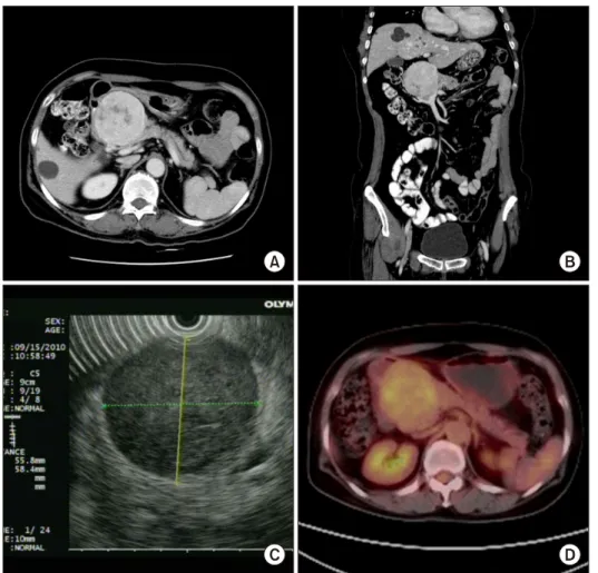
Fig. 1. Imaging study findings.
(A and B) An abdominal com- puted tomography (CT) scan showing a well-enhanced mass measuring at 6.5 cm×6.5 cm with central low dense lesion ap- pearing as necrosis (axial & co- ronal). (C) Positron emission to- mography (PET) showing a hy- permetabolic lesion measured at 7 cm (SUVmax 4.0) in the pan- creatic head without evidence of abnormal hypermetabolic lesion, suggesting lymph node or distant metastasis. (D) Endoscopic ul- trasonography (EUS) revealing a well-defined homogenous hypo- echoic mass like lesion (EUS- guided biopsy was done) without posterior acoustic shadowing at the pancreatic head. The main pancreatic duct was mildly dilated.
performed, yielding pathological diagnosis of gastro- intestinal stromal tumor. Positron emission tomography (PET) detected hypermetabolic lesion measured at 7 cm (SUVmax 4.0) in the pancreatic head without evidence of abnormal hypermetabolic lesion, suggesting LN or distant metastasis (Fig. 1D).
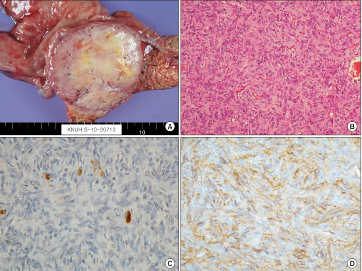
Under a preoperative diagnosis of a duodenal GIST or pancreatic neuroendocrine tumor, we performed a pylorus preserving pancreatoduodenectomy for this lesion. On pathologic gross examination, the tumor measured at 7 cm in its greatest dimension almost entirely involved the pan- creatic head. It was easily separated from duodenum. Its cut surface was rubbery and white. Surrounded by a thin pseudocapsule, it was well-demarcated (Fig. 2A).
Microscopically, the tumor was composed of spindle cells (Fig. 2B). Mitotic count was 5/50 high-power fields (HPF). Immunohistochemically, neoplastic cells were diffuse. They were weakly positive for antibodies against C-KIT(CD117) but positive for CD 34 and vimentin (Fig.
2C and D). However, smooth-muscle actin reactions with
antibodies against S-100 or desmin were negative. Based on above findings, the tumor was finally diagnosed as EGIST originating from the pancreas.
She had an uneventful postoperative course. The patient was discharged home on postoperative day 20. Adjuvant therapy was not started due to the low mitotic rate of the tumor and its intermediate size. She is free of recurrence of EGIST after the surgery.
DISCUSSION
EGISTs arising in the pancreas are extremely rare.
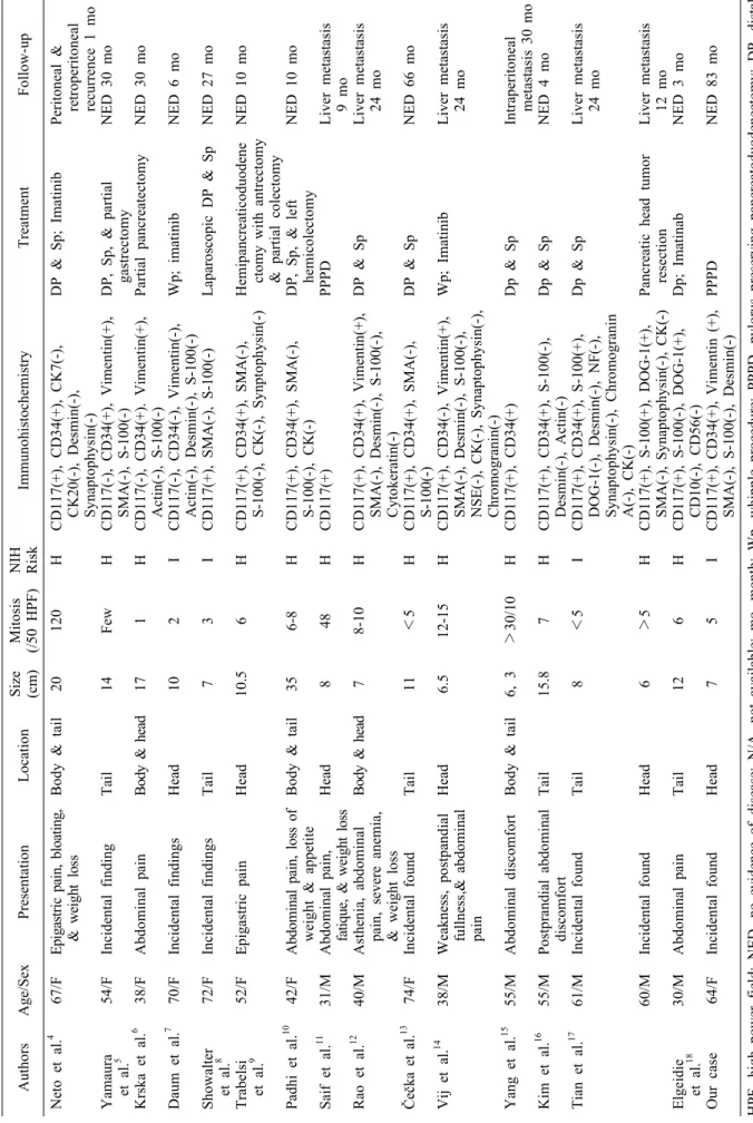
Reports of pancreatic EGISTs are limited to individual case reports and small number of case series. A total of 17 patients who were confirmed as pancreatic GISTs after resection were collected in this study.4-18 The clin- icopathological features and outcomes of these cases are summarized in Table 1. Age of these patients ranged from 30 to 74 years (median, 54.7 years; mean, 53.1 years).
Eight patients were males and nine were females. Most
Fig. 2. Pathologic study findings. (A) On pathologic gross examination, the tumor measured at 7 cm in its greatest dimension involving the pancreatic head. Its cut surface was rubbery and white. Surrounded by a thin pseudocapsule, it was well-demarcated, infiltrating the duodenal wall. (B) The tumor was composed of spindle cells (H&E, ×200). (C) Immunohistochemical examination revealing diffuse and weak CD 117 positivity. (D) Tumor cells showing positive immunoreactivity for CD 34.
clinical presentations included vague abdominal pain or discomfort, weight loss, and fatigue. However, seven (41.2%) cases were found incidentally during a workup for unrelated conditions. These tumors ranged from 6 to 35 cm in maximum diameter (median, 10.0 cm; mean, 11.9 cm). These tumors could occur in any part of the pancreas. Their most common location was head (6/17, 35.3%) and tail (6/17, 35.3%), followed by body & tail (3/17, 17.6%) and head & body (2/17, 11.8%) in the pancreas.
Characteristic immunohistochemical profile of GIST has been proven to be very helpful in its diagnosis. On immunohistochemistry, positivity of c-Kit (CD117) con- firms the diagnosis of GIST. C-kit gene is expressed in 95% of cases. CD 34 is also expressed in 60-70% of GIST cases. The frequency of CD 34 positivity depends
on the location of GISTs. The frequency of positive CD34 is high in GISTs of the esophagus and colon (95%).
However, it is relatively low in small bowel and ex- tra-gastrointestinal sites. GISTs may also show variable positivities for other immune markers, such as SMA (30-40%), S-100 (5%), and desmin (1-25%).19 In our re- view, 14 (82.4%) of 17 pancreatic EGISTs showed pos- itivity for CD117 while 9 (52.9%) cases were positive for CD 34.
GIST exhibits a broad spectrum of clinical behaviors.
The prognosis of patients with GIST depends on the bio- logical behavior of GIST. Fletcher et al.1 have defined the criteria to estimate risks of aggressive behavior and meta- stasis of GIST using tumor dimension (cm) and mitotic count (/50 HPF). According to their criteria, GISTs are divided into risk categories of very low (<2 cm, <5/50
Table 1. Reported cases of pancreatic extra-gastrointestinal stromal tumors AuthorsAge/SexPresentationLocationSize (cm)Mitosis (/50 HPF)NIH RiskImmunohistochemistryTreatmentFo Neto et al.4 Yamaura et al.5 Krska et al.6 Daum et al.7 Showalter et al.8 Trabelsi et al.9 Padhi et al.10 Saif et al.11 Rao et al.12 Čečka et al.13 Vij et al.14 Yang et al.15 Kim et al.16 Tian et al.17 Elgeidie et al.18 Our case
67/F 54/F 38/F 70/F 72/F 52/F 42/F 31/M 40/M 74/F 38/M 55/M 55/M 61/M 60/M 30/M 64/F
Epigastric pain, bloating, & weight loss Incidental finding Abdominal pain Incidental findings Incidental findings Epigastric pain Abdominal pain, loss of weight & appetite Abdominal pain, fatique, & weight loss Asthenia, abdominal pain, severe anemia, & weight loss Incidental found Weakness, postpandial fullness,& abdominal pain Abdominal discomfort Postprandial abdominal discomfort Incidental found Incidental found Abdominal pain Incidental found
Body & tail Tail Body & head Head Tail Head Body & tail Head Body & head Tail Head Body & tail Tail Tail Head Tail Head
20 14 17 10 7 10.5 35 8 7 11 6.5 6, 3 15.8 8 6 12 7
120 Few 1 2 3 6 6-8 48 8-10 <5 12-15 >30/10 7 <5 >5 6 5
H H H I I H H H H H H H H I H H I
CD117(+), CD34(+), CK7(-), CK20(-), Desmin(-), Synaptophysin(-) CD117(-), CD34(+), Vimentin(+), SMA(-), S-100(-) CD117(-), CD34(+). Vimentin(+), Actin(-), S-100(-) CD117(-), CD34(-), Vimentin(-), Actin(-), Desmin(-), S-100(-) CD117(+), SMA(-), S-100(-) CD117(+), CD34(+), SMA(-), S-100(-), CK(-), Synptophysin(-) CD117(+), CD34(+), SMA(-), S-100(-), CK(-) CD117(+) CD117(+), CD34(+), Vimentin(+), SMA(-), Desmin(-), S-100(-), Cytokeratin(-) CD117(+), CD34(+), SMA(-), S-100(-) CD117(+), CD34(-), Vimentin(+), SMA(-), Desmin(-), S-100(-), NSE(-), CK(-), Synaptophysin(-), Chromogranin(-) CD117(+), CD34(+) CD117(+), CD34(+), S-100(-), Desmin(-), Actin(-) CD117(+), CD34(+), S-100(+), DOG-1(-), Desmin(-), NF(-), Synaptophysin(-), Chromogranin A(-), CK(-) CD117(+), S-100(+), DOG-1(+), SMA(-), Synaptophysin(-), CK(-) CD117(+), S-100(-), DOG-1(+), CD10(-), CD56(-) CD117(+), CD34(+), Vimentin (+), SMA(-), S-100(-), Desmin(-)
DP & Sp; Imatinib DP, Sp, & partial gastrectomy Partial pancreatectomy Wp; imatinib Laparoscopic DP & Sp Hemipancreaticoduodene ctomy with antrectomy & partial colectomy DP, Sp, & left hemicolectomy PPPD DP & Sp DP & Sp Wp; Imatinib Dp & Sp Dp & Sp Dp & Sp Pancreatic head tumor resection Dp; Imatinab PPPD
Peritonea retroperitoneal recurrence 1 NED 3 NED 3 NED 6 NED 2 NED 1 NED 1 Liver meta 9 mo Liver meta 24 mo NED 6 Liver meta 24 mo Intraperi meta NED 4 Liver meta 24 mo Liver meta 12 mo NED 3 NED 8 HPF, high power field; NED, no evidence of disease; N/A, not available; mo, month; Wp, whipple procedure; PPPD, pylorus preserving pancreatoduodenectom pancreatectomy; Sp, splenectomy; VL, very low; L, low; I,intermediate; H, high
HPF), low (2-5 cm, <5/50 HPF), intermediate (<5 cm, 6-10/50 HPF or 5-10 cm, <5/50 HPF), and high risk (>5 cm, >5/50 HPF or >10 cm, any mitotic count) of metastasis.1 In our review, the risk of aggressive behavior according to Fletcher criteria was high in 13 (76.5%) cases. The remaining three (11.8%) cases had intermediate risk. The frequency of high risk aggressive behavior tend- ed to be high for GIST.
It is well-known that resection should be considered as the first treatment choice for resectable localized GIST because high potential of malignancy is associated with cases of GIST. Complete surgical resection with negative microscopic margins is the standard treatment for GISTs.20 Systemic regional lymphadenectomy is not gen- erally considered because lymph node metastasis is rare (prevalence of about 1%).21
Response rates of GISTs to conventional chemotherapy and radiotherapy are 10% and 5%, respectively.9,13 Adjuvant treatment with imatinib can be provided in order to enhance the possibility of curing the cancer by eradicat- ing microscopic lesions that might remain after complete resection of visual tumors.19
DeMattero et al.22 have reported that most recurrence of GIST will occur within 24 months after resection, with liver and peritoneum being the most common site of recurrence. Reith et al.23 reported that 39% of patients with EGIST developed metastatic disease or died from tu- mors within a short period, suggesting that EGIST was an aggressive type of stromal tumor. In our review, ma- jority (76.5%) of pancreatic EGISTs are classified to the high risk category at the time of diagnosis. Within 30 months after resection, recurrence occurred in 7 (7/17, 41.2%) patients, with hepatic metastasis being more fre- quent (6/7, 85.7%) than other sites.
Thus, complete surgical resection with negative micro- scopic margin should be considered as first-line treatment for pancreatic EGIST. Intense surveillance after surgical treatment is advocated. Adjuvant treatment following sur- gical treatment should be considered to enhance the possi- bility of curing the cancer. It can prevent recurrences or metastasis because pancreatic EGIST has high potential for malignancy or recurrence.
Our review on pancreatic EGISTs is limited and in- sufficient to make a conclusion regarding their clinical features. These manifested large masses tended to have
aggressive biological and clinical behavior. Therefore, pancreatic EGISTs need to be carefully differentiated from their gastrointestinal counterparts. Adequate surgical intervention and postoperative adjuvant treatment are nec- essary for EGISTs.
REFERENCES
1. Fletcher CD, Berman JJ, Corless C, Gorstein F, Lasota J, Longley BJ, et al. Diagnosis of gastrointestinal stromal tumors:
A consensus approach. Hum Pathol 2002;33:459-465.
2. Agaimy A, Wünsch PH. Gastrointestinal stromal tumours: a reg- ular origin in the muscularis propria, but an extremely diverse gross presentation. A review of 200 cases to critically re-evaluate the concept of so-called extra-gastrointestinal stromal tumours.
Langenbecks Arch Surg 2006;391:322-329.
3. Park JK, Choi SH, Lee S, Min KO, Yun SS, Jeon HM.
Malignant gastrointestinal stromal tumor of the gallbladder. J Korean Med Sci 2004;19:763-767.
4. Neto MR, Machuca TN, Pinho RV, Yuasa LD, Bleggi-Torres LF. Gastrointestinal stromal tumor: report of two unusual cases.
Virchows Arch 2004;444:594-596.
5. Yamaura K, Kato K, Miyazawa M, Haba Y, Muramatsu A, Miyata K, et al. Stromal tumor of the pancreas with expression of c-kit protein: report of a case. J Gastroenterol Hepatol 2004;
19:467-470.
6. Krska Z, Pesková M, Povýsil C, Horejs J, Sedlácková E, Kudrnová Z. GIST of pancreas. Prague Med Rep 2005;106:201-208.
7. Daum O, Klecka J, Ferda J, Treska V, Vanecek T, Sima R, et al. Gastrointestinal stromal tumor of the pancreas: case report with documentation of KIT gene mutation. Virchows Arch 2005;
446:470-472.
8. Showalter SL, Lloyd JM, Glassman DT, Berger AC. Extra-gas- trointestinal stromal tumor of the pancreas: case report and a re- view of the literature. Arch Surg 2008;143:305-308.
9. Trabelsi A, Yacoub-Abid LB, Mtimet A, Abdelkrim SB, Hammedi F, Ali AB, et al. Gastrointestinal stromal tumor of the pancreas: a case report and review of the literature. N Am J Med Sci 2009;1:324-326.
10. Padhi S, Kongara R, Uppin SG, Uppin MS, Prayaga AK, Challa S, et al. Extragastrointestinal stromal tumor arising in the pan- creas: a case report with a review of the literature. JOP 2010;
11:244-248.
11. Saif MW, Hotchkiss S, Kaley K. Gastrointestinal stromal tumors of the pancreas. JOP 2010;11:405-406; author reply 412.
12. Rao RN, Vij M, Singla N, Kumar A. Malignant pancreatic ex- tra-gastrointestinal stromal tumor diagnosed by ultrasound guid- ed fine needle aspiration cytology. A case report with a review of the literature. JOP 2011;12:283-286.
13. Čečka F, Jon B, Ferko A, Šubrt Z, Nikolov DH, Tyčová V.
Long-term survival of a patient after resection of a gastro- intestinal stromal tumor arising from the pancreas. Hepatobiliary Pancreat Dis Int 2011;10:330-332.
14. Vij M, Agrawal V, Pandey R. Malignant extra-gastrointestinal stromal tumor of the pancreas. A case report and review of literature. JOP 2011;12:200-204.
15. Yang F, Jin C, Fu D, Ni Q. Extra-gastrointestinal stromal tumor of the pancreas: clinical characteristics, diagnosis, treatment, and outcome. J Surg Oncol 2011;103:739-740.
16. Kim HH, Koh YS, Park EK, Seoung JS, Hur YH, Kim JC, et al. Primary extragastrointestinal stromal tumor arising in the pan-
18. Elgeidie A, El-Magd EA, El-Maaty SRA, El-Hawary AK.
Pancreatic gastrointestinal stromal tumor: a case report. Int J Surg Case Rep 2016;29:67-70.
19. Kang YK, Kang HJ, Kim KM, Sohn T, Choi D, Ryu MH, et al. Clinical practice guideline for accurate diagnosis and effec- tive treatment of gastrointestinal stromal tumor in Korea. Cancer Res Treat 2012;44:85-96.
20. Valsangkar N, Sehdev A, Misra S, Zimmers TA, O'Neil BH,
22. DeMatteo RP, Lewis JJ, Leung D, Mudan SS, Woodruff JM, Brennan MF. Two hundred gastrointestinal stromal tumors: re- currence patterns and prognostic factors for survival. Ann Surg 2000;231:51-58.
23. Reith JD, Goldblum JR, Lyles RH, Weiss SW. Extragastrointes- tinal (soft tissue) stromal tumors: an analysis of 48 cases with emphasis on histologic predictors of outcome. Mod Pathol 2000;13:577-585.