Neurofibromatosis are cancer-prone hamar- tomatoses that involve a variety of tissues and cell types1. Gastrointestinal stromal tumors are fre- quently associated with this condition, and are usually multiple benign tumors showing diverse histopathological features2. Malignant stromal tumors of gastrointestinal tract are rarely associated with von Recklinghausen’s disease2. We herein present a case of solitary malignant jejunal stromal tumor in a 50-year-old woman with NF1.
CASE REPORT
A 50-year-old Korean woman with a history of in- termittent abdominal pain of two years’ duration consulted for her skin lesions. Her entire body was
covered with multiple, discrete, variable sized freckles and several caf au lait spots (Fig. 1A).
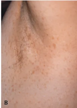
Physical examinations revealed mutiple freckles on the whole body including axillary and inguinal re- gions with no subcutaneous nodules of skin (Fig.
1B). Her brother and her son had the same ap- pearance in addition to multiple neurofibromas.
She was diagnosed to have neurofibromatosis type I.
Esophagography and gastroscopy revealed no polyps. Pelvic MRI revealed intestinal mass (Fig.
2). At exploratory laparotomy, a jejunal segment with an attached mass on the serosal surface was re- sected. The mass, 12.0×7.0×6.0 cm and 250g, is roughly ovid and firm, with a smooth brown external surface. On sectioning the mass showed firm, pale brown whorled cut surfaces with foci of hemor- rhage. Histologic examinations of the lesions re- vealed involvement of serosal surface. The tumor consisted of multiple interweaving bundles of spindle cells with indistinct cell borders. The nuclei were, for the most part, oval with blunted ends (Fig. 3A). In some areas, the nuclei were more pleomorphic, becoming larger and more rectangular, but mitotic figures were rarely observed (Fig. 3B).
Multiple foci of necrosis were observed. Immuno- histochemical stains for actin, desmin, S-100 protein, neuron-specific enolase (NSE), neurofilament, and synaptophysin were performed. The lesion
Solitary Malignant Gastrointestinal Stromal Tumor Associated with a Neurofibromatosis Type I
Hyun Jin Mo, M.D., Hyung Ok Kim, M.D., Chung Won Kim, M.D., and Tae Yoon Kim, M.D.
From the Department of Dermatology, College of Medicine, The Catholic University of Korea, Kangnam St. Mary’s Hospital, 505, Banpo-Dong, Seocho-Gu, 137-040, Seoul, Korea,
Gastrointestinal stromal tumors are usually late manifestations of neurofibromatosis (von Recklinghausen”s disease) and most become clinically apparent in middle-aged patients as mul- tiple benign tumors. To our review of the literature, solitary malignant stromal tumor of gas- trointestinal tract is exceptionally rare in von Recklinghausen”s disease. We herein present a case of solitary jejunal stromal tumor in a 50-year-old woman with NF1, which histopatho- logically showed a malignant change and combined smooth muscle-neural type.
(Ann Dermatol 15(1) 12~14, 2003).
Key Words : Neurofibromatosis Type I, Gastrointestinal Stromal Tumor
* This case was presented at the 53th Annual Meeting of the Korean Dermatological Association on April 18,2001.
Received January 9, 2002
Accepted for publication June 11, 2002
Reprint request to: Tae Yoon Kim, MD. Department of Dermatology, Kangnam St. Mary’s Hospital, College of Medicine, The Catholic University of Korea, 505, Banpo-Dong, Seocho-Gu, Seoul, 137-040, Korea Tel. 02-590-2626, Fax: 02-594-3255
E-mail. [email protected] 12
was strong positive for NSE , focally positive for actin, and had scatterd S-100 protein-positive cells, but negative for desmin, neurofilament, synaptophysin.
DISCUSSION
Neurofibromatosis are a heterogenous set of genetic disorders that involve a variety of tissues and cell types. The most frequent form of neurofibromatosis is type 1 (NF1), also known as von Reckling-
hausen’s disease, the gene of which is located at 17q11.2.
The diagnostic criteria of the National Institute of Health for NF12are met in an individual if two or more of the following are found: six or more caf -au- lait macules; multiple freckles in the axillary or in- guinal regions; optic glioma; two or more Lisch nodules; a distinct osseous lesion, such as sphenoid dysplasia or thinning of the bone cortex with or without pseudoarthrosis; or a first degree relative (parent, sibling, or offspring) who meets these criteria for NF1. Our patient meets this criteria in that she shows ten caf -au-lait macules and multiple freckles in the axillary and inguinal regions, and her brother and her son meet the diagnostic criteria.
Gastrointestinal manifestations of NF1 were re- viewed by Fuller and Williams3. The most com- mon gastrointestinal finding is a stromal tumor with varying degrees of neural or smooth muscle differentiation ; such a tumor occurs in middle- aged patients as multiple benign tumors. A minori- ty of such tumors show convincing evidence of neural or muscle differentiation either on elec- tronmicroscopy or on immunohistochemistry but many defy classification and can only be designated as gastrointestinal “stromal” tumors3. The small number of stromal tumors from NF1 patients studied by Fuller and Williams showed focal strong S-100 Solitary Malignant Gastrointestinal Stromal Tumor Associated with a Neurofibromatosis Type I 13
Fig. 1. (A) Multiple caf au lait spots on the back. (B) Numerous freckles are noted on the left side of axilla.
Fig. 2. Pelvic MRI reveals about a 10×7cm sized, lob- ulated mass of small intestine. The mass has an irregu- lar thick wall and some solid portion with hemorrhage.
positivity, but no other unequivocal evidence of neural origin. Our case was similar to leiomyoma with hematoxylin-eosin stain but was negative for desmin, focally positive for actin, strong positive with NSE, and had scattered S-100 protein-posi- tive cells. We finally diagnosed this tumor as com- bined smooth muscle-neural tumor. Neoplasms re- sembling leiomyomas in the gastrointestinal tract of patients with neurofibromatosis were first reported by Lukash4. While some might argue that such cases represent the coincidental occurrence of leiomy- omas in the gastrointestinal tract of patients with neurofibromatosis, the relative rarity of both gas- trointestinal leiomyomas and neurofibromatosis, plus the presence of gastrointestinal involvement in as many as one fourth of patients with neurofi- bromatosis, would argue against this5.
Malignant stromal tumors of gastrointestinal tract is exceptionally rare in von Recklinghausen’s disease3. Few reported cases have led to metastatic spread6 and most have been regarded as malignant on the basis of conventional criteria of malignancy, namely tumor size and mitotic activity. Although scant mitotic figures, our case could be regarded as malignancy due to multiple foci of necrosis, large tu- mor size, and moderate cellular pleomorphism.
The risk of developing malignant tumors and early death is increased in patients with neurofi- bromatosis1. These risks need to consider gastroin- testinal tumors when gastrointestinal symptoms develop in NF1 patients although clinical surveys of symptomatic gut tumors in NF1 patients are much
less and affect less than 5% of patients7,8. Also, we should know that von Recklinghausen’s neurofi- bromatosis denotes more than the dermatologic curiosity of caf au lait spots.
REFERENCES
1. Minna Poyhonen, Solja Niemela, Riitta Herva.
Risk of malignancy and death in neurofibromatosis.
Arch Pathol Lab Med 1997; 121: 139-143.
2. Neurofibromatosis conference statement: National Institute of Health consensus development confer- ence. Arch Neurol 1988; 45: 575-578.
3. Fuller CE, Williams GT. Gastrointestinal manifes- tations of type 1 neurofibrosis (von Reckling- hausen’s disease). Histopathology 1991; 19: 1-11.
4. Lukash WM, Johnson BB. Gastointestinal neo- plasms in von Recklinghausen’s disease. South Med J 1969; 62: 1237.
5. Schaldenbrand JD, Appelman HD. Solitary solid stromal gastrointestinal tumors in von Reckling- hausen’s disease with minimal smooth muscle dif- ferentiation. Human pathol 1984; 15: 229-232.
6. Gennatas CS, Exarhakos G, Sutton DR. Malignant schwannoma of the stomach in a patient with neu- rofibromatosis. Eur J Surg Oncol 1988; 14: 261-264.
7. Huson SM, Harper PS, Compston DAS. Von Recklinghausen neurofibromatosis: a clinical and population study in South-East Wales. Brain 1988;111:1355-1381.
8. Riccardi VM. Von Recklinghausen neurofibro- matosis. N Engl J Med 1981;305: 1617-1627.
14 HJ Mo, et al.
Annals of Dermatology Vol. 15, No. 1, January 2003
Fig. 3. (A) This field shows cigar-shaped spindle cells with oval, blunt-ended nuclei, suggestive of smooth muscle tu- mor (hematoxylin and eosin staining, ×200). (B) In some areas, pleomorphic, hyperchromatic nuclei are observed (hematoxylin and eosin staining, ×200).
A B