Copyrights © 2015 by The Korean Gastric Cancer Association www.jgc-online.org This is an open-access article distributed under the terms of the Creative Commons Attribution Non-Commercial License (http://creativecommons.org/ licenses/by-nc/4.0) which permits unrestricted noncommercial use, distribution, and reproduction in any medium, provided the original work is properly cited.
Introduction
Gastrointestinal stromal tumors (GISTs) occur most fre-quently in the stomach.1-4 Wedge resection of the stomach with
R0 resection is regarded as a standard treatment because of the low risk of lymph node metastasis from the tumor.2,5-7 Relatively
small sized tumors have been safely treated by various
laparo-scopic approaches since the first report of laparolaparo-scopic wedge resection (LWR) of a gastric submucosal tumor in 1991.8-11
Although laparoscopic gastric wedge resection for extralumi-nal tumors can be performed easily, intralumiextralumi-nal or small tumors are difficult to localize laparoscopically, requiring an intragastric approach or gastrotomy for tumor resection.12-16 Consequently,
the possibility of spreading cancer cells in the abdominal cav-ity arises due to the additional manipulation of the tumor and luminal exposure during these procedures. Thus, it could be a risk factor for peritoneal recurrence. In addition, if the tumors have ulcerations, the risk of cancer cell dissemination might in-crease.17,18
So far, there is no report regarding the long-term outcomes of laparoscopic gastric wedge resection with gastrotomy for the Correspondence to: Taeil Son
Department of Surgery, Yonsei University College of Medicine, 50 Yonsei-ro, Seodaemun-gu, Seoul 03722, Korea
Tel: +82-2-2228-2100, Fax: +82-2-313-8289 E-mail: [email protected]
Received October 6, 2015 Revised October 17, 2015 Accepted October 19, 2015
Oncologic Safety of Laparoscopic Wedge Resection with
Gastrotomy for Gastric Gastrointestinal Stromal Tumor:
Comparison with Conventional Laparoscopic Wedge Resection
Sejin Lee1, You Na Kim1, Taeil Son1, Hyoung-Il Kim1, Jae-Ho Cheong1, Woo Jin Hyung1,2, and Sung Hoon Noh1
1
Department of Surgery, 2
Robot and Minimally Invasive Surgery Center, Yonsei University College of Medicine, Seoul, Korea
Purpose: Various laparoscopic wedge resection (LWR) techniques requiring gastrotomy for gastrointestinal stromal tumors (GISTs) of the stomach have been applied to facilitate tumor resection and preserve the remnant gastric volume. However, there is the possibility of cancer cell dissemination during these procedures. The aim of this study was to assess the oncologic safety of LWR with gastrotomy (LWR-G) compared to LWR without luminal exposure.
Materials and Methods: Clinicopathologic and operative results of 193 patients who underwent LWR for gastric GIST were retrospec-tively analyzed from 2003 to 2013. We stratified the patients into two groups: LWR-G and LWR without gastrotomy (LWR-C). Clinico-pathologic features, short-term outcomes, and long-term outcomes were compared.
Results: A total of 26 patients underwent LWR-G, and 167 patients underwent LWR-C. The LWR-G group showed significantly more an-terior wall-located (n=10, 38.5%), intraluminal (n=20, 76.9%), and ulcerative (n=13, 50.0%) tumors than the LWR-C group (n=33, 19.8%; n=96, 57.5%; n=46, 27.5%, respectively). Postoperative short-term outcomes did not differ between the two groups. When tumor staging was compared, no statistical difference was noted. There was no recurrence in the LWR-G group, while 2 patients in the LWR-C group experienced recurrence. The two recurrences in the LWR-C group were found in the liver and in the remnant stomach at 63 and 12 months after the operation, respectively. No gastric GIST-related death was recorded in any group during the study period. Conclusions: LWR-G for gastric GIST is an oncologically safe procedure even for masses with ulcerations.
treatment of gastric GISTs. In the current study, we compared the oncologic safety of LWR with gastrotomy (LWR-G) and LWR without luminal exposure. Specifically, the long-term consequences for ulcerative GISTs requiring luminal exposure during operation were investigated. Recurrence patterns were also analyzed.
Materials and Methods
1. PatientsBetween March 2003 and December 2013, 205 LWRs of the stomach were performed in patients with histologically con-firmed gastric GISTs at the Department of Surgery at Yonsei University College of Medicine. The study included 193 patients who underwent LWR of the stomach. Of the 193 patients, 26 underwent LWR-G, while the other 167 underwent LWR with-out gastrotomy (LWR-C). Patients were excluded if they had a ruptured tumor at the time of diagnosis, underwent palliative re-section or an endoscopic procedure before the operation, or had insufficient data regarding mitotic rate for proper staging (Fig. 1). On the basis of preoperative endoscopy, endoscopic ultrasound, and abdominopelvic computed tomography scanning, LWR was generally indicated for relatively small gastric submucosal tumors up to 5 cm in the early period, and later on, the indica-tion was expanded to include tumors larger than 5 cm. The type
of resection was selected at the surgeon’s discretion according to the tumor location and size. Patients who were pathologically classified as high risk according to the National Institutes of Health-Fletcher classification were recommended for treatment with imatinib mesylate (Gleevec®; Novartis, Basel, Switzerland), whenever possible. Clinicopathologic characteristics, short-term outcomes, and long-term outcomes, including recurrence and survival status, were analyzed retrospectively.
This retrospective study to compare outcomes with another surgical techniques was approved by the Institutional Review Board (IRB) of Severance Hospital, Yonsei University College of Medicine (4-2015-0865).
2. Surgical technique
Various laparoscopic gastric wedge resection techniques have been described in the literature.1,5,12,13,15,18 When tumors grow
outward from the stomach toward the peritoneal cavity, wedge resection using endolinear staplers can be performed easily without considerable manipulation of the tumor.12-14 In cases of
intraluminal tumors in the posterior wall of the stomach, trans-gastric tumor-everting methods followed by gastrotomy of the anterior wall of the stomach facilitate tumor resection.13,14,19,20
When performing this procedure, the lesion was identified by endoscopy or laparoscopic ultrasonography, and the optimal site of the anterior stomach wall for gastrotomy was chosen. After an incision was made, the tumor was removed by transecting the inverted posterior wall using endolinear staplers. The anterior wall was closed with endolinear staplers or with a laparoscopic suture technique.14,19 When relatively larger intraluminal tumors
are located in the anterior wall, the eversion method can fa-cilitate tumor resection and minimize excessive resection of the normal gastric wall.15 For this procedure, a gastrotomy was
cre-ated about 1 cm from the tumor margin by using intraoperative laparoscopic ultrasonography. Then, the tumor was exteriorized via the incision and resected by endolinear staplers. The advan-tage of this procedure was that the gastrotomy was closed at the same time the endolinear staplers were applied.15 For
intralu-minal tumors near the cardia or the pylorus, the preferred pro-cedure may be intragastric wedge resection with single incision intragastric or conventional intragastric procedures to minimize deformity of the esophagogastric junction or the pylorus.18,21-23
For the single incision intragastric procedure, two wound pro-tectors were used. The anterior gastrotomy was made and pulled out of the abdominal incision, and then another wound protector 205 patients underwent laparoscopic wedge
resection for gastric GIST
202 3 with ruptured tumor at diagnosisor during endoscopic diagnosis
200 2 underwent palliative(R1) resection
199 1 with preoperative endoscopic
submucosal dissection
193 6 with insufficient data
of mitotic rate
LWR-C (n=26)
LWR-C (n=167)
Fig. 1. Patient disposition. GIST = gastrointestinal stromal tumor; LWR-G = laparoscopic wedge resection with gastrotomy; LWR-C = laparoscopic wedge resection without gastrotomy.
was applied via gastrotomy. After laparoscopic removal of the endoluminal tumor, the anterior gastrotomy was closed with en-dolinear staplers.23 For small intraluminal tumors, intraoperative
endoscopic guidance was sometimes required.16,18
3. Statistical analysis
Clinicopathologic features, short-term outcomes, and long-term outcomes were analyzed using the IBM SPSS statistical software ver. 20 (IBM Co., Armonk, NY, USA). Categorical and continuous variables were analyzed by the χ2 (or Fisher’s exact
test) and Student’s t test, respectively. Survival curves were de-picted by the Kaplan-Meier method and compared by log-rank test. Two-sided P-values of less than 0.05 were considered to be
statistically significant.
Results
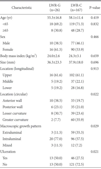
The clinicopathologic features are shown in Table 1. Of a total of 193, 26 patients (13.5%) underwent LWR-G, and 167 patients (86.5%) underwent LWR-C. The mean age did not dif-fer between the two groups (P=0.419). The mean body mass in-dex was similar between the two groups (P=0.659). Mean tumor size and longitudinal location between the G and LWR-C groups were comparable (P=0.696 and P=0.913, respectively). However, more anterior wall-located, intraluminal tumors were found in the LWR-G group compared to the LWR-C group (P=0.022 and P=0.029, respectively). A significantly larger num-ber of tumors (n=13, 50.0%) in the LWR-G group than in the LWR-C group had ulcerations (P=0.021).
Operative outcomes are shown in Table 2. All patients in-cluded in the current study underwent complete tumor resection without gross spillage, tumor rupture, or microscopic margin in-volvement. No open conversion was noted in any patients. The mean operation time for LWR-G was 64.0 minutes compared with a mean of 55.9 minutes for LWR-C (P=0.313). Resumption of soft diet and postoperative hospital stay did not differ between the two groups. No postoperative complications were noted in the LWR-G group, while 3 patients (1.8%) in the LWR-C group
Table 1. Comparison of clinicopathologic features of patients who underwent LWR-G and LWR-C (n=193) Characteristic LWR-G (n=26) (n=167)LWR-C P-value Age (yr) 55.3±16.8 58.1±11.4 0.419 <65 18 (69.2) 119 (71.3) 0.832 ≥65 8 (30.8) 48 (28.7) Sex 0.466 Male 10 (38.5) 77 (46.1) Female 16 (61.5) 90 (53.9)
Body mass index (kg/m2) 24.0±2.6 24.3±3.1 0.659 Size (mm) 36.3±23.3 37.9±18.0 0.696 Location (longitudinal) 0.913 Upper 16 (61.6) 102 (61.1) Middle 5 (19.2) 37 (22.1) Lower 5 (19.2) 28 (16.8) Location (circular) 0.022 Anterior wall 10 (38.5) 33 (19.7) Posterior wall 6 (23.1) 35 (21.0) Lesser curvature 8 (30.7) 39 (23.4) Greater curvature 2 (7.7) 60 (35.9)
Macroscopic growth pattern 0.029
Extraluminal 3 (11.5) 59 (35.3) Intraluminal 20 (77.0) 96 (57.5) Mixed 3 (11.5) 12 (7.2) Ulceration 0.021 Yes 13 (50.0) 46 (27.5) No 13 (50.0) 121 (72.5)
Values are presented as mean±standard deviation or number (%). LWR-G = laparoscopic wedge resection with gastrotomy; LWR-C = laparoscopic wedge resection without gastrotomy.
Table 2. Comparison of short-term operative outcomes
Variable LWR-G (n=26) (n=167)LWR-C P-value
Open conversion 0 0
Operation time (min) 64.0±29.9 55.9±33.4 0.313 Completeness of resection
R0 26 167
R1 0 0
Resumption of soft diet (POD) 1.5±1.2 1.5±1.6 0.958 Hospital stay (POD) 2.6±2.1 2.7±2.1 0.748
Complication >0.999
Yes 0 3 (1.8)
No 26 164 (98.2)
Mortality 0 0
Values are presented as number only, mean±standard deviation, or number (%). LWR-G = laparoscopic wedge resection with gastrotomy; LWR-C = laparoscopic wedge resection without gastrotomy; POD = postoperative day.
Ta bl e 4. C lini ca l fe atur es o f p ati en ts w ith r ec urr ence Pa tien t No. Ag e (y r) Sex Lo ca tio n (L/C) Gr owt h typ e U lcer O pera tio n Size (mm) M ito tic ra te (mi tos es/HP Fs) NIH- Fletch er TNM* Re cur ren ce sit e Tim e t o re cur ren ce (m o) Sur viva l stat us 1 44 Ma le U/GC In tra lumin al Ye s LWR -C 40 >10/50 Hi gh II Li ve r 63 Al ive 2 57 Ma le U/GC In tra lumin al No LWR -C 15 2/50 Ver y lo w IA Remn an t s to m ac h 12 Al ive L/C = lo ng itudin al/cir cu lar ; HP Fs = hig h p ow er f ield s; NIH = N at io na l I ns tit ut es o f H ea lth; U = u pp er ; GC = g re ater c ur va tur e; L WR -C = l ap ar os co pic w edg e r es ec tio n w ith ou t ga str ot om y. *Th e se ven th e di tio n o f t he A m er ica n J oin t C ommi tte e o n C an cer S ta gin g S ys tem.
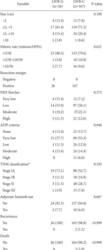
Table 3. Comparison of pathologic outcomes
Variable LWR-G (n=26) (n=167)LWR-C P-value Size (cm) 0.190 <2 4 (15.4) 13 (7.8) ≥2, <5 17 (65.4) 119 (71.2) ≥5, ≤10 4 (15.4) 34 (20.4) >10 1 (3.8) 1 (0.6)
Mitotic rate (mitoses/HPFs) 0.621
≤5/50 23 (88.5) 133 (79.6) >5/50 ≤10/50 1 (3.8) 18 (10.8) >10/50 2 (7.7) 16 (9.6) Resection margin Negative 0 0 Positive 26 167 NIH-fletcher 0.573 Very low 4 (15.4) 12 (7.2) Low 14 (53.9) 97 (58.1) Moderate 5 (19.2) 37(22.1) High 3 (11.5) 21 (12.6) AFIP criteria 0.846 None 4 (15.4) 23 (13.7) Very low 15 (57.7) 89 (53.3) Low 3 (11.5) 20 (12.0) Moderate 4 (15.4) 24 (14.4) High 0 11 (6.6) TNM classification* 0.193 Stage IA 19 (73.1) 88 (52.7) Stage IB 3 (11.5) 18 (10.8) Stage II 3 (11.5) 48 (28.7) Stage III 1 (3.9) 13 (7.8)
Adjuvant Imatinib use 0.667
No 24 (92.3) 157 (94.0) Yes 2 (7.7) 10 (6.0) Recurrence No 26 (100) 165 (98.8) >0.999 Yes 0 2 (1.2) Death No 26 (100) 164 (98.2) >0.999 Yes 0 3 (1.8)
Values are presented as number (%) or number only. LWR-G = laparoscopic wedge resection with gastrotomy; LWR-C = laparoscopic wedge resection without gastrotomy; HPFs = high power fields; NIH = National Institutes of Health; AFIP = American Forces Institute of Pathology. *The seventh edition of the American Joint Committee on Cancer Staging System.
had complications.
Pathologic and long-term outcomes are shown in Table 3. There was no statistically significant difference in the mean tu-mor size (P=0.696). When the tutu-mor size and mitotic rate were stratified according to the current risk criteria of gastric GIST, there was no difference in proportion of the size and mitotic rate between the two groups (P=0.190 and P=0.621, respectively). Consequently, risk based on National Institutes of Health-Fletcher, American Forces Institute of Pathology criteria, and TNM classification did not differ between the groups (P=0.573, P=0.846, and P=0.193, respectively). Adjuvant therapy with imatinib mesylate was administered to 2 patients (7.7%) and 10 patients (6.0%) in the LWR-G and LWR-C groups, respectively. During the median follow-up period of 36 months, 2 patients in the LWR-C group had recurrence in the liver and in the rem-nant stomach at 63 and 12 months after surgery, respectively. No patient in the LWR-G group had recurrence. The characteristics of the patients with recurrence are depicted in Table 4. During the follow-up period, 3 patients in the LWR-C group died, but there were no gastric GIST-related deaths.
Discussion
In the current study, we found that peritoneal recurrence due to potential spillage of cancer cells may not happen during LWR-G for gastric GIST. In addition, ulcerative GIST treated with the same procedures did not increase the risk of peritoneal recurrence. Gastrotomy with everting/eversion methods or in-tragastric procedures may not increase the rate of recurrence due to tumor spillage by luminal exposure during LWR. In addition, we observed that the pattern of recurrence, especially peritoneal recurrence, was significantly low after LWR-G or LWR-C for relatively small GISTs.
GISTs are rare tumors and a distinctive histopathological group of intestinal neoplasms of mesenchymal origin.3 They
comprise fewer than 3% of all gastrointestinal cancers, and the most frequently involved site is the stomach, followed by the small intestine.3,24 Complete R0 resection without
lymphad-enectomy for primary non-metastatic GIST remains the only curative treatment.2,6 Since the first report of LWR of a gastric
submucosal tumor, laparoscopic resection of GIST is considered to be feasible and safe from both the technical and the oncologic point of view.6,8,17
Although LWR for gastric GIST has demonstrated
accept-able oncologic outcomes, the current indication is limited to relatively small tumors due to possible rupture of the tumor into the peritoneal cavity during the procedure.10,11,25-28 The
Eu-ropean Society for Medical Oncology guidelines recommend laparoscopic gastric wedge resection for tumors less than 2 cm in size, while the National Comprehensive Cancer Network and Japanese guidelines recommend the procedure for tumors less than 5 cm by experienced surgeons.10,27,28 The risk of possible
tumor cell dissemination into the peritoneal cavity can be greatly increased when it is performed in conjunction with more com-plicated procedures such as those requiring transgastric or intra-gastric approaches. Transintra-gastric everting for intraluminal masses in the posterior wall of the stomach and eversion methods for intraluminal tumors in the anterior wall have shown satisfactory short-term outcomes.14,15 However, during these procedures,
tumors can be manipulated more vigorously, and gastric luminal contents can be spilled out into the peritoneal cavity.17,18
Fur-thermore, for tumors with ulcerative lesions, the potential hazard is expected to worsen.18 If the GIST ruptures into the peritoneal
cavity, the recurrence rate increases by almost 100%.29
However, we experienced only 2 cases of recurrence in the LWR-C group during the median follow-up period of 36 months. The overall incidence of recurrence was 1.0% in the studied patients. The results were comparable to other reported series in the literature.25,26 Even with ulcerative lesions, which
comprised 50% of the lesions in the LWR-G group and had low to intermediate risk in most cases (data not shown), we did not observe any recurrence during the follow-up period. The low incidence of recurrence in the current study might have been achieved by careful manipulation of the tumors to avoid tumor rupture and significant spillage of gastric contents into the peritoneal cavity. There was no intraoperative tumor rup-ture in the current study. Second, the indications were limited to relatively small tumors so that only a small portion of patients were classified as high-risk. Finally, by using wound protectors to avoid direct contact of the tumors with the surgical wound when retrieving the specimens, the possibility of cancer cell dis-semination to the surgical wound was minimized. In addition, all patients who received adjuvant imatinib treatment showed favorable outcomes without recurrence in the study period.
To our knowledge, this is the first study in the literature to investigate the long-term outcomes of LWR-G for gastric GISTs compared to conventional LWR. Our study revealed that gastric lumen exposure during procedures, which facilitate
tumor localization and resection while avoiding excessive resec-tion of the remnant stomach to prevent funcresec-tional and structural deformities can be safely performed even in ulcerative lesions, given meticulous handling of the tumors and properly indicated patients. However, the study has several limitations. First, the current study was conducted retrospectively in a single center. Second, long-term outcomes, especially recurrence patterns, could not be properly assessed because the number of high-risk patients was small in both groups. Therefore, the results of the current study should be compared to a larger number of cases to determine the exact impact of luminal exposure during LWR for ulcerative lesions in high-risk patients.
In conclusion, LWR-G did not increase overall or peritoneal recurrence for the selected patients. This technique might be safely performed even for ulcerative gastric GISTs.
Conflicts of Interest
No potential conflict of interest relevant to this article was reported.
References
1. Matthews BD, Joels CS, Kercher KW, Heniford BT. Gas-trointestinal stromal tumors of the stomach. Minerva Chir 2004;59:219-231.
2. Das A, Wilson R, Biankin AV, Merrett ND. Surgical therapy for gastrointestinal stromal tumours of the upper gastrointestinal tract. J Gastrointest Surg 2009;13:1220-1225.
3. Pidhorecky I, Cheney RT, Kraybill WG, Gibbs JF. Gastrointes-tinal stromal tumors: current diagnosis, biologic behavior, and management. Ann Surg Oncol 2000;7:705-712.
4. Emory TS, Sobin LH, Lukes L, Lee DH, O'Leary TJ. Prognosis of gastrointestinal smooth-muscle (stromal) tumors: depen-dence on anatomic site. Am J Surg Pathol 1999;23:82-87. 5. Privette A, McCahill L, Borrazzo E, Single RM, Zubarik R.
Laparoscopic approaches to resection of suspected gastric gastrointestinal stromal tumors based on tumor location. Surg Endosc 2008;22:487-494.
6. DeMatteo RP, Lewis JJ, Leung D, Mudan SS, Woodruff JM, Brennan MF. Two hundred gastrointestinal stromal tumors: recurrence patterns and prognostic factors for survival. Ann Surg 2000;231:51-58.
7. Hassan I, You YN, Shyyan R, Dozois EJ, Smyrk TC, Okuno
SH, et al. Surgically managed gastrointestinal stromal tumors: a comparative and prognostic analysis. Ann Surg Oncol 2008;15:52-59.
8. Fowler DL, White SA. Laparoscopic resection of a submucosal gastric lipoma: a case report. J Laparoendosc Surg 1991;1:303-306.
9. Berindoague R, Targarona EM, Feliu X, Artigas V, Balagué C, Aldeano A, et al. Laparoscopic resection of clinically suspected gastric stromal tumors. Surg Innov 2006;13:231-237.
10. Nishida T, Hirota S, Yanagisawa A, Sugino Y, Minami M, Yamamura Y, et al; GIST Guideline Subcommittee. Clinical practice guidelines for gastrointestinal stromal tumor (GIST) in Japan: English version. Int J Clin Oncol 2008;13:416-430. 11. Mochizuki Y, Kodera Y, Fujiwara M, Ito S, Yamamura Y,
Sawa-ki A, et al. Laparoscopic wedge resection for gastrointestinal stromal tumors of the stomach: initial experience. Surg Today 2006;36:341-347.
12. Choi YB, Oh ST. Laparoscopy in the management of gastric submucosal tumors. Surg Endosc 2000;14:741-745.
13. Basso N, Rosato P, De Leo A, Picconi T, Trentino P, Fantini A, et al. Laparoscopic treatment of gastric stromal tumors. Surg Endosc 2000;14:524-526.
14. Avital S, Brasesco O, Szomstein S, Liberman M, Rosenthal R. Technical considerations in laparoscopic resection of gastric neoplasms. Surg Endosc 2003;17:763-765.
15. Hyung WJ, Lim JS, Cheong JH, Kim J, Choi SH, Noh SH. Lap-aroscopic resection of a huge intraluminal gastric submucosal tumor located in the anterior wall: eversion method. J Surg Oncol 2005;89:95-98.
16. Tangoku A, Yamamoto K, Hirazawa K, Takao T, Mori N, Tada K, et al. Laparoscopic resection of large leiomyomas of the gas-tric fundus. Surg Endosc 1999;13:1050-1052.
17. Novitsky YW, Kercher KW, Sing RF, Heniford BT. Long-term outcomes of laparoscopic resection of gastric gastrointestinal stromal tumors. Ann Surg 2006;243:738-745.
18. Kiyozaki H, Saito M, Chiba H, Takata O, Rikiyama T. Laparo-scopic wedge resection of the stomach for gastrointestinal stro-mal tumor (GIST): non-touch lesion lifting method. Gastric Cancer 2014;17:337-340.
19. Motson RW, Fisher PW, Dawson JW. Laparoscopic resection of a benign intragastric stromal tumour. Br J Surg 1995;82:1670. 20. Ibrahim IM, Silvestri F, Zingler B. Laparoscopic resection of
posterior gastric leiomyoma. Surg Endosc 1997;11:277-279. 21. Hwang SH, Park do J, Kim YH, Lee KH, Lee HS, Kim HH,
et al. Laparoscopic surgery for submucosal tumors located at the esophagogastric junction and the prepylorus. Surg Endosc 2009;23:1980-1987.
22. Na JU, Lee SI, Noh SM. The single incision laparoscopic intra-gastric wedge resection of intra-gastric submucosal tumor. J Gastric Cancer 2011;11:225-229.
23. Choi CI, Lee SH, Hwang SH, Kim DH, Jeon TY, Kim DH, et al. Single-incision intragastric resection for upper and mid gastric submucosal tumors: a case-series study. Ann Surg Treat Res 2014;87:304-310.
24. Miettinen M, Majidi M, Lasota J. Pathology and diagnostic cri-teria of gastrointestinal stromal tumors (GISTs): a review. Eur J Cancer 2002;38 Suppl 5:S39-S51.
25. Kim KH, Kim MC, Jung GJ, Kim SJ, Jang JS, Kwon HC. Long term survival results for gastric GIST: is laparoscopic surgery for large gastric GIST feasible? World J Surg Oncol 2012;10:230.
26. Honda M, Hiki N, Nunobe S, Ohashi M, Kiyokawa T, Sano T,
et al. Long-term and surgical outcomes of laparoscopic sur-gery for gastric gastrointestinal stromal tumors. Surg Endosc 2014;28:2317-2322.
27. Blay JY, Bonvalot S, Casali P, Choi H, Debiec-Richter M, Dei Tos AP, et al; GIST consensus meeting panelists. Consen-sus meeting for the management of gastrointestinal stromal tumors. Report of the GIST Consensus Conference of 20-21 March 2004, under the auspices of ESMO. Ann Oncol 2005;16:566-578.
28. Demetri GD, Benjamin RS, Blanke CD, Blay JY, Casali P, Choi H, et al; NCCN Task Force. NCCN Task Force report: manage-ment of patients with gastrointestinal stromal tumor (GIST): update of the NCCN clinical practice guidelines. J Natl Compr Canc Netw 2007;5 Suppl 2:S1-S29.
29. Hohenberger P, Ronellenfitsch U, Oladeji O, Pink D, Ströbel P, Wardelmann E, et al. Pattern of recurrence in patients with ruptured primary gastrointestinal stromal tumour. Br J Surg 2010;97:1854-1859.