J. of Korean Bone & Joint Tumor Soc.
Volume 11, Number 2, December, 2005
※통신저자: 김 한 수
서울특별시 종로구 연건동 2 8 서울대학교 의과대학 정형외과학교실
Tel: 02) 2072-2362, Fax: 02) 764-2718, E-mail: [email protected]
✽ 본 연구는 서울대학교병원 일반연구과제 지원을 받음.
골육종에서 세포 사멸 관련 유전자 s u rvivin, bcl-2, bax의 발현과 임상적 의의
서울대학교 의과대학 정형외과학교실, 서울대학교 의과대학 병리학교실†, 서울대학교병원 임상의학연구소*
강현귀・김한수・이미라*・설소미*・오주한・이상훈・강경훈†
목적: 골육종에서 세포 사멸과 관련된 survivin, bcl-2, bax 유전자의 발현을 조사하여 임 상적 결과와의 연관성에 대해 알아 보고자 하였다.
대상 및 방법: 항암 약물 치료 전 절개 생검으로 얻은 5 0예의 골육종 조직을 면역조직화학 염색법을 통하여 survivin, bcl-2, bax의 발현을 관찰하였다. 임상적으로 항암 약물 치료에 대한 반응율, 국소재발, 원격전이, 종양학적 결과 등을 연구하여 surviving, bcl-2, bax 유 전자 각각의 발현 또는 이들의 복합적인 발현과의 연관성에 대해 통계적으로 분석하였다.
결과: 면역조직화학염색법으로 검사한 survivin 유전자의 발현은 2 6예 ( 5 2 % )에서 관찰되 었고, bcl-2는 2 3예 (46%), bax는 2 1예 ( 4 2 % )에서 관찰되었다. Survivin과 b c l - 2의 공동 발현은 1 9예 (38%), survivin과 b a x의 공동 발현은 1 3예 (26%), bcl-2와 b a x의 공동 발 현은 8예 (16%), 그리고 이들 3가지 모두의 발현은 총 8예 ( 1 6 % )에서 관찰되었다. 검사한 3 가지 세포 사멸 관련 유전자의 발현과 여러 임상적 변수와의 상관 관계를 조사 하였을 때 조 직학적 분류, 나이, 성별, 국소 재발 등과는 유의한 연관성이 없었다. 항암 약물 반응율과 통 계적으로 유의한 연관성을 보인 인자는 bcl-2 (P=0.04), 그리고 s u r v i v i n과 b c l - 2가 동시 발 현된 경우 ( P = 0 . 0 4 4 )였으며 나쁜 항암 약물 반응율을 나타냈다. 종양학적 결과 중 질병으로 인한 사망과의 연관성을 보인 인자 역시 bcl-2 (P=0.001), 그리고 s u r v i v i n과 b c l - 2가 동시 발현된 경우 ( P = 0 . 0 2 7 )였으며, 이들의 발현은 나쁜 종양학적 결과를 나타냈다. Kaplan- Meier 생존율 분석에서 bcl-2 (P=0.0012)의 발현과 survivin, bcl-2의 동시 발현 ( P = 0 . 0 0 7 5 )은 생존율과 역의 상관관계를 보였다.
결론: 골육종에서 세포 사멸 관련 유전자의 발현은 비교적 높게 보였으며, bcl-2의 발현은 항암 약물치료에 대한 불량한 반응과 낮은 생존율에 의미 있는 연관성을 보이며, survivin은 b c l - 2와 동 시에 발현되는 경우에만 이러한 종양학적 결과와 의미 있는 관련이 있었다. 따라서 세포 사멸 관련 유전자들의 면역조직화학염색법을 통한 관찰이 골육종의 예후 판정에 도움이 될 것으로 사료된다.
색인 단어: 골육종, 세포 사멸 유전자, survivin, bcl-2, bax, 임상적 연관성
서 론
세포 사멸은 프로그램 되어있는 세포의 죽음으로 정의될 수 있는데 이는 핵의 응축 ( c o n d e n s a t i o n ) , D N A의 분절화 (fragmentation), 그리고 세포의 위축 ( s h r i n k a g e )으로 특징지어지는 에너지 의존 과정이다. 싸이토카인이나 암유전인자, 방사선 조 사, 바이러스 감염 등 많은 다양한 자극들에 의해 세 포의 사멸은 유도될 수 있다. 세포 사멸을 일으키는 다양한 과정들이 있는데 이들 중 시스테인 단백분해 효소 (cystein protease) 분자들은 세포 사멸에 필 수적인 효소적 파괴를 일으키게 된다.
세포의 자연사멸이 손상된 유전자는 암유전자로 작용하거나 종양 억제 유전자로 작용할 수 있는데, s u r v i v i n은 세포 사멸을 억제하는 유전자이며 정상 적인 태아발달 과정에서 보일 뿐만 아니라 다양한 암 조직에서 발현되지만 정상적인 성인 조직이나 양 성 종양에서는 보이지 않는다고 보고되고 있다2 ). 이 러한 s u r v i v i n은 폐, 대장, 췌장, 전립선, 유방암 등에서 과발현되며 나쁜 예후와 밀접한 관계가 있다 고 보고 되었다1 , 2 , 8 , 1 6 , 2 1 , 2 3 ).
B c l - 2는 사람의 소포 B -세포 (follicular B-cell) 림프종에서 14:18 염색체 전위 ( t r a n s l o c a t i o n )에 서 발견되었으며 생리적, 병태적, 약리적 등 다양한 자극에 의해 세포 사멸을 억제하는 암 유발 유전자 의 하나로 알려졌으나 세포증식과는 직접적인 연관 이 없다고 하였다2 5 , 2 6 ). 이들 bcl-2 유전자는 전립선 암, 대장-직장암, 폐의 상피세포암, 유방암, 비인후 악성종양 등 다양한 고형암에서 높은 발현을 보인다 고 보고되고 있다3 , 6 , 9 , 1 4 , 1 5 ).
B a x는 1 9번 염색체에 위치되며 세포 사멸 신호에 의해 조절되는 p 5 3 -결합 영역 ( d o m a i n )을 가져 초 기에 반응하는 유전자이다. Bax는 b c l - 2와 21% 상 동성 ( h o m o l o g y )을 가지며 세포 내 동반자적인 관 계를 가지는데, 이는 21-kd 단백질로 면역학적인 연 구로 처음 밝혀졌다. Bax의 발현은 세포 사멸을 억 제하지 않고 bcl-2/bax 이형복합체를 형성하거나 bcl-2 타겟에 경쟁적으로 작용하여 bcl-2 기능을 억 제하는 것으로 보고되고 있다1 7 , 1 9 ).
세포 사멸의 조절은 암세포의 증식과 종양의 진행 에 큰 영향을 줄 수 있으며 이미 사람의 많은 종류의 암에서 이들과 관련된 유전자의 과발현이 보고되고
있다. 하지만 사람의 골육종에서는 그에 따른 연구 와 보고된 예가 매우 적고 부분적이다.
이에 저자들은 골육종 환자에서 종양 조직을 채취 하여 세포 사멸과 관계된 survivin, bcl-2, bax 유 전자들의 발현 정도를 조사하고, 그 결과를 임상적 자료와 비교 분석하여 세포 사멸 관련 유전자들이 골육종에서 종양의 증식, 전이, 그리고 종양학적 결 과와 어떠한 연관성을 갖는지 연구하고자 하였다.
연구대상 및 방법 1. 연구 대상
1 9 9 6년 3월부터 1 9 9 9년 8월 사이에 원발성 골육 종으로 진단, 치료 받았던 환자 중에서 항암약물치 료 이전의 절개 생검에서 얻어진 종양 조직을 파라 핀에 포매하여 보관할 수 있었던 5 0예를 대상으로 하였다. 남자 3 2예, 여자 1 8예로 평균연령은 2 1 . 7 세 ( 4 ~ 8 1세)였다. 골육종의 위치는 대퇴골 3 4예, 경골 9예, 상완골 3예, 골반골 2예, 천골 1예, 비골 1예 순이였고, 병리적 구분으로 골모세포성 골육종 이 3 4예, 연골모세포성 7예, 혼합성 3예, 섬유모세 포성 3예였으며 기타 소세포성, 방골성, 모세혈관 확장성이 각각 1예였다. 술 전 Enneking 방법의 단계 분류로 stage IB 1예, IIA 1예, IIB 44예, 그 리고 처음 진단 당시에 폐 전이가 있었던 I I I가 4예 였다. 방골성 골육종으로 진단된 1예를 제외한 전례 에서 수술 전 화학요법을 시행하였으며 화학요법에 대한 반응은 Huvos grade III과 I V가 2 0예, I과 I I 가 2 9예였다. 화학 요법에 사용된 약제는 고농도 메 톡스렉쎄이트와 아드리아마이신, 시스플라틴으로 전 례에서 동일하였다.
골육종의 수술 시 절제연은 변연부 절제연 ( m a r- ginal margin)이 1예, 부적절한 광범위 절제술 (inadequate wide margin)이 3예, 적절한 광범위 절제술 (adequate margin) 이상이 4 6예였다. 술 후 추시관찰 동안 2 6예에서 원격 전이가 발생되어 수술 전후 총 3 0예의 원격 전이가 관찰되었으며, 8 예에서는 국소 재발이 관찰되었다.
5 0명의 골육종 환자에 대한 평균 추시 기간은 3 6 . 9개월 ( 6 ~ 1 0 0개월)이었다. 지속적 무병 생존 2 6예, 실질적 무병 생존 8예, 사망 1 6예였다.
2. 연구 방법
골육종 조직에 대해서는 면역조직화학염색법 중 특이성과 민감도가 높은 A B C법 (avidin biotin peroxidase complex method)를 이용하여 s u r- vivin, bcl-2, bax 유전자의 발현 정도를 분석하 고, 임상적으로는 환자의 항암 화학요법에 대한 반 응, 원격전이 여부, 국소재발 여부, 5년 생존율 등 의 예후를 분석하여, 세포 사멸 관련 유전자의 발현 정도와 임상적인 예후와의 관계를 비교 분석하고자 하였다.
1) 면역조직화학염색
포르말린으로 고정시킨 후 파라핀에 포매된 종양 조직 절편을 5 μm 두께로 자른 후 슬라이드 위에 놓고 x y l e n e으로 탈파라핀한 후 e t h a n o l로 수화시 켰다. Phosphate buffered saline (PBS)으로 세 척한 뒤 endogenous peroxidase의 활성을 억제하 기 위하여 0.3% hydrogen peroxide로 1 5분간 처 리한 후 다시 P B S로 세척하였다. 비특이적 항원-항 체 반응을 억제하기 위하여 정상 혈청으로 4℃에서 1시간 반응시킨 후 1차 항체로 4℃에서 o v e r n i g h t 하였다. 본 연구에서는 H i s t o s t a i nⓇ-Plus Kits (Zymed Laboratories Inc., San Francisco, CA, USA)를 사용하였고, 1차 항체로는 s u r v i v i n (Abcam, Cambridgeshire, UK), bcl-2 (Santa-Cruz Biotechnology, Santa-Cruz, CA, USA), bax (Santa-Cruz Biotechnology)를 각 각 1 : 1 0 0으로 희석하여 사용하였다. PBS로 세척한
후 b i o t i n이 결합된 2차 항체로 실온에서 1시간 반 응시켰다. 다시 P B S로 세척한 뒤 p e r o x i d a s e - c o n- jugated streptavidin을 사용하여 실온에서 3 0분간 반응시켰다. PBS로 세척하고 DAB (3,3- diaminobenzidine tetrahydrochloride)로 염색하 여 현미경으로 관찰하였다. 대조염색으로 h e m a- toxylin (Sigma, St. Louis, MO, USA)을 사용 하였다.
2) 슬라이드의 검경 관찰
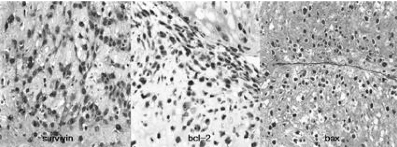
면역조직화학염색 결과는 환자의 임상 특징을 모 르는 병리의사에 의하여 판독되었다. 각 슬라이드 당 일차 항체에 염색된 암세포를 전체 암세포에 대 한 비율로 양성여부를 판별하였고, 10 배 고배율 시 야를 검색하여 전체 세포 중 염색된 세포의 평균이 10% 이상일 때 양성으로 정하였다(Fig. 1).
3) 통계학적 분석
SPSS-statistical software (SPSS Inc., Chicago, Illinois, USA) version 12.0을 사용하 였다. 세포 사멸 관련 유전자의 발현과 여러 임상적 변수와의 비교 및 유의성 판정에는 카이제곱 ( ( 2 ) 검정법을 사용하였으며, 환자의 생존율 분석에는 Kaplan-Meier method를 사용하였고 군간 생존율 비교에는 log-rank test를 사용하였다.
결 과
면역조직화학염색법으로 검사한 survivin 유전자
Fig. 1. Immunohistochemical staining of survivin, bcl-2 and bax shows an example of positive staining (×200).
의 발현은 2 6예 ( 5 2 % )에서 관찰되었고, bcl-2는 2 3 예 (46%), bax는 2 1예 ( 4 2 % )에서 관찰되었다.
S u r v i v i n과 b c l - 2의 공동 발현은 1 9예 ( 3 8 % ) , s u r v i v i n과 b a x의 공동 발현은 1 3예 (26%), bcl- 2와 b a x의 공동 발현은 8예 (16%), 그리고 이들 3 가지 모두의 발현은 총 8예 ( 1 6 % )에서 관찰되었다.
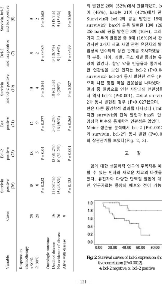
검사한 3가지 세포 사멸 관련 유전자의 발현과 여러 임상적 변수와의 상관 관계를 조사하였을 때 조직학 적 분류, 나이, 성별, 국소 재발 등과는 유의한 연관 성이 없었다. 항암 약물 반응율과 통계적으로 유의 한 연관성을 보인 인자는 bcl-2 (P=0.04) 그리고 s u r v i v i n과 b c l - 2가 동시 발현된 경우 ( P = 0 . 0 4 4 )였 으며 나쁜 항암 약물 반응율을 나타냈다. 종양학적 결과 중 질병으로 인한 사망과의 연관성을 보인 인 자 역시 bcl-2 (P=0.001), 그리고 s u r v i v i n과 b c l - 2가 동시 발현된 경우 ( P = 0 . 0 2 7 )였으며, 이들의 발 현은 나쁜 종양학적 결과를 나타냈다 (Table 1). 하 지만 s u r v i v i n의 단독 발현과 b a x의 단독 발현은 임상적 변수와 통계학적 연관성은 없었다. Kaplan- Meier 생존율 분석에서 bcl-2 (P=0.0012)의 발현 과 survivin, bcl-2의 동시 발현 ( P = 0 . 0 0 7 5 )은 역 의 상관관계를 보였다(Fig. 2, 3).
고 찰
암에 대한 생물학적 연구의 주목적은 예후를 예측 할 수 있는 인자와 새로운 치료의 타겟을 밝히는데 있다. 유전자와 다양한 단백질 발현에 대한 체계적 인 연구자료는 종양의 예후와 전이 가능성 그리고
Fig. 2. Survival curves of bcl-2 expression showed nega- tive correlation (P=0.0012).
+: bcl-2 negative, x: bcl-2 positive
치료를 위한 선택적 종양 타겟을 정하는데 정형화된 정보를 제공할 수 있을 것으로 기대된다. 저자들의 연구는 세포 사멸과 관련된 유전자의 발현과 사람의 골육종의 임상적 결과의 상관관계를 알아내고자 하 는 것이며, 이들 세포 사멸 관련 유전자는 전술한 바 와 같이 사람의 다양한 내장기관의 악성 종양에서 주로 나쁜 예후와 관련되어 발현되고 있다. 특히 s u r v i v i n은 폐의 선암, 상피세포암1 2 ), 췌장의 선암, 대장의 선암, 유방암, 식도 상피세포암2 0 ), 비 호드킨 스 림프종2 )에서 그들의 진행과 연관되어 면역조직화 학염색에서 관찰되었다. 또한 s u r v i v i n의 발현은 대 장, 직장, 위, 폐, 방광암과 흑색종에서 나쁜 예후와 관련되어 높게 발현되었다1 0 , 1 3 , 2 2 ).
S u r v i v i n은 8 0 %가 세포질에서 나머지 2 0 %가 핵 내에서 위치하는 것으로 아세포 단위에서 구분할 수 있다고 하였다4 ). Triebs 등2 4 )은 다른 종양에서와 달 리 골육종에서 s u r v i v i n이 핵 내에 발현되면 높은 5 년 생존율을 보여 좋은 예후와 연관성이 있다고 하 였고, 반대로 골육종에서 s u r v i v i n의 위치가 핵 내 로 국한되지 않으면 임상적인 면과 생물학적인 면에 서 좀더 공격적인 양상으로 보인다고 보고하면서, 면역조직화학염색상 구분 되는 s u r v i v i n의 세포 내 위치를 증명하는 것은 골육종 환자의 생존율을 예측 하는 하나의 중요한 인자가 될 수 있다고 하였다. 이 와 같이 세포 사멸 유전자들이 암의 종류와 위치에 따라서 서로 정반대의 기능으로 인지되곤 하는데, 즉 위암에서의 핵 내 발현되는 s u r v i v i n은 좋은 예 후와 연관되어 있고1 8 ), 방광의 이형세포암11) ( t r a n s i- tional cell carcinoma)에서도 치료 후 종양의 증
거가 없는 생존율이 높은 것과 밀접한 관련이 있다.
본 연구에서도 s u r v i v i n의 위치에 대한 구분으로 핵 내 또는 세포질 내 발현으로 구분 하려고 하였으나 경험이 많은 병리과 의사의 현미경학적 판단 시에 명확히 어느 쪽에 양성 판정을 해야할지 어려움이 많았고, 이전 판독 결과를 모른 채 판독 시에 다른 판정이 나오는 경우도 있어 이러한 s u r v i v i n의 핵 내 또는 세포질 내 위치의 구분은 위 양성 또는 위 음성의 잘못된 판독 가능성이 높다고 생각되어졌다.
따라서 survivin 하나만의 세포 사멸 유전자보다 bcl-2, bax를 연구에 포함시켜 이들의 단일 발현 또 는 공동 발현과 임상적 상관 관계를 조사하여 좀더 신뢰성을 높게 하고자 한 것이다. 본 연구에서는 골 육종에서 s u r v i v i n의 단독 발현은 임상적 및 종양학 적 결과와 의미있는 통계학적 결과는 보이지 않았 다. 하지만 s u r v i v i n과 bcl-2 공동 발현은 나쁜 항 암 약물 반응율 및 높은 질병으로 인한 사망률을 보 였으며 낮은 5년 생존율과 상관관계를 보였다.
골육종에서 bcl-2 유전자의 조사는 보고된 예가 없으나 그 역할은 전이를 일으킨 흑색종에서 조사되 었다. 또한 b c l - 2와 bax 사이에 발현 비율이 이들 중 하나의 발현만 보는 것보다 세포 사멸을 예측하 는데 더 좋은 결정 방법이라고 제시되어 왔다4 , 2 7 ). 본 연구에서 b c l - 2의 단독 발현은 나쁜 항암 약물 반응 율 및 높은 질병으로 인한 사망률을 보였으며 낮은 5년 생존율과 상관관계를 보였다. 하지만 b c l - 2와 b a x의 공동 발현은 이들과 의미 있는 통계학적 연관 성은 보이지 않았다.
B a x는 b c l - 2를 억제하여 세포사멸을 촉진하는 것 으로 알려져 있으나1 5 ), 골육종에서의 보고는 없었다.
본 연구결과 골육종에서는 bax 유전자의 발현과 통 계적으로 의미있는 상관관계가 없었다.
결 론
골육종에서 세포 사멸 관련 유전자의 발현은 비교 적 높게 보였으며, bcl-2의 발현은 항암 약물치료에 대한 불량한 반응과 낮은 생존율에 의미 있는 연관 성을 보이며, survivin은 b c l - 2와 동시에 발현되는 경우에만 이러한 종양학적 결과와 의미 있는 관련이 있었다. 따라서 세포 사멸 관련 유전자들의 면역조 직화학염색법을 통한 관찰이 골육종의 예후 판정에 Fig. 3. Survival curves of survivin and bcl-2 coexpres-
sion showed negative correlation (P=0.0075).
+: survivin and bcl-2 negative, x: survivin and bcl-2 positive
도움이 될 것으로 사료된다.
REFERENCES
01) Adida C, Haioun C, Gaulard P, et al: Prognostic significance of survivin expression in diffuse large B-cell lymphomas. Blood, 96:1921-1925, 2000.
02) Ambrosini G, Adida C and Altieri DC: A novel anti-apoptosis gene, survivin, expressed in cancer and lymphoma. Nat Med, 3:917-921, 1997.
03) Ben-Ezra JM, Kornstein MJ, Grimess MM, et a l: Small-cell carcinoma of the lung express the Bcl-2 protein. Am J Pathol, 145:1036-1040, 1994.
04) Gazzaniga P, Gradilone A, Vercillo R, et al: Bcl- 2/bax mRNA expression ratio as prognostic factor in low grade urinary bladder cancer. Int J Cancer, 69:100-104, 1996.
05) Gradilone A, Greco C, Gazzaniga P, et al: BAX gene expression in melanoma metastases. J Invest Dermatol, 106:382, 1996.
06) Hague A, Moorghen M, Hicks D, et al: Bcl-2 expression in human colorectal adenomas and carci- nomas. Oncogene, 9:3367-3370, 1994.
07) Huvos AG, Rosen G and Marcove RC: Primary osteogenic sarcoma: Pathologic aspects in 20 patients after treatment with chemotherapy, en bloc resection, and prosthetic bone replacement. A r c h Pathol Lab Med, 101:14-18, 1977.
08) Islam A, Kageyama H, Takada N, et al: High expression of survivin, mapped to 17q25, is signifi- cantly associated with poor prognostic factors and promotes cell survival in human neuroblastoma.
Oncogene, 19:617-623, 2000.
09) Joensuu H, Pylkkanen L and Tokkanan S: Bcl-2 protein expression and long-term survival in breast cancer. Am J Pathol, 145:1191-1198, 1994.
10) Kawasaki H, Altieri CD, Lu CD, et al: Inhibition of apoptosis by survivin predicts shorter survival rates in colorectal cancer. Cancer Res, 58:5071- 5074, 1998.
11) Lehner R. Lucia MS, Jarboe EA, et al : Immunohistochemical localization of the IAP protein survivin in bladder mucosa and transitional cell sar- coma. Appl Immun Mol Morphol, 10:134-138, 2002.
12) Lo Muzio L, Staibano S, Pannone G, et al : Expression of the apoptosis inhibitor survivin in aggressive squamous cell carcinoma. Exp Mol Pathol, 70(3):249-254, 2001.
13) Lu CD, Altieri DC and Tanagawa N: Expression
of a novel apoptosis gene, survivin, correlates with tumor cell apoptosis and p53 accumulation in gas- tric carcinoma. Cancer Res, 1:1808-1812, 1998.
14) Lu QL, Elia G, Lucas S, et al: Bcl-2 proto-onco- gene expression in Epstein-Barr-associated nasopha- ryngeal carcinoma. Int J Cancer, 53:29-35, 1993.
15) McDonnell TJ, Troncoso P, Brisbay SM, et al:
Expression of the proto-oncogene bcl-2 in the prostate and its association with emergence of androgen-independent prostate cancer. Cancer Res, 52:6940-6944, 1992.
16) Monzo M, Rosell R, Felip E, et al: A novel anti- apotosis gene: re-expression of survivin messenger RNA as a prognosis marker in non-small-cell lung cancers. J Clin Oncol, 17:2100-2104, 1999.
17) Miyashita T: Transcriptional control of bax by p53.
Exp Med, 13:1873-5, 1995.
18) Okada E. Murai Y, Matsui K, et al : Survivin expression in tumor cells nuclei is predictive of a favorable prognosis in gastric cancer patients.
Cancer Res, 163:109-116, 2001.
19) Oltvai Z, Milliman C and Korsmeyer SJ: Bcl-2 heterodimerizes in vivo with a conserved homolog bax, that accelerates programmed cell death. C e l l , 774:609-619, 1993.
20) P Grabowsk, T Ku ¨ hnel, F Mu ¨ hr-Wilkenshoff, et al: Prognostic value of nuclear surviving expression in oesophageal squamous cell carcinoma. British J of Cancer, 88:115-119, 2003.
21) Sarela AI, Macadam RC, Farmery SM, Markham AF and Guillou PJ: Expression of the antiapoptosis gene, survivin, predicts death from recurrent cororec- tal carcinoma. G u t, 46:645-650, 2000.
22) Swana HS, Grossman D, Anthony JN, et al: Tumor content of the antiapoptotic molecule survivin and recur- rence of bladder cancer. J A M A, 341:452-453, 1999.
23) Tanaka K, Iwamoto S, Gon G, Nohara T, Iwamoto M and Tanigawa N: Expression of sur- vivin and its relationship to loss of apoptosis in breast carcinomas. Clin Cancer Res, 6:127-134, 2000.
24) Trieb K, Lehner R, Stulnig T, et al: Survivin expression in human osteosarcoma is a marker for survival. Euro J Surg Oncol, 29:379-382, 2003.
25) T s u j i m o t o Y , C o s s m a n n J , J a f f e E , e t a l : Involvement of the bcl-2 gene in human follicular lymphoma. Science, 228:1440-1443, 1985.
26) Vaux DL, Cory S and Adams JM: Bcl-2 gene promotes haemopoietic cell survival and cooperates with c-myc to immortalize pre-B cells. N a t u r e ,
335:440-442, 1988.
27) Zhang SL and Zhao CQ: Expression of survivin gene and its relation with the expression of bcl-2
and bax protein in epithelial ovarian cancer.
Zhonghua Fu Chan KezaZhi, 38(4) 203-206, 2003.
Expressions of Apoptotic Genes (survivin, bcl-2, bax) and Clinical Relevance in Osteosarcoma
Hyun Guy Kang, M.D., Han-Soo Kim, M.D., Mi-Ra Lee*, So Mi Seol*, Joo Han Oh, M.D., Sang-Hoon Lee, M.D., Gyeong Hoon Kang, M.D.†
Department of Orthopaedic Surgery, Department of Pathology
†Seoul National University College of Medicine,
Clinical Research Institute of Seoul National University Hospital*, Seoul, Korea
Purpose: The expression of apoptosis-related genes, such as survivin, bcl-2, and bax has been examined in the human osteosarcoma and then evaluated the correlation with clinical data of patients.
Materials and Methods: Fifty human osteosarcoma specimens were established from inci- sional biopsy and examination of survivin, bcl-2, and bax by immunohistochemical study was performed. We investigated the correlation of survivin, bcl-2, bax and their two or three com- bined expressions with clinical data including the response of chemotherapy, local recurrence, distance metastasis, and oncologic outcome.
Results: Survivin was showed in 26 cases (52%), bcl-2 in 23 cases (46%), and bax in 21 cases (42%) osteosarcoma. And coexpression of survivin and bcl-2 was showed in 19 cases (38%), survivin and bax in 13 cases (26%), bcl-2 and bax in 8 cases (16%), and all three expression was showed in 8 cases (16%).
There was no correlation between their apoptosis related gene and histologic difference, the presence of local recurrence and distant metastasis. Whereas neoadjuvant chemotherapy response correlated with bcl-2 expression (P=0.04), and survivin and bcl-2 coexpression (P=0.044) with poor chemoresponse. The rate of died of disease was correlated with bcl-2 (P=0.001), survivin and bcl-2 coexpression (P=0.027) with bad outcome. Survival curves of bcl- 2 (P=0.0075), survivin and bcl-2 (P=0.0012) was showed negative correlation in the Kaplan- Meier method.
Conclusion: The apoptosis related gene expression was relatively high in osteosarcoma, bcl-2 expression was correlated with poor chemotherapy response and poor survival rate, but survivin was correlated with this oncologic outcome only in the bcl-2 coexpression. The examination of immunohistochemical stain of apoptosis related gene in osteosarcoma could be helpful in the judgment of osteosarcoma prognosis.
Key Words: Osteosarcoma, Apoptosis related gene, survivin, bcl-2, bax, Clinical correlation
Address reprint requests to Han-Soo Kim, M.D.
Department of Orthopaedic Surgery, Seoul National University College of Medicine, 28 Yongon-Dong, Jongno-Gu Seoul, 110-744, Korea
TEL: 82-2-2072-2362, Fax: 82-2-764-2718, E-mail: [email protected]
Abstract