Effects of Exercise Intensity on Bcl-2, Bax, Caspase-3 Protein Level and DNA Fragmentation in Soleus and EDL Muscle on 60 wk SD Rats
Ji Yung Lee and Yong An Kim
1*
Department of Physical Education, Dankook University, Yongsan 609-735, Korea
1
Institute of Biotechnology, Chungnam Nat'l University, Daejeon 305-764, Korea Received February 18, 2010 /Accepted April 22, 2010
The purpose of this study was to find out the Bcl-2 (B-cell leukemia/lymphoma-2), Bax, and caspase-3 (cysteine-aspartic proteases-3) protein expression in soleus and EDL muscle according to treadmill ex- ercise intensity in 60 week-old SD rats. The SD rats were randomly divided into four groups (n=10 in each group): control (CON), low intensity exercise (LE), moderate intensity exercise (ME), and high intensity exercise (HE). The exercise was given to the rats for 8 wk, 5 day/wk. The animals under- went treadmill exercise at intensities of 30 min at 8 m/min for the LE group, 15 min at 16 m/min for the ME group, and 9 min at 24 m/min for the HE group. The results were as follows: the ex- pression of Bcl-2 protein was lowest in the HE group and the expression of Bax protein was highest in the HE group. The expression of caspase-3 (cleaved form) protein was observed in the HE group.
For the different types of muscle fiber, Bcl-2 protein expression in the soleus muscle was decreased in all groups. Bax protein expression in the soleus muscle was increased in the HE group only. Bcl-2 protein expression in the EDL muscle was decreased in the HE group, and Bax protein expression in the EDL muscle was increased in the ME and HE groups. Consequently, the protein expression related to the aged rats shows a difference according to the intensity of exercise. In addition, caspase-3 protein expression appeared in the HE group; however, in all amounts of intensity, DNA fragmenta- tion was not observed. Therefore, apoptosis on skeletal muscles of aged mice can be intervened with optimal exercise. On the other hand, high intensity exercise can potentially accelerate the apoptosis of muscle fiber in aged rats.
Key words : Exercise intensity, apoptosis, Bcl-2, Bax, caspase-3
*Corresponding author
*Tel:+82-42-821-7523, Fax:+82-42-822-7548
*E-mail : [email protected]
서 론
근육감소증(Sarcopenia)은 자연스런 노화로 인해 50세 이후 부터 매년 약 1~2%의 근육량과 근력이 감소하는 근위축현상 이다[5]. 근육감소증은 70세 미만은 25%, 80세 이상은 40%로 발생빈도가 높으며[3], 독립적인 생활을 할수 없을뿐만 아니라 사회적으로도 부정적인 영향을 미치는 것으로 보고되고 있다 [9]. 선행연구들은 근육감소증의 원인으로 신경계의 변화[34], 호르몬의 변화, 신체적 비활동[31], 불충분한 영양 상태[12] 등 을 제안하였다. 그러나 최근 노화에 의한 근육양의 감소가 근 육세포의 ubiqutination system과 autophagy, apoptosis에 의 한 근위축에 기인한다는 연구가 보고되고 있다[11,13,15].
apoptosis는 예정된 세포의 사멸과정으로 anti-apoptosis에 해 당하는 Bcl-2 (members of the B-cell leukemia/lymphoma-2) 에 의해 억제되고, pro-apoptosis에 해당하는 Bax에 의해 촉 진될 수 있다. 이들 균형이 무너져 Bax가 증가하게되면 cas- pase-3가 cleaved form을 형성하여 apoptosis가 최종 실행된
다[8]. 이 과정은 형태적으로 세포의 비중 감소, 세포막의 파괴, 염색체의 응축과 세포내부의 물질들이 사멸체(apoptotic body)라는 포낭을 형성하면서 식세포 작용을 거치는 작용과 함께 진행된다[35].
이러한 불균형은 자연스런 노화 과정에서도 보고되었다. 노 후된 개체의 가자미근에서 caspase-3의 증가[10], 비복근에서 Bcl-2의 감소와 Bax의 증가를 보고하였다[6].
또한, 노후된 동물들에서의 규칙적인 운동수행은 apoptosis 의 가능성을 줄임으로써 근손실을 방해하여 잠재적으로 근육 감소증의 시작을 지연시키고 진행을 약화시킨다고 보고되었 다[26,27,28]. 12주간의 트레드밀 운동 훈련을 실시한 노후된 쥐의 비복근과 가자미근에서의 Bax의 발현은 감소되었고 동 일한 근육에서 Bcl-2의 발현이 증가되었다[6]. 그러나 고강도 의 운동은 근섬유에서 apoptosis를 유발한다고 보고하였다 [17]. 이것은 apoptosis를 인위적으로 유도하기 위해 탈진적인 운동을 적용한것으로 근손상 과정에서의 apoptosis 반응일 것 으로 사료된다.
또한, 노화가 진행됨에 따라 근육 형태별로 노화에 대한 각
기 다른 감수성을 나타낸다는 연구가 발표되었다[18]. 즉, 노화
에 따라 apoptosis 관련 단백질인 Bax, Bcl-2 및 caspase-3의
발현이 가자미근(Soleus)과 장지신근(EDL)에서 차이가 난다
고 보고되었다[23]. 따라서 규칙적인 운동과 근섬유의 형태에 따라 apoptosis관련 인자의 차이가 있다는 가설이 성립된다.
그러나 이 가설을 검증하기 위해 노후된 개체를 대상으로 근 섬유 유형에 따른 apoptosis와 운동 강도 효과를 다룬 연구는 극히 제한적이다.
따라서 이 연구는 노화된 고령 쥐를 대상으로 8주간 저, 중, 고강도 운동후 가자미근과 장지신근에서 Bcl-2, Bax, caspase-3 의 변화와 DNA fragmentation을 연구하고자 한다.
재료 및 방법
실험 대상
실험동물은 60 주령의 Sprague Dawley계열의 흰쥐 수컷 (678.2±9.1 g)을 이용하여, 대조군(control; CON, n=10), 저강도 (low-exercise intensity; LE, n=10), 중강도(moderate-exercise intensity; ME, n=10), 고강도(High-exercise intensity; HE, n=10)으로 분류하였다. 1주일간 적응기를 가진후 본실험을 수 행하였고, 사료와 물은 자유스럽게 섭취하도록 하였다. 온도 (22±1
oC), 습도(55±3%), 밤낮은 자연주기 조건에서 사육하였다.
운동 방법
모든 실험용 운동그룹은 트레드밀 속도 8 m/min, 경사도 0%, 10분 2일간의 적응기를 실시하였으며 1일 경과된 후 8주 간 주당 5회 운동을 실시하였다. 트레드밀 속도의 설정은 Bedford 등[4]의 방법을 변형하여 적용하였다. 트레드밀의 경 사도는 0%로 고정하고 저강도 운동군은 8 m/min의 속도로 30분간 지속하였고, 중강도 운동군은 16 m/min의 속도로 15 분, 고강도 운동군은 24 m/min의 속도로 9분간 지속하였다.
처치기간동안 동일한 시간대에 운동을 수행하였다.
조직적출 및 분석
마지막 운동 종료 48시간 이후에 장지신근(extensor dig- itirum longus, EDL)과 가자미근(Soleus)을 적출한 후, 액화질 소에 급속 냉동시켜 분석 시까지 -70도에서 보관하였다. 통제 집단 동물들도 같은 시간에 동일한 방법을 실시하였다.
Western Blotting
각 조직에서 cell lysis buffer를 이용하여 2 ug/1 ul 단백질 을 얻었다. 단백질 분리하기 위해 10% polyacrylamide one-Gel을 사용하여 SDS-PAGE를 시행한 후 PVDF mem- brane (polyvinylidine difluoride)로 gel transfer 하였다.
Membrane을 TBS-T로 1회 세척한 후 3% bovine serum albu- min/TBS-T로 2시간 이상 blocking한 후, TBS-T로 1회 세척하 였다. 일차 항체는 tubulin antibody (1:5,000 dilution), Bax an- tibody (1:1,000 dilution), Bcl-2 antibody (1:1,000 dilution), 그 리고 caspase-3 antibody (1:1,000 dilution)를 각각 첨가 후
membrane과 4
oC에서 overnight 반응시킨 후 TBS-T 용액으로 5분씩 6회 세척하였다. 이차 항체로는 horseradish peroxidase (HRP) 결합된 anti-mouse IgG antibody (1:15,000 dilution) 또는 anti-rabbit IgG antibody (1:20,000 dilution)를 mem- brane과 4
oC에서 1시간 반응시킨 후 TBS-T 용액으로 5분씩 6회 세척하였다. 반응 후, HRP의 기질인 ECL용액 으로 발색 반응을 일으켜 X-ray film에 감광하여 분석하였다.
DNA fragmentation
적출한 조직을 막자사발에 곱게 갈아서 PBS 용액으로 세척 하였다. 다시 1,500 rpm에서 5분 동안 원심분리하고, PBS 용액 으로 세척하였다. 세포의 pellet을 잘 풀어준 뒤 1 ml의 ex- trection buffer [10 mM Tris-Cl (pH 8.0), 0.1 M EDTA, 0,5%
SDS]를 첨가하여 혼합하였다. 여기에 40 μg/ml의 RNase A를 반응시킨 용액에 100 μg/ml 농도의 proteinase K를 혼합하여 55°C에서 3시간동안 반응시켰다. 효소 반응 시에는 30분에 한 번씩 용액을 잘 흔들어 주었다. 3시간 반응이 끝나면 phe- nol/cholroform extraction을 통하여 단백질을 제거하고, DNA를 2-4 volume의 99% 에탄올로 침전시킨 후 원심분리하 여 멸균수에 녹였다. 분리된 DNA는 260 mm에서 흡광도 측정 하여 양을 결정한 후, 2% agarose gel에서 20 volt로 1시간 동안 전기 영동하여 DNA 절편화를 분석하였다.
자료 처리
본 실험의 지료는 SPSS 12.0 통계프로그램을 이용하였으며, 각 종속변인의 기술통계치 평균과 표준편차를 산출한 후, 그 룹 간 차이를 분석하기 위해 one-way ANOVA를 이용하였다.
사후검증은 duncan을 이용하였고, 유의수준은 p<0.05로 하였다.
결 과
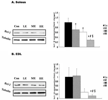
운동강도별 Soleus와 EDL에서 Bcl-2 단백질 변화
운동강도별 8주간 트레이닝 후 Bcl-2의 발현은 Soleus근에 서 대조군과 비교하여 저강도(p=0.010), 중강도(p=0.002), 고강 도(p=0.000) 운동군에서 모두 유의하게 낮았다(p<0.05). 또한 고강도 운동군과 비교하여 저강도(p=0.001)와 중강도 운동군 (p=0.006)에서 유의한 차이로 낮게 나타났다(p<0.05). 그러나 저강도와 중강도 운동군 사이에는 유의한 차이가 없었다(Fig.
1A). EDL근의 Bcl-2의 발현은 대조군과 비교하여 고강도 (p=0.034)운동군에서만 유의하게 낮았다(p<0.05). 또한 저강도 운동군과 비교하여 고강도(p=0.023) 운동군에서 유의한 차이 를 나타냈다(p<0.05). 그러나 대조군과 저강도, 중강도 운동군 사이에는 유의한 차이가 없었다(Fig. 1B).
운동강도별 Soleus와 EDL에서 Bax 단백질 변화
운동강도별 8주간 트레이닝 후 Bax의 발현은 Soleus근에서
Fig. 1. Bcl-2 protein expression by exercise in- tensity in Soleus (A) and EDL (B) muscle. Bcl-2 protein expression was determined by Western blot analysis.
The inset shows are representative blot for bcl-2 in Soleus and EDL muscles isolated from CON, LE, ME and HE animals. The data are presented as means±SE. CON, sedentary control;
LE, low intensity exercise trained; ME, moderate intensity exercise trained;
HE, high intensity exercise trained.
*
Significantly different from sedentary control (CON) muscles (p<0.05).
#
Significantly different from low in- tensity exercise trained (LE) muscles (p<0.05).
$Significantly different from moderate intensity exercise trained (ME) muscles (p<0.05).
대조군과 비교하여 고강도(p=0.045)에서 유의하게 높았다 (p<0.05). 또 저강도(p=0.049), 중강도(p=0.027 운동군과의 비 교에서도 고강도 운동군(p=0.049)이 유의하게 높았다. 그러나 대조군, 저강도, 중강도 운동군 간의 유의한 차이는 없다(Fig.
2A). EDL근에서는 대조군과 비교하여 중강도(p=0.035), 고강 도(p=0.003) 운동군은 유의하게 높게 나타났다(p<0.05). 그러 나 대조군과 저강도 운동군, 저강도 운동군과 중강도, 고강도 운동군, 중강도와 고강도 운동군 간의 유의한 차이는 없었다 (Fig. 2B).
운동강도별 Soleus와 EDL에서 caspase-3 단백질 변화 Fig. 3에서 보는 바와 같이 고강도 운동군(HE)의 Soleus와 EDL 근에서 모두 caspase-3의 활성화가 이루어짐을 관찰할 수 있었다. 그러나 대조군(Con)과 저강도(LE), 중강도 운동군 (ME)에서는 두 근섬유 유형에서 모두 caspase-3의 활성화를 관찰할 수 없었다.
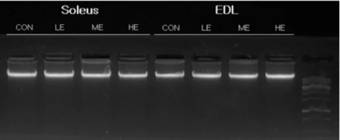
운동강도별 Soleus와 EDL에서 DNA Fragmentation
운동강도에 따른 apoptosis를 관찰하기 위한 일반적인 생화 학적 지표인 gel electrohoresis에 의한 DNA 절편화 분석을 실시한 결과, 모든 그룹과 근유형에서 절편화 현상은 나타나 지 않았다(Fig. 4).
고 찰
60 주령된 고령쥐를 대상으로 8주간 저, 중, 고 강도 운동을
Fig. 2. Bax protein expression by exercise intensity in Soleus (A) and EDL (B) muscle. The inset shows are representa- tive blot for Bax protein in Soleus and EDL muscles iso- lated from CON, LE, ME and HE animals. The data are presented as means±SE. CON, sedentary control; LE, low intensity exercise trained; ME, moderate intensity exercise trained; HE, high intensity exercise trained.
*
Significantly different from sedentary control (CON) mus-
cles (p<0.05).
#Significantly different from low intensity ex-
ercise trained (LE) muscles (p<0.05).
$Significantly different
from moderate intensity exercise trained (ME) muscles
(p<0.05).
Fig. 3. Caspase-3 protein expression by exercise intensity in sol- eus and EDL muscle fibers. Caspase-3 protein expression was determined by Western blot analysis. The inset shows are representative blot for caspase-3 protein in sol- eus and EDL muscles isolated from CON, LE, ME and HE animals.
Fig. 4. DNA fragmentation. Qualitative analysis of DNA frag- mentation by DNA gel electrophoresis in soleus and EDL muscles of CON, LE, ME and HE animals. DNA was extracted from soleus and EDL muscles, and DNA was loaded on 2% agarose gel.
실시하여 apoptosis관련 단백질의 변화를 검토하였다.
모든 운동강도에서 8주간의 규칙적인 운동수행은 노후된 근육에서 운동강도가 증가할수록 caspase-3가 높아지는 경향 은 나타났으나 apoptosis는 관찰되지 않았다. Koctürk 등[17]
은 24 주령된 수컷 wistar albino rat을 대상으로 경사도 5 도에 서 25 m/min의 속도로 탈진시까지 수행한 결과 대조군에 비 해 운동직후뿐만 아니라 회복기 48 hr 후에도 apoptosis가 유 의하게 증가하는 것으로 보고하였다. 이러한 결과는 apopto- sis를 유도하기 위해 탈진시까지 인위적으로 유도한 것으로 본 연구에서 적용한 고강도 운동방법은 24 m/min 속도로 9분 간 지속한 방법으로 고강도에서 apoptosis가 유도될 수 있는 환경의 지속 또는 역치 이상에 노출되지 않았기 때문에 DNA 절편화는 나타나지 않은 것으로 판단된다. 그러므로 트레드밀 운동에서 근육세포의 apoptosis는 운동의 지속 시간과 빈도에 의존적일 것으로 사료된다. Soleus에서 Bcl-2 발현은 대조군과 비교하여 운동강도가 높아질수록 발현이 모두 유의하게 감소 하였고 Bax의 발현은 고강도에서만 유의하게 증가하였다.
EDL에서 Bcl-2는 고강도에서만 유의하게 감소하였다. Bax는 중강도와 고강도에서 유의하게 증가한 것을 발견할 수 있었 다. 즉 고강도의 운동은 노화된 쥐의 근조직의 apoptosis 유발 단백질의 활성을 촉진시키고 억제단백질 발현의 저하는, 선행 연구에서 고강도 운동에 의해 골격근의 apoptosis가 유발된다
는 연구결과와 일치한다[20,24]. 그 원인은 근형질세망의 Ca
2+조절의 실패로 인한 ER stress와 유리기의 증가, 글리코켄의 감소, 체온의 상승 등이 유발되며 이러한 변화는 세포질과 미 토콘드리아의 안정성을 저하시키고 단백질의 변성을 일으킬 뿐만 아니라 세포 사멸을 경유한 근섬유 손상을 유발하기 때 문이라고 제안하였다[14,32]. 본 연구에서 운동강도가 높아짐 에 따라 soleus와 EDL에서 Bax의 발현양은 증가했고, Bcl-2의 양은 감소하는 것을 관찰할 수 있었다. 그러나 apoptosis의 최종 수행결정자인 caspase-3의 활성이 고강도에서만 일어났 으며 저강도와 중강도에서는 활성화 되지 않았음을 관찰할 수 있었다. 이는 저강도와 중강도에서는 apoptosis를 최종적 으로 실행될 만큼의 발현이 일어나지 않았기 때문이다. 또한 두 가지 타입의 근육에서 동일한 결과를 나타내었다. 더욱이 DNA절편화 현상은 모든 그룹에서 나타나지 않았다. 규칙적 인 고강도 운동 수행 시 Soleus와 EDL에서 caspase-3의 활성 화가 관찰되었으나, apoptosis를 유도할 만큼의 자극이 크지 않았던 것으로 판단된다. 이는 훈련 형태에는 차이가 있으나 Sandri 등[25]이 고강도 운동에 참여한 영양실조에 걸린 실험 쥐의 골격근에서 Bcl-2는 감소하였으나 Bad의 발현이 증가되 었다는 연구 결과와 일치한다[24]. 그러나 개체의 생체내 환경 에 따라 노후된 근육에서 고강도의 운동은 잠재적인 위험성을 가지고 있다. 이러한 현상은 격렬한 운동이 Glucocorticoid (GO) 분비, 세포 내 Ca
2+, reactive oxygen specise (ROS) 등이 증가하며 gluthatione 감소, thiol oxidative, DNA 손상, 저산 소(hypoxia) 등이 다양한 신호를 자극하여 잠재적인 apopto- sis를 유발한다는 이론에서 그 원인을 찾을 수 있을 것이다 [7,21,29,33].
그러나 최근 12주간 트레드밀 운동을 시킨 늙은 쥐의 white gastrocnemius와 Soleus 근에서 DNA분절, cleaved caspase-3, Bax, Bax/Bcl-2 비율이 현저하게 감소하는 동안 Bcl-2는 증가 되었다고 보고하였고[30], Siu 등은 8 주간 주 5회 젊은 쥐를 대상으로 트레드밀을 이용한 중강도의 지구성 운동을 실시한 결과 운동을 시키지 않은 쥐의 집단과 비교하였을 때 운동한 집단의 Soleus에서 Bax mRNA는 감소하였고, Bcl-2 단백질의 발현의 증가를 보고하였다[26]. 그러나 본 연구결과는 대조군 과 비교했을 때 저강도와 중강도 운동집단의 Soleus에서 Bax 의 발현은 유의한 차이가 없었으나 Bcl-2의 경우 두 집단 모두 유의하게 감소되었다. 이러한 apoptosis 관련 단백질의 발현 에도 불구하고 caspase-3의 활성화는 이루어지지 않았다. 이는 아마도 Bax나 Bcl-2가 apoptosis를 일으킬 만큼 발현량이 충분 하지 않았던 것으로 사료된다.
골격근에서 노화는 근세포, 특히 TypeⅡ 근섬유에서의 횡
단면적의 감소와 근세포의 손실로 특정지어진다. 골격근에서
의 apoptosis는 근위축 뿐 아니라 근섬유의 손실에도 영향을
미친다. 최근의 연구는 미토콘드리아의 Bcl-2 family 경로가
노화의 핵심이라고 제안한다. 노화가 진행됨에 따라 쥐의 골
격근에서 DNA분절, cleaved caspase-3, pro-apoptosis가 증가
된다는 것을 입증하였다[30]. Soleus 근은 현저하게 typeⅠ 근
섬유로 구성되어 있으며 많은 양의 미토콘드리아를 가지고 있는 반면 gastronemius 근은 원래 typeⅡ 섬유로 구성되어 있으며 적은 양의 미토콘드리아를 가지고 있다. 따라서 이 근 육들은 미토콘드리아의 양의 차이와 약 3-4배의 산화대사량의 차이로 두 개의 중요한 근섬유 형태를 설명하였고[2,22], 최근 연구에서는 노화된 쥐의 plantaris와 gastrocnemius에서 apoptosis의 조절에 대한 연구에서 apoptosis의 기전이 근육 의 특수성일지도 모른다고 제안하였다[1,19].
Rice 등[23]은 노화가 진행됨에 따라 근타입별로 apoptosis 조절자의 발현이 다르게 타나난다는 것을 증명하였다. 예를 들어 EDL에서 Bax는 노화가 진행됨에 따라 증가하지만 Soleus 에서 Bax는 변화하지 않았다. 또 EDL은 30개월과 36개월에 Bcl-2가 유의하게 증가되었고 Soleus에서는 변화가 없었다[23].
이 실험에서 중강도 운동수행 시에 대조군과 비교하여 EDL 에서 Bax는 증가하였고 Soleus에서는 변화하지 않았다. Bcl-2 의 경우는 운동강도에 따라 변화하지 않았음을 관찰할 수 있 었다. 이러한 결과는 노후된 근육에서 EDL의 pro-apoptosis의 발현이 Soleus보다 더 쉽게 발현됨을 시사한다. 이는 TypeⅠ 섬유에 비해 상대적으로 적은 양의 미토콘드리아 수와 산화대 사량으로 인하여 운동과 같은 외부 자극으로부터 apoptosis 유발단백질의 유도가 빠르게 반응한 것으로 사료된다. 그러나 apoptosis를 일으킬 만큼의 발현은 되지는 않았다.
본 실험에 사용된 60 주령의 SD계열의 쥐에서 caspase-3의 발현은 근섬유 타입별로 비운동군을 비교했을 때 선행연구의 결과와 마찬가지로 TypeⅡ 근섬유인 EDL에서 활성화가 많이 이루어짐을 확인할 수 있었으나 유의성은 나타나지 않았다 (Fig. 3). 이는 노화된 근육에서의 apoptosis는 근육 타입에 따 라 다르게 나타나며 특히 typeⅡ (fast twich)의 근육세포들은 typeⅠ (slow twich) 근육세포에 비해 apoptosis 현상에 더욱 민감하기 때문이다[20].
그러나 장기간 규칙적인 고강도 운동을 수행했을 때 EDL과 Soleus에서 근섬유 유형에 관계없이 cleaved caspase-3가 관찰 되었으나 DNA 절편화는 일어나지 않았다. typeⅠ섬유 (Soleus)는 typeⅡ근에 비하여 다량의 미토콘드리아를 가지고 산화 대사를 하게 되는데[2,22] 대부분 산소스트레스가 증가됨 에 의해 apoptosis가 이루어지는 것 같다고 보고한다[17]. 노화 된 EDL은 이미 기능이 약화되어 있는 상태에서 장기간 고강 도 운동 수행으로 노화된 개체의 apoptosis를 더욱 유발시킨 것으로 사료된다. 또한 고강도의 운동으로 유발된 근 손상은 대개 근섬유의 기계적 과부화로 시작된다. 즉 근섬유와 기저막 과 관련된 복합적인 반응의 결과이다. apoptosis의 유발기전 중 하나는 유리기(free radical)에 의한 손상일 것이다[16,17].
감사의 글
이 논문은 2009 년도 정부(교육과학기술부)의 재원으로 한 국연구재단의 지원을 받아 수행된 기초연구사업임(No.
2009-0077015).
References