성상교세포종에서 Apoptosis와 Bcl-2 발현 *
연세대학교 원주의과대학 신경외과학교실, 해부병리학교실
**장연규·황 금·홍순원
**
= Abstract =
Apoptosis and Bcl-2 in Astrocytic Tumors
Yeon Gyoe Jang, M.D., Kum Whang, M.D., Soon-Won Hong, M.D.**
Department of Neurosurgery and Pathology,** Wonju College of Medicine, Yonsei University, Wonju, Korea
bjective:To study the expression of apoptosis and bcl-2 in the astrocytic tumors.
Patients and Methods:A total of thirty-eight astrocytomas(9 cases in low grade astrocytoma, 12 cases in anaplastic astrocytoma and 17 cases in glioblastoma) are included in this study. Immunohistochemical stain for bcl-2 using monoclonal antibody, in situ end labelling technique for apoptosis were used.
Results:The malignant group(anaplastic astrocytoma and glioblastoma) showed significantly higher apoptosis positive index(PI) compared to the benign group(low grade astrocytoma)(1.35 vs 0.14). However apoptosis PI and bcl-2 PI were not significantly different among three groups. Correlation between apoptosis PI and bcl-2 PI was not statistically significant(p=0.58).
Conclusion:This result suggest that apoptosis PI and bcl-2 PI are not related the degree of malignancy in astrocytic neoplasm, but apoptosis PI in malignant group was higher possibly due to greater DNA damage.
KEY WORDS:Astrocytic neoplasm・bcl-2・Apoptosis・Immunohistochemical stain.
서 론
종양의 성장을 결정짓는 데는 두 가지 요소가 있는데, 우 선은 세포의 증식이며, 다른 면은 세포의 사멸이다. 교세포종 에서 세포의 증식은 다양한 면에서 연구되고 있는데 대표적 으로 p53 유전자의 변이
5)29), bcl-2 원시종양유전자(pr- otooncogene)의 발현
8), PCNA(proliferating cell nuclear antigen)발현
14)28)등이 관련 있다고 알려져 있다. 세포의 사멸과 종양의 성장과의 관계는 잘 알려져 있지 않으나 ap- optosis와의 관계가 여러 저자들에 의해 알려지기 시작하였다.
Apoptosis(programmed cell death)는 태생학적 발생 및 생리학적 과정으로 발생하며, 종양발생에서 나타나기도
한다
7)33). Apoptotic세포의 사멸은 괴사(necrosis)와는 달리,
특징적인 세포내 현상이 나타나는데, 세포질의 수축(shrin-
kage)과 nucleosomesize(50~300kb) 분절 내에서 특이 한 endonuclease에 의해 DNA의 분활이 야기되며
5), 이것 이 apoptosis의 생화학적 특이성이다. 최근 보고에 의하면 p53 유전자의 역할이 손상 받은 세포를 G
1기에서 정지시키 며, apoptosis를 유도한다고 알려져 있다
5)29).
반면에 원시종양유전자, bcl-2의 산물인 26-kd 단백은 apoptosis를 억제하는 것으로 세포 주기중 G
0/G
1기에 세포 를 정지시켜 오랫동안 생존케 한다
8).
bcl-2 종양단백이 첫 번째 알려진 것은 t(14;18) 전이 가 B-cell 백혈병과 여포성 림프종(follicular lymphoma) 에서 있었다
30). 이후 점차 비혈액성 악성종양에서도 발견되 어 현재 전립선
6), 직장
21), 인후부
11), 유방
13), 폐종양
2)및 흑 색종양
31)등에서 보고되고 있다. 또한 bcl-2의 발현이 신 경모세포종, 수아세포종, 교모세포종 같은 악성 신경세포계 에서 발견되었다
26).
또한 최근 연구에 의하면 bcl-2 발현이 뇌종양 조직 중 신경교종 및 수아세포종에서 다양하게 발현됨이 보고되고
OOOO
*본 논문은 1997년도 연세대학교 학술연구비에 의해 이루어졌음.
있다
23).
저자는 성상교세포종의 증식과 사멸에 관계된 인자 중 증 식에 관계된 bcl-2 유전자의 산물인 종양 단백의 발현과 사멸에 관계된 apoptosis의 상관관계를 통해서 성상교세포 종의 악성도와 어떤 연관성이 있는지 알아보고자 하였다.
대상 및 방법
1. 대 상
1986년 1월부터 1997년 12월까지 본원 신경외과에서 수술적 처치를 하였던 성상교세포종 환자에서 후향적으로 추적조사가 가능하였던 38예를 대상으로 하였고, 저등급 성 상세포종(low grade astrocytoma) 9예, 역행성 성상세포 종(anaplastic astrocytoma) 12예, 교모세포종(glioblast- oma)은 17예가 있었다(WHO Classification). 악성과 양성 종양의 구별은 저등급 성상세포종을 양성 종양군으로, 역행 성 성상세포종 및 교모세포종을 악성종양군으로 분류하였다.
2. 방 법
1) Apoptosis, bcl-2 면역조직화학염색
수술시 적출된 조직을 이용하여, 연속절편의 일부는 Apo- pTagTM In situ apoptosis detection kit(Oncor, Gai-
thersburg, MD, USA)로 TUNEL(terminal deoxynucl- eotidyl transferase dUTP Nick End Labeling) assay를 하여 apoptosis 여부를 검색하였다. 연속 절편이 도말된 sl- ide를 trypsin(1g/L, Sigma Chemical Co., St. Louis, MO, USA)으로 37℃에서 30분간 반응시키고, terminal deo- xynucelotidyl transferase(TdT)를 처리한 후 37℃에서 1시간 반응시키고 반응 정지 및 수세 완충 용액을 첨가하여 37℃에서 30분간 방치하고 반응을 정지시켰다. 그후 pe- roxidase conjugated anti-Digoxigenin Antibody를 처리 하고 실온에 30분간 방치하고 3, 3'-diaminobenzidine(Zy- med Laboratoried Inc., South San Francisco, CA, USA) 으로 발색시킨 후 methylgreen으로 대조염색후 검경하였다.
bcl-2는 면역조직화학 염색을 약간 수정하여 microwave oven에서 98℃로 10분간 끓이고 식힌 후 증류수로 수세하 고, Bcl-2 antibody를 처리하고 4℃에 하룻밤동안 반응시 킨 후, biotinylated anti-mouse IgG, biotinylated anti- rabbit IgG(DAKO Corp., Carpinteria, CA, USA)와 1시간, peroxidase conjugated streptavidin(DAKO Corp., Car- pinteria, CA, USA)에 1시간 동안 차례로 반응시키고 PBS 로 세척한 후 3-amino-9-ethylcarbazole로 발색시키고 hematoxylin으로 대조 염색하여 광학 현미경하에서 검색하 였다.
2) 양성지수(positive index:PI)
400배의 고배율 광학현미경 시야에서 염색이 비교적 잘 된 부위를 5군데 이상 선택하여 최소한 1,000개이상의 종 양세포의 핵 또는 세포질의 숫자를 세어 전체 종양세포수중 갈색으로 염색된 세포핵 또는 세포질수의 비율을 구하였고 그 백분율을 양성 지수(positive index)로 정의하였다.
Table 1. Apoptosis PI(positive index) according to the WHO astrocytoma classification
Tumor group No. of
positive case Min-max Mean±SD Astrocytoma 3/ 9(33%) 0-1.8 0.41±0.72 Anaplastic astrocytoma 6/12(50%) 0-9.1 1.41±2.60 Glioblastoma 11/17(64.7%) 0-6.1 1.31±1.82 SD:standard deviation, Min:minimum, Max:maximum
Fig. 1. Immunohistochemical staining photomicrographs of low grade astrocytoma showing higher(A) apoptosis PI(1.8) as compared to(B) bcl-2 PI(0).
A A A
A B B B B
3) 통계분석
세 군간의 apoptosis, bcl-2 염색 차이의 검증을 위하여 Oneway Anova test를 사용하였고, apoptosis, bcl-2의 상 관관계는 Pearson correlation과 Linear regression을, 양 성군과 악성군 종양 사이의 관계는 Student T test를 사용 하였다.
결 과
1. 성상세포종의 WHO분류에 따른 apoptosis 양성지수(PI) Apoptosis 면역조직화학염색에서 저등급 성상세포종군의 경우 33.3%의 양성반응을 보였고, 평균 양성 지수는 0.41 (0~1.8)이었으며, 역행성 성상세포종군은 50%의 양성반 응을 보였고, 평균 양성 지수는 1.41(0~9.1)이고, 교모세 포종군은 64.7%의 양성반응을 보였고, 평균 양성 지수는 1.31(0~6.1)이었다. 세 군간에 통계학적으로 의미 있는 차
이가 없었다(p=0.449)(Table 1, Fig. 1, 2, 3).
2. 성상세포종의 WHO분류에 따른 bcl-2 양성지수(PI) bcl-2 면역조직화학염색에서 저등급 성상세포종군의 경 우 33.3%의 양성반응을 보였고, 평균 양성 지수는 2.81(0
~12.6)이었으며, 역행성 성상세포종군은 91.7%의 양성반 응을 보였고, 평균 양성 지수는 4.58(0~11.2)이고, 교모세포 종군은 58.8%의 양성반응을 보였고, 평균 양성 지수는 4.71 (0~44.8)이었다. 세 군간에 통계학적으로 의미 있는 차이
Table 2. Bcl-2 PI(positive index) according to the WHO as- trocytoma classification
Tumor group No. of
positive case Min-max Mean±SD Astrocytoma 3/ 9(33%) 0-12.6 2.81± 4.73 Anaplastic astrocytoma 11/12(91.7%) 0-11.2 4.58± 3.87 Glioblastoma 10/17(58.8%) 0-44.8 4.71±10.94 SD:standard deviation, Min:minimum, Max:maximum p=0.834
Fig. 2. Immunohistochemical staining photomicrographs of anaplastic astrocytoma showing higher(A)apoptosis PI(1.0) as compared to(B) bcl-2 PI(0.6).
Fig. 3. Immunohistochemical staining photomicrographs of glioblastoma showing higher(B) bcl-2PI(44.8) as compared to(A) apoptosis PI(0.2).
A A A
A B B B B
A A A
A B B B B
가 없었다(p=0.834)(Table 2, Fig. 1, 2, 3).
3. 양성 성상세포종군과 악성 성상세포종군간의 apopto- sis 양성지수(PI) 차이 및 bcl-2 양성지수(PI) 차이 Apoptosis염색은 양성 성상세포종군의 평균 양성지수는 0.41이고, 악성 성상세포종군의 평균 양성지수는 1.35였으 며 두 군간의 통계학적 유의성이 있었다(p<0.05)(Table 3).
bcl-2염색은 양성 성상세포종군의 평균 양성지수는 2.81이 고, 악성 성상세포종군의 평균 양성지수는 4.66이었으며 두 군간의 통계학적 유의성이 없었다(p=0.42)(Table 4).
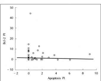
4. Apoptosis와 bcl-2와의 상관관계
연구대상 전체 군에서 apoptosis와 bcl-2와의 상관관계 를 관찰한 결과 apoptosis와 bcl-2 사이에는 통계학적으로 의미 있는 상관관계가 없었다(p=0.58)(Fig. 4).
고 찰
1974년에 Lockshin 등
20)이 특이한 호르몬 및 신경의 신 호들에 의해 metamorphosis에 삽입된 세포의 자연사를
apoptosis라고 명명하였다. apoptosis는 능동적이고 계획된 세포사로 정상세포에서 생체의 항상성을 유지하기 위해 일 어나며 후천성 면역결핍증 바이러스에 의한 뇌염 및 치료받 지 않은 악성종양에서도 자연적으로 발생할 수 있는 것으로 보고되고 있다
9)17)24). 또한 모든 뇌종양에서 일어날 수 있으 며, 그 외 방사선, 항암치료제, 호르몬, 세포독성 T 임파구, mild hyperthermia(43℃ for 30min), APO-1/Fas 항원 에 대한 항체나 protein kinase C inhibitor에 의해서도 유 도된다고 보고되고 있다
17). 또한 촉진요소로 growth factor deprivation, glucocorticoid, retinoid, cytotoxic chemic- als, toxins, physical damage, cytokines 등도 알려져 있다
1). 감지방법으로는 광학현미경검사, 전자현미경검사, 전기영동 법 및 in-situ end labeling 방법이 있으며 그중 전자현미경 검사가 현재까지는 가장 신뢰적인 방법으로 여겨지고 있다.
그러나 전자현미경검사를 위해서는 신선한 조직절편이 필 요하며 정량분석은 어렵다
16). 본 연구에서 사용한 방법인 in-situ end labeling technique은 1995년 Gavrieli 등
3)에 의해 TUNEL(terminal deoxynucleotidyl transferase- mediated dUTP-biotin nick end labeling) method로 처 음 소개되었는데, 방법이 쉬우며 파라핀 조직에서의 검출이 가능하여 후향적 검사가 가능하고 정량분석이 가능하다는 장 점이 있는 반면 괴사세포에 의한 위양성을 완전히 배제할 수 없다는 것과 조직의 고정과정 중 생길 수 있는 DNA 손상이 정확한 결과분석에 영향을 줄 수 있다는 단점이 있다
15)32). 1995년 Nakagawa 등
22)이 62명의 뇌종양 환자의 종양조직 에서 종양세포의 증식속도가 빠를수록 apoptosis index도 높 은 경향을 보인다고 하였다. 그러나 1995년 Schiffer 등
27)은 신경상피종양에서 apoptotic index와 세포증식능 사이에 통 계학적 유의성이 없다고 보고하였다.
본 연구에서는 연구대상 전체군에서 53%(20/38)의 양 성율을 보였고 apoptosis의 양성지수는 0에서 9.1의 범위 를 보였다. 종양세포의 증식지표에 대한 연구는 동반되지 않았으나 종양의 악성도가 증가할수록 apoptosis의 양성지 수가 의미 있게 증가되지는 않았다. 그러나 조직학적 악성 도가 심해질수록 양성반응의 백분율이 증가함을 볼 수 있어 Schiffer 등과 같은 결과를 얻었다. 그러나 양성종양과 악성 종양군으로 나눴을 때의 apoptosis 양성지수는 통계학적으 로 의미 있는 차이가 있고, 악성종양군에서 현저히 높음을 알 수 있었다.
bcl-2 원시종양유전자는 염색체(chromosome) 18의 long arm에 위치하여 26-KD의 단백을 합성하며 이것이 mit- ochondria의 내측막, perinuclear막 및 endoplasmic reti- culum에 위치하여
21)23), apoptotic cell death의 potential
Table 4. Bcl-2 PI in benign and malignant groups
Tumor group No. of positive Min-max Mean±SD Benign 3/ 9(33.3%) 0-12.6 2.81±4.73 Malignant 21/29(72.4) 0-44.8 4.66±8.62 SD:standard deviation, Min:minimum, Max:maximum p=0.42
Table 3. Apoptosis PI in benign and malignant groups Tumor group No. of positive Min-max Mean±SD Benign 3/ 9(33.3%) 0-1.8 0.41±0.72 Malignant 17/29(58.6) 0-9.1 1.35±2.13 SD:standard deviation, Min:minimum, Max:maximum p<0.05
Fig. 4. Linear regression graph of correlation between bcl-2
PI and apoptosis PI(p=0.58)
blocker로서 apoptosis 최종단계에 작용하고 종양세포를 세포주기의 G0/G1 phase에 정지시키거나 APO-1/Fas 항 체를 통한 apoptosis를 막아 생존을 연장시키는 것으로 알 려져 있다
8). 또한 bcl-2 family로 bcl-x, box,mcl-1과 A1 등도 apoptosis에 관여하는 것으로 알려져 있다
19). 일 부 저자에 의하면 bcl-2 단백은 태생기 동안에 신경기원을 포함한 체세포에 널리 발현된다고 하며, 성숙한 조직 내에 서는 활발히 분화하는 세포에서 강력하게 발현되는데 이것 은 조직의 morphogenesis와 clonal selection에 작용한다 고 한다
21).
면역조직화학염색상 bcl-2 유전자의 발현정도가 신경교 종의 경우 조직학적 악성도가 높은 종양에서 낮은 종양에 비해 더 높다는 보고가 있는 반면, 종양의 조직학적 악성도 는 관계가 없으며 반응성 성상세포(reactive astrocyte)에 서도 발현된다는 보고도 있다
12)19). 또한 정상세포에서는 발 현이 없으며 악성화될 때 작용한다는 보고도 있다
12). Kri- shna 등
26)에 의하면 성상교 세포에서 bcl-2가 악성종양 발생에 유일한 방편은 아니며, 중추 신경계 손상에 대한 반 응으로 bcl-2가 발현되기도 한다. bcl-2의 과잉 발현은 성 장인자 결핍에 의한 apoptosis 및 화학적 요법 치료나 방사 선에 의한 apoptosis를 억제시키고, 저등급 성상세포종 및 반응성 성상세포에서는 기능적으로 세포사(cell death)에 저항적이라 하였다. 본 연구결과에서는 bcl-2 유전자의 발 현정도가 종양의 조직학적 악성도와 통계학적으로 의미 있 는 차이가 없었지만 악성군으로 갈수록 bcl-2 발현이 증가 하는 것을 볼 수 있었다.
최근에는 bcl-2 family중 bax, bad, bak, bcl-X-s는 apoptosis를 촉진하는 쪽으로, bcl-2, bcl-X-
L은 억제하는 쪽으로 작용하며 그 중 특히 bax:bcl-2 비율이 더 중요한 것 으로 보고되고 있으며, 일부 종양에서는 bcl-2유전자의 발현 이 많을수록 약물치료에 반응이 낮고 재발기간이 짧으며 생존 기간이 짧아 임상적 경과가 나쁜 것으로 알려져 있어 뇌종양의 예후 판정에 의의가 있음을 알 수 있다는 보고도 있으므로 추 후 bcl-2 family에 대한 연구가 진행되어야 하겠다
4)10)25).
김 등
18)의 보고에 의하면, p53변이와 apoptosis사이에는 의미 있는 양적 상관관계(positive correlation)가 있으나 bcl-2 유전자와 apoptosis사이에는 의미 있는 상관관계가 없었다 한다. 본 연구에서도 bcl-2 유전자와 apoptosis사 이에는 의미 있는 상관관계가 없었다.
결 론
원시종양유전자이며 apoptosis의 최종단계에서 blocker로
작용하는 bcl-2는 면역조직화학염색상 성상세포종의 WHO 분류에 의한 3군간에 통계학적 유의성은 없었으며, 양성군 (평균양성지수=2.81)에 비해 악성군(평균양성지수=4.65) 에서 현저히 높았으나 통계학적 유의성은 없었다(p=0.42).
Apoptosis의 면역조직화학적 염색상 WHO분류에 의한 grade II, III, IV의 3군간에 통계학적 유의성은 없었으며, 양성군(평균양성지수=0.41)에 비해 악성군(평균양성지수
=1.35)에서 현저히 높았으며 통계학적 유의성이 있었다 (p<0.05). 또한 bcl-2 유전자의 발현정도와 apoptosis 사 이에는 의미 있는 상관관계는 없었다.
•
논문접수일:1999년 11월 15일•
심사완료일:2000년 1월 14일•
책임저자:황 금220-070 강원도 원주시 일산동 162 연세대학교 원주의과대학 신경외과학교실 전화:033) 741-1330, 전송:033) 746-2287 E-mail:[email protected]
References
1) Ashkenazi A, Dixit VM:Death receptors:signaling and
modulation. Science 28
;281
(5381
):1305-8, 1998
2) Ben-Ezra JM, Kornstein MJ, Grimes MM, et al:Small cell
carcinomas of the lung express the bcl-2 protein. Am J Pathol 145
:1036-1040, 1994
3) Ben-Sasson SA, Sherman Y, Gavrieli Y:Identification of dying
cells-In situ staining, in Schwartz LM
:Cell death., Osborne BA
(eds
):Academic Press, pp29-40, 1995
4) Chresta CM, Masters JRW, Hickman JA:Hypersensitivity of
human testicular tumors to Etoposide-induced apoptosis is associated with functional p53 and a high Bax
:Bcl-2 ratio.
Cancer Res 56
:1834-1841, 1996
5) Clarke AR, Purdic CA, Harrison DJ, et al:Thymocyte apop-
tosis induced by p53 dependent and independent pathways.
Nature 362
:849-852, 1993
6) Colombel M, Symmans F, Gil S, et al:Detection of the
apoptosis-suppressing oncoprotein bel-2 inn hormone refrac- tory human prostate cancer AM J Pathol 143
:390-400, 1993
7) Cotter TG, Lennon SV, Glynn JG, et al:Cell death via apo-ptosis and its relationship to growth, development and differ- entiation of both tumour and normal cells. Anticancer Res 10
:1153-1159, 1990
8) de Jong D, Prins FA, Mason DY, et al:Subcellular local-
ization of the bcl-2 protein in malignant and normal lymphoid cells. Cancer Res 54
:256-260, 1994
9) Dickson DW:Apoptosis in the brain physiology and path-
ology Am J Pathol 146
(5
):1040, 1995
10) Gillardon F Wickert H, Zimmermann M:Up-regulation of
bax and own-regulation of bcl-2 is associated with kainate
induced apoptosis in mouse brain. Neurosci Letters 192
:85- 88, 1995
11) Hague A, Moorghen M, Hicks D, et al:Bcl-2 expression in
human colorectal adenomas and carcinomas. Oncogene 9
:3367-3370, 1994
12) Ikeda H, Hirato J, Akami M, et al:Bcl-2 oncoprotein ex-
pression and apoptosis in neuroblastoma. J Pediatr Surg 30
(6
):805-808, 1995
13) Joensuu H. Pylkknen L, Toikkanen S:Bcl-2 protein expre-
ssion and long-term survival in breast cancer. Am J Pathol 145
:1191-1198, 1994
14) Karamitoopoulou E, Perentes E, Melachrinou M, et al:Pr-
oliferating cell nuclear antingen immunoreactivity in human central nervous system neoplasms. Acta Neuropathol 85
:316-322, 1993
15) Kasagi N, Gomyo Y, Shirai H, et al:Apoptotic cell death in
human gastric carcinoma
:Analysis by terminal deoxymucle- otidyl transferase-mediated dUTP-biotin nickend labeling. Jpn J Cancer Res 85
:939-945, 1994
16) Kerr JFR, Gobe GC, Winterford CM, et al:Anatomical
methods in cell death, in Schwartz LM
:Cell death. San Diego, Osborne BA
(eds
):Academic Press, pp1-27, 1995
17) Kerr JFR, Winterford CM, Harmon BV:Apoptosis. Its sig-
nificance in cancer and cancer therpy. Cacer 73
(8
):2013- 2026, 1994
18) Kim SH, Cho KG, Yoon SH, et al:Relationship between p53,
Bcl-2, Apoptosis and Histologic Grade of Brain tumors. J Kor Neurosur Soc 26
:40-47, 1997
19) Krishna M, Smith TW, Echt LD:Expression on bcl-2 in
reactive and neoplastic astrocytes
:Lack on correlation with presence or degree of malignancy. J Neurosurg 83
:1017- 1022, 1995
20) Lockshin RA, Beaulaton J:Programmed cell death. Life-Sci
15
(9
):1549-65, 1974
21) Lu QL, Elia G, Lucas S, et al:Bcl-2 proto-oncogene expre-
ssion in Epstein-Barr-virus-associated nasopharyngeal car- cinoma. Int J Cancer 53
:29-35, 1993
22) Nakagawa S, Shiraishi T, Kihara S, et al:Detection of DNA
strand breaks associated with apoptosis in human brain tuors.
Virchows Arch 427
:175-179, 1995
23) Nakasu S, Nakasu Y, Nioka H, et al:bcl-2 protein expression
in tumors of the central nervous system. Acta Neuropathol 88
:520-526, 1994
24) Petito CK Roberts B:Evidence of apoptotic cell death in
HIV encephalitis Am J Pathol 146
(5
):1121-1128, 1995
25) Reed JC:Regulation of apoptosis by bcl-2 family proteinsand its role in cancer and chemoresistance. Oncolgy 7
:541- 546, 1995
26) Reed JC, Meister L, Tanaka S, et al:Differential expression
of bcl-2 protooncogene in neuroblastoma and other human tumor cell lines of neural origin Cancer Res 51
:6529-6538, 1991
27) Schiffer D, Cavalla P, Migheli A, et al:Apoptosis and cell
proliferation in human neuroepithelial tumors. Neurosci letters 195
:81-84, 1995
28) Schiffer D, Chio A, Giordana MT, Pezzulo T, Vighani MC:
Proliferating cell nuclear antigen expression in brain tumors, and its prognostic role in ependymomas
:an immunohis-to- chemical study. Acta neuropathol 85
:495-502, 1993
29) Symonds H, Krall L, Remington L, et al:p53 dependent ap-optosis suppresses tumor growthand progression in vivo. Cell 78
:703-711, 1994
30) Tsujimoto Y, Croce CM:Analysis of the structure, transcr-
ipts, and protein products of bcl-2, the gene involved in human follicular lymphoma. Proc Natl Acad Sci USA 83
:5214-5218.
1986
31) van den Oord JJ, vandeghinste N, Del Ley M, et al:Bcl-2
expression in human melanocytes and melanocytic tumors. Am J Pathol 145
:294-300, 1994
32) Wijsman JH, Jonker RR, Keijzer R, et al:A new method to
detect apoptosis in paraffin sections
:In situ end-labeling on fragmented DNA. J Histo Cyto 41
(1
):7-12, 1993
33) Wyllic AH:The biology of cell death in tumours. Anticancer