297
Outcomes of open versus closed treatment in the management of mandibular subcondylar fractures
Seong-Yong Kim, Jae-Young Ryu, Jin-Yong Cho, Hyeon-Min Kim
Department of Oral and Maxillofacial Surgery, Gachon University Gil Medical Center, Incheon, Korea
Abstract(J Korean Assoc Oral Maxillofac Surg 2014;40:297-300)
Objectives: To compare the clinical and radiological outcomes after closed reduction (CR) and open reduction and internal fixation (ORIF) in the management of subcondylar fractures.
Materials and Methods: Forty-eight patients presenting with subcondylar fracture between January 2010 and March 2013 were evaluated retrospec- tively. Fifteen patients were treated with CR and 33 patients with ORIF. The clinical and radiologic parameters were evaluated during follow-up (mean, 7.06 months; range, 3 to 36 months).
Results: In the CR group, no patients had any problems with regard to the clinical parameters. The average period of maxillomandibular fixation (MMF) was 5.47 days. The preoperative average tangential angulation of the fractured fragment was 3.67º, and loss of ramus height was 2.44 mm. In the ORIF group, no clinical problems were observed, and the average period of MMF was 6.33 days. The preoperative average tangential angulation of the sub- condylar fragment was 8.66º, and loss of ramus height was 3.61 mm.
Conclusion: CR provided satisfactory clinical results, though ORIF provided more accurate reduction of the fractured fragment. So there is no dis- tinct displacement of fractured fragment, CR should be selected than ORIF because of no need for surgery.
Key words: Subcondylar fracture, Closed reduction, Open reduction and internal fixation
[paper submitted 2014. 8. 12 / revised 2014. 9. 16 / accepted 2014. 11. 10]
II. Materials and Methods
Forty-eight patients presenting with subcondylar fracture between January 2010 and March 2013 at the Department of Oral and Maxillofacial Surgery, Gachon University Gil Medical Center were included in this retrospective study. The treatment methods were decided according to the surgeons’
preferences and experiences. Fifteen patients (14 males and 1 female) with an age range of 6 to 52 years (mean, 42 years) were treated with CR, and 33 patients (28 males and 5 females) with an age range of 17 to 62 years (mean, 34 years) underwent an ORIF operation. Patients with CR were followed-up for an average of 10.7 months (range, 3 to 36 months). Those with ORIF were followed-up for an average of 3.8 months (range, 3 to 24 months). All patients included in this study had more than three months of follow-up. A telephone survey was performed for the 6 CR patients who did not visit the clinic for three months after the first visit.
Chart review was performed, and occlusion, maximal mouth opening, deviation on mouth opening, and nerve injury were evaluated. Radiologic assessment was also performed to
I. Introduction
The subcondylar area is the most common site of man- dibular condylar fracture1,2. The treatment of subcondylar fractures can be divided into two major treatment methods:
closed treatment or closed reduction (CR) and open reduc- tion and internal fixation (ORIF). CR requires a period of maxillomandibular fixation (MMF), followed by active physiotherapy. ORIF allows good anatomical repositioning and immediate functional movement of the jaw. This study compares the clinical and radiological outcomes after CR and ORIF management of subcondylar fractures.
ORIGINAL ARTICLE
Jae-Young Ryu
Department of Oral and Maxillofacial Surgery, Gachon University Gil Medical Center, 21 Namdong-daero 774 Beon-gil, Namdong-gu, Incheon 405-760, Korea TEL: +82-32-460-3372 FAX: +82-32-460-3101
E-mail: [email protected]
This is an open-access article distributed under the terms of the Creative Commons Attribution Non-Commercial License (http://creativecommons.org/licenses/by-nc/3.0/), which permits unrestricted non-commercial use, distribution, and reproduction in any medium, provided the original work is properly cited.
CC
Copyright Ⓒ 2014 The Korean Association of Oral and Maxillofacial Surgeons. All rights reserved.
http://dx.doi.org/10.5125/jkaoms.2014.40.6.297 pISSN 2234-7550·eISSN 2234-5930
J Korean Assoc Oral Maxillofac Surg 2014;40:297-300
298
±2.18 mm, and tangential angulation was 3.67º±2.53º at the first visit. Three months after CR, the average loss of ramus height was 1.99±0.99 mm, and tangential angulation was 2.35º±2.23º. The difference between the first visit and the three-month follow-up was 1.25±1.61 mm in loss of ramus height and 0.32º±1.56º in tangential angulation.
In the ORIF group, the average loss of ramus height was 3.61±2.33 mm, and tangential angulation was 8.66º±5.12º at the first visit. Three months after ORIF, the average loss of ramus height was 1.01±1.19 mm, and tangential angulation was 1.74º±0.89º. The difference between the first visit and three-month follow-up was 2.60±2.02 mm in loss of ramus height and 6.92º±4.86º in tangential angulation.
There was a statistically significant difference between the evaluate the tangential angulation of the displaced fragment
and loss of ramus height, as described in Palmieri et al.3. The amount of condyle fragment displacement (tangential angula- tion and loss of ramus height) was evaluated on panoramic radiographs at the first visit and three months later.(Figs. 1, 2)
The data were analyzed using IBM SPSS Statistics for Windows, version 19.0 (IBM Co., Armonk, NY, USA). Mea- surements are given as the mean±standard deviation. Com- parisons between the two groups were performed using the Mann-Whitney U test.
III. Results
1. Patient data
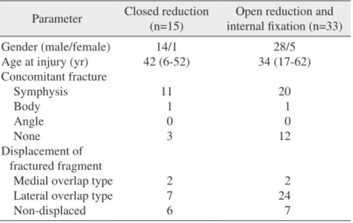
In the CR group, 2 condylar fragments were displaced medially, 7 laterally, and 6 were non-displaced. In the ORIF group, 2 condylar fragments were displaced medially, 24 laterally, and 7 were non-displaced. The average period of MMF was 5.47 days in the CR group and 6.33 days in the ORIF group. Associated mandibular fractures occurred in 68.75% of cases, especially symphysis fractures (64.58%).
Twelve patients (11 symphysis, 1 body) from the CR group and 21 patients (20 symphysis, 1 body) from the ORIF group had concomitant mandibular fractures. All concomitant frac- tures were treated with ORIF.(Table 1)
2. Radiologic results
In the CR group, the average loss of ramus height was 2.44
Fig. 1. Illustration showing the method by which loss of ramus height was measured on the panoramic view. A reference line was drawn through both gonial angles. A perpendicular line between the most superior point of the condyle and the reference line was drawn on the panoramic radiograph. The difference between the non-fractured and fractured side was used as a measure of differ- ence in ramus length.
Seong-Yong Kim et al: Outcomes of open versus closed treatment in the management of mandibular subcondylar fractures. J Korean Assoc Oral Maxillofac Surg 2014
Fig. 2. Illustration showing the method by which tangential dis- placement was quantified on the panoramic view. A reference line was drawn through both gonial angles, and another line was drawn tangential to the posterior border of the condylar process on each side. The angle between the intersection of the tangent and the condylar process was calculated. The difference in this angle between the non-fractured and fractured sides was used as a measurement of tangential angulation.
Seong-Yong Kim et al: Outcomes of open versus closed treatment in the management of mandibular subcondylar fractures. J Korean Assoc Oral Maxillofac Surg 2014
Table 1. Patient’s data
Parameter Closed reduction
(n=15) Open reduction and internal fixation (n=33) Gender (male/female)
Age at injury (yr) Concomitant fracture Symphysis Body Angle None Displacement of
fractured fragment Medial overlap type Lateral overlap type Non-displaced
14/1 42 (6-52)
11 1 0 3
2 7 6
28/5 34 (17-62)
20 1 0 12
2 24 7 Values are presented as number or mean (range).
Seong-Yong Kim et al: Outcomes of open versus closed treatment in the management of mandibular subcondylar fractures. J Korean Assoc Oral Maxillofac Surg 2014
Outcomes of open versus closed treatment in the management of mandibular subcondylar fractures
299
IV. Discussion
Despite a plethora of treatment guidelines, management of subcondylar fracture of the mandible remains controversial.
During the past few decades, CR has been the preferred treat- ment4; however, closed treatment requires a period of MMF, followed by active physiotherapy5. Also, long-term complica- tions such as pain, arthritis, open bite, deviation of the man- dible on opening, inadequate restoration of vertical height of the ramus leading to malocclusion, and ankylosis can result from the CR method6. However, we found no significant dif- ferences between groups in the average period of MMF (5.47 days in CR group and 6.33 days in ORIF group) and no seri- ous complications.
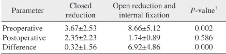
Surgical treatment allows proper anatomical repositioning and immediate functional movement of the jaw7. There is consensus that correct anatomical repositioning of the man- dibular condyle process is an important prerequisite for re- establishing function8. In cases of severe displacement or dis- location, surgical management is preferred6,9. In our study, all treatment methods were decided by the treating surgeons. In our clinics, doctors tend to provide CR for patients with less displaced fractures and ORIF to those with more displaced fractures. The preoperative tangential angulation in the CR group (3.67º±2.53º) was significantly less (P=0.002) than that in ORIF group (8.66º±5.12º). However, the difference between the groups in preoperative loss of ramus height was not significant.
Palmieri et al.3 reported that open reduction might produce functional benefits for patients with severely dislocated con- dylar process fractures, and Undt et al.7 reported that ORIF allows appropriate anatomical repositioning and immedi- ate functional movement of the mandible. Similarly, in our study, the difference between preoperative and postoperative loss of ramus height in the ORIF group (2.60±2.02 mm) was statistically greater than that in the CR group (1.25±1.61 mm;
P=0.008). The difference between preoperative and postop- erative tangential angulation in the ORIF group (6.92º±4.86º) was statistically greater than that in the CR group (0.32º±
1.56º; P=0.000).
Haug and Assael10 showed no differences for maximum interincisal opening, deviation on opening, and occlusion be- tween closed and open management groups after treatment.
Likewise, we found no clinical differences, such as occlusion or interincisal mouth opening, between the CR and ORIF groups. However, 6 patients (40%) in the CR group and 11 patients (33%) in the ORIF group showed deviation on maxi- groups in the loss of ramus height (P=0.008) and tangential
angulation (P=0.000).(Tables 2, 3) There was also a statisti- cally significant difference between the groups in preopera- tive tangential angulation (P=0.002).(Table 3)
3. Clinical results
The clinical parameters were observed three months after treatment or by telephone survey. Neither group had any pa- tients with post-treatment malocclusion or permanent nerve injury. The mouth opening of all patients was greater than 40 mm. However, 6 of 15 patients (40%) in the CR group showed deviation on mouth opening. Similarly, 11 of 33 pa- tients (33%) in the ORIF group showed deviation on mouth opening.(Table 4)
Table 2. Radiologic parameters for loss of ramus height (mm) Parameter Closed
reduction Open reduction and
internal fixation P-value1 Preoperative
Postoperative Difference
2.44±2.18 1.99±0.99 1.25±1.61
3.61±2.33 1.01±1.19 2.60±2.02
0.079 0.301 0.008
1Mann-Whitney U test.
Values are presented as mean±standard deviation.
Seong-Yong Kim et al: Outcomes of open versus closed treatment in the management of mandibular subcondylar fractures. J Korean Assoc Oral Maxillofac Surg 2014
Table 3. Radiologic parameters for tangential angulation of the fractured fragment ( ˚ )
Parameter Closed
reduction Open reduction and
internal fixation P-value1 Preoperative
Postoperative Difference
3.67±2.53 2.35±2.23 0.32±1.56
8.66±5.12 1.74±0.89 6.92±4.86
0.002 0.586 0.000
1Mann-Whitney U test.
Values are presented as mean±standard deviation.
Seong-Yong Kim et al: Outcomes of open versus closed treatment in the management of mandibular subcondylar fractures. J Korean Assoc Oral Maxillofac Surg 2014
Table 4. Clinical parameters
Parameter Closed reduction (n=15)
Open reduction and internal fixation (n=33) Malocclusion
Mouth opening limitation Deviation on opening Nerve injury
0 0 6 0
0 0 11 0 Values are presented as number.
Seong-Yong Kim et al: Outcomes of open versus closed treatment in the management of mandibular subcondylar fractures. J Korean Assoc Oral Maxillofac Surg 2014
J Korean Assoc Oral Maxillofac Surg 2014;40:297-300
300
2. Lindahl L. Condylar fractures of the mandible. I. Classification and relation to age, occlusion, and concomitant injuries of teeth and teeth-supporting structures, and fractures of the mandibular body.
Int J Oral Surg 1977;6:12-21.
3. Palmieri C, Ellis E 3rd, Throckmorton G. Mandibular motion after closed and open treatment of unilateral mandibular condylar pro- cess fractures. J Oral Maxillofac Surg 1999;57:764-75.
4. Brandt MT, Haug RH. Open versus closed reduction of adult man- dibular condyle fractures: a review of the literature regarding the evolution of current thoughts on management. J Oral Maxillofac Surg 2003;61:1324-32.
5. Suzuki T, Kawamura H, Kasahara T, Nagasaka H. Resorbable poly-L-lactide plates and screws for the treatment of mandibular condylar process fractures: a clinical and radiologic follow-up study. J Oral Maxillofac Surg 2004;62:919-24.
6. Iizuka T, Lädrach K, Geering AH, Raveh J. Open reduction without fixation of dislocated condylar process fractures: long-term clinical and radiologic analysis. J Oral Maxillofac Surg 1998;56:553-61.
7. Undt G, Kermer C, Rasse M, Sinko K, Ewers R. Transoral mini- plate osteosynthesis of condylar neck fractures. Oral Surg Oral Med Oral Pathol Oral Radiol Endod 1999;88:534-43.
8. Baker AW, McMahon J, Moos KF. Current consensus on the man- agement of fractures of the mandibular condyle. A method by ques- tionnaire. Int J Oral Maxillofac Surg 1998;27:258-66.
9. Sugiura T, Yamamoto K, Murakami K, Sugimura M. A compara- tive evaluation of osteosynthesis with lag screws, miniplates, or Kirschner wires for mandibular condylar process fractures. J Oral Maxillofac Surg 2001;59:1161-8.
10. Haug RH, Assael LA. Outcomes of open versus closed treatment of mandibular subcondylar fractures. J Oral Maxillofac Surg 2001;59:370-5.
mal opening of the lower jaw three months after treatment.
Our study used only 48 patients and a relatively short follow- up period. Future research should compare the outcomes of many more cases with long-term follow-up.
V. Conclusion
In conclusion, CR had clinically satisfactory results, al- though ORIF produced more accurate reduction of fractured fragments. In the absence of distinct displacement of a frac- tured fragment, therefore, CR should be selected over ORIF to prevent the need for an operation.
Conflict of Interest
No potential conflict of interest relevant to this article was reported.
References
1. Lee SC, Kim YG, Ryu DM, Lee BS, Yoon OB, Jin TH. A clinical and statistical study of condylar fracture of mandible. J Korean As- soc Oral Maxillofac Surg 1998;24:326-9.