Treatment of Intra-articular Calcaneal Fracture with Open Reduction and Internal Fixation
Jun-Won Choi, M.D., Joon-Cheol Choi, M.D., Young-Sang Lee, M.D., Hwa-Yeop Na, M.D., Woo-Sung Kim, M.D., Sang-Ho Han, M.D.
Department of Orthopedic Surgery, Bundang Jesaeng General Hospital, Seongnam, Korea
=Abstract=
Purpose: To evaluate the clinical outcomes and radiographic results of open reduction and internal fixation for intra- articular calcaneal fractures.
Materials and Methods: We reviewed 20 cases of calcaneal fractures managed with open reduction and internal fixation from March 2003 to January 2005. We used the computed tomographic classification system proposed by Sanders et al to classify these fractures. Preoperative and postoperative Bohler’s angle, heel height (calcaneal facet height) and calcaneal length, calcaneal width were measured. The Creighton-Nebraska Health Foundation Assessment score was used for clinical evaluation.
Results: There were 12 cases of type II fractures, 5 of type III fractures and 3 of type IV fractures. The mean clinical score was 84.3 for type II, 82.6 for type III and 56.1 for type IV. The mean preoperative Böhler angle was 6.1˚ and final was 22.8˚. The mean preoperative calcaneal facet height was 76.6 mm and final was 80.3 mm (The mean calcaneal facet height was changed from preop 76.6 mm to postop 80.3 mm). The mean preoperative calcaneal length was 88.2 mm and final was 92.6 mm. The mean preoperative width was 38.1 mm and final was 35.6 mm.
Conclusion: Open reduction and internal fixation showed good results for type II and III fractures, but for type IV fractures the clinical result was significantly worse than the other types. However, type IV fractures still had restoration of (should be restored in) Böhler’s angle, calcaneal facet height, calcaneal length and width which may be helpful in later subtalar fusion.
Key Words: Calcaneus, Intraarticular fracture, Open reduction and internal fixation
∙Address for correspondence Joon-Cheol Choi, M.D.
Department of Orthopedic Surgery, Bundang Jesaeng General hospital, 255-2, Seohyun-dong, Bundang-gu, Seongnam-si, Gyeonggi-do, 463-774, Korea
Tel: +82-31-779–0179 Fax: +82-31-779-0176 E-mail: [email protected]
* 본 논문의 요지는 2006년도 제 50차 정형외과 추계학술대회에서 구연 되었음.
서 론
종골은 그 해부학적 구조와 골절 양상의 복잡성으로 인 하여 치료가 어려운 골절 중 하나로 특히 거골하 관절을 침 범하는 종골 골절의 경우 수술적 치료와 보존적 치료 사이 에 많은 논란이 있다.
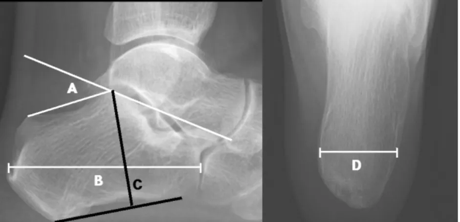
Figure 1. Radiologic parameter of lateral image of the normal calcaneus; angle A shows Bohler’s angle, line B indicates the calcaneal length, line C describes the calcaneal facet height and line D indicates width of calcaneus.
최근 여러 저자들에 의해 거골하 관절을 침범하는 종골 골절에 대하여 관혈적 정복술 및 내고정술을 실시하여 좋은 결과를 얻었다는 보고들이 늘고 있다. 하지만 Randle 등15) 은 수술적 치료가 보존적 치료에 비해 더 좋은 결과를 보이 는 경향이 있으나 꼭 수술이 좋다는 결론을 내리기는 힘들 다고 하였으며, Parmar 등13)은 전향적 무작위 선택을 한 연구에서 관절내 종골 골절에서 수술적 치료 및 비수술적 치료의 1년 추시 결과 두 군 사이에 차이가 없다고 하였다.
또한 Buckley 등3)은 수술적 치료 결과와 보존적 치료 결과 가 차이가 없으나 산재 환자를 제외한 어떤 그룹에서는 수 술적 치료 결과가 더 좋았다고 보고하고 있다.
이에 저자들은 골절의 양상에 따른 선택적 관혈적 정복 술 및 내고정술 보다는 골절의 양상에 관계없이 적극적인 관혈적 정복술 및 내고정술을 시행하는 것이 임상적, 방사 선적으로 도움이 될 것으로 생각하고 후향적 방법으로 결과 를 분석하여 보고자 한다.
대상 및 방법
2003년 3월부터 2005년 1월까지 관절내 종골 골절로 수 술적 치료를 받은 환자 중 최소 1년 이상 추시가 가능하였던 18명(20예)을 대상으로 후향적 방법으로 분석하였다.
1. 연령 및 성별 분포
남자가 17명, 여자가 1명이었으며 수상당시 평균 연령은 37.5세(23~64세)였으며, 부위는 우측이 13예, 좌측이 7예 였다.
2. 손상 기전 및 동반 손상
총 18명 중 추락사고로 인한 수상이 16명이었고, 실족사 고는 1명, 그리고 교통사고는 1명이었다. 동반 손상은 총 18명 중 6명에서 발생하였으며, 요골 골절이 2명, 족부 및 족근 관절 골절이 3명, 뇌출혈이 1명이었다.
3. 수술전 검사
모든 환자에 있어서 단순 방사선 검사를 시행하여 Bӧhler 씨 각, 종골 소면 높이(calcaneal facet height), 종골 길이 (calcaneal length) 및 너비(width)를 측정하였다(Fig. 1)16). 수술 전 컴퓨터 단층 촬영을 시행하여 후방돌기 관절에 전위가 없는 경우를 Type Ⅰ으로, 1개의 전위된 골절선이 있는 경우를 Type Ⅱ로, 2개의 골절선이 있는 경우를 Type
Ⅲ로, 3개 이상의 골절선이 있는 경우를 Type Ⅳ로 분류 하는 Sanders 분류7,8,17,18)를 이용하여 분류하였다.
4. 치료방법
수술 시기는 부종이 심하지 않는 경우 수상 후 2-3일 내 바로 시행하였으며, 부종이 심한 경우에는 급성 부종이 소실된 후 수술을 시행하였다.
수술은 광범위 외측 도달법4)을 사용하였으며, 피부 절개 후 피하 연부 조직과 골막을 함께 박리하고 골막하 거상을 시행하였다. 수술시 과도한 견인을 피하고 골절면의 용이한 노출을 위하여 3개의 K-강선들을 비골 외과, 거골 및 입방 골에 삽입 후 수직으로 휘어 피부편을 보호하였다. 골절 부 위를 노출한 후 후관절면을 포함하고 있는 내, 외 골편을 거
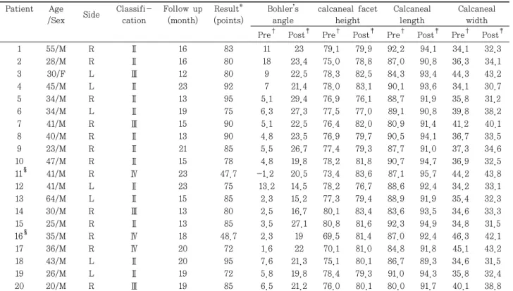
Table 1. Summary of Cases Patient Age
/Sex Side Classifi- cation
Follow up (month)
Result*
(points)
Bohler’s angle
calcaneal facet height
Calcaneal length
Calcaneal width Pre
†Post
‡Pre
†Post
‡Pre
†Post
‡Pre
†Post
‡1 55/M R Ⅱ 16 83 11 23 79.1 79.9 92.2 94.1 34.1 32.3
2 28/M R Ⅱ 16 80 18 23.4 75.0 78.8 87.0 90.8 36.3 34.1
3 30/F L Ⅲ 12 80 9 22.5 78.3 82.5 84.3 93.4 44.3 43.2
4 45/M L Ⅱ 23 92 7 21.4 78.0 83.1 90.1 93.6 34.1 30.7
5 34/M R Ⅱ 13 95 5.1 29.4 76.9 76.1 88.7 91.9 35.8 31.2
6 34/M L Ⅱ 19 75 6.3 27.3 77.5 77.0 89.1 90.8 39.8 38.2
7 41/M R Ⅲ 15 90 5.1 22.5 76.4 82.0 80.9 91.4 41.2 40.1
8 40/M R Ⅱ 13 90 4.8 23.5 76.9 79.7 90.5 94.1 36.7 33.5
9 23/M R Ⅱ 21 85 5.5 26.7 77.4 79.3 87.7 91.0 37.3 34.6
10 47/M R Ⅱ 15 78 4.8 19.8 78.2 81.8 90.7 94.7 36.9 32.5
11
§41/M R Ⅳ 23 47.7 -1.2 20.5 73.4 83.6 87.1 95.7 44.2 43.8
12 41/M L Ⅱ 23 75 13.2 14.5 78.2 76.7 88.6 92.4 34.2 33.1
13 64/M L Ⅱ 15 85 2.3 15.2 77.3 79.4 88.9 91.9 35.4 32.3
14 30/M R Ⅲ 13 80 2.5 16.7 80.1 83.4 83.6 93.5 34.6 33.3
15 25/M R Ⅱ 13 85 3.5 27.1 80.8 81.6 92.3 94.9 34.8 31.5
16
§35/M R Ⅳ 18 48.7 2.3 19 69.5 81.4 87.0 92.4 46.3 42.1
17 36/M R Ⅳ 20 72 1.6 22 70.1 81.0 84.8 91.8 45.1 43.2
18 43/M L Ⅱ 20 95 7.6 21.3 75.1 80.1 86.7 89.3 34.6 31.5
19 26/M L Ⅱ 19 72 5.8 19.8 78.4 79.3 91.0 94.3 35.8 32.4
20 20/M R Ⅲ 19 85 6.5 21.2 76.0 80.1 80.0 91.7 40.1 38.8
*Result, Creighton-Nebraska health foundation assessment score;
†Pre, Pre-operation;
‡Post, Post-operation;
§11, 16, After subtalar fusion.
5. 치료평가
임상 결과에 대한 평가는 Creighton-Nebraska health foundation의 종골 골절 평가 방법을 이용하여 동통, 활동 성, 관절운동 범위, 직업의 복귀여부, 신발 크기 변화, 종창 의 정도를 점수로 환산하여 90점 이상은 우수, 80점 이상 -89점 이하는 양호, 65점 이상-79점 이하는 보통, 64점 이하는 불량으로 분류하였다.
방사선적인 결과는 비체중 부하시 종골 측면상에서 Böhler씨 각, 종골 소면 높이 및 종골 길이를 측정하고, 비
평가 결과 우수 5예, 양호 6예, 보통 2예였고, Creighton- Nebraska health foundation에 의한 임상 평가 점수는 평균 84.3점이었다. Bӧhler씨 각은 수술 전 7.3도에서 수술 후 22.5도로, 종골 소면 높이는 수술 전 77.6 mm에서 수술 후 79.6 mm, 종골 길이는 수술 전 89.5 mm에서 수술 후 92.6 mm, 너비는 수술 전 평균 38.1 mm에서 수술 후 35.6 mm로 측정되었다(Table 1).
Sander 분류 Type Ⅲ는 4예였으며, 임상 평가 결과 우수 1예, 양호 3예였고, 평균 점수는 82.6점이었다. Bӧhler씨 각은 수술 전 5.7도에서 수술 후 20.7도로, 종골 소면 높이 는 수술 전 77.7 mm에서 수술 후 81.5 mm, 종골 길이는
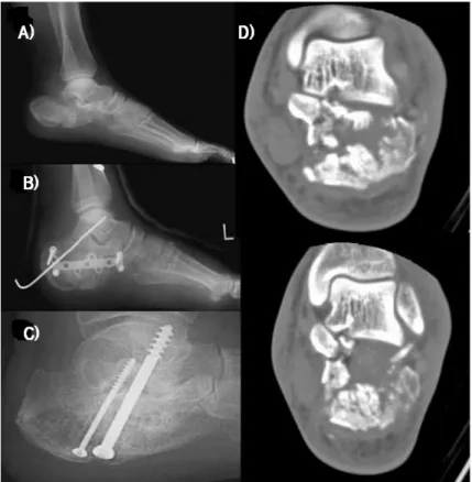
Figure 2. Radiographs showed 41 year old male patient with intra-articular calcaneus fracture.
(A) Preoperative radograph of calcaneus fracture with comminution.
(B) Postoperative radiograph showing reduction of the posterior facet ant Bohler angle of 20.5 degrees.
(C) Postoperative 14 months radiograph showing subtalar arthrodesis for arthritis.
(D) Preoperative calcaneal CT with semicoronal view.
Severe comminution was noted. It was classified Sander’s type lV calcaneal fracture.
수술 전 85.2 mm에서 수술 후 92.5 mm, 너비는 수술 전 평균 40.1 mm에서 수술 후 38.9 mm로 측정되었다.
Sander Type Ⅳ는 3예였으며, 임상 평가 결과 보통 1예, 불량 2예였고, 평균 점수는 56.1점이었다. Bӧhler씨 각은 수술 전 2.8도에서 수술 후 20.5도로, 종골 소면 높이는 수술 전 71.0 mm에서 수술 후 82.0 mm, 종골 길이는 수술 전 86.0 mm에서 수술 후 93.3 mm, 너비는 수술 전 평균 45.2 mm에서 수술 후 43.0 mm로 측정되었다. 불량 2예에 대해서는 관혈적 정복술 및 내고정술 후 14개월 및 16개월 에 내고정물을 이용한 거골하 관절의 압박 고정술로 거골하 관절 유합술을 시행하여 마지막 추시에서 각각 임상 평가 결과 81점, 78점으로 양호와 보통의 결과를 얻을 수 있었다.
합병증으로는 창상 감염이 1예, 창상 파열 1예, 거골하 관절염이 2예였다. 창상 감염은 지속적인 항생제 투여 및 소독으로 치유되었으며, 창상 파열은 재봉합으로 치유되 었다. Sander Type Ⅳ에서 발생한 거골하 관절염은 거골 하 관절 유합술을 시행하였다.
Type Ⅱ와 Ⅲ 골절에서 관혈적 정복술 및 내고정술을 시 행하였을 경우, Type Ⅳ 골절에 비하여 임상적으로 통계상 유의한 결과를 보였지만(p<0.05), 방사선적으로는 유의한 차이를 보이지 않았다(p>0.05). Type Ⅱ와 Type Ⅲ 골절간 의 비교에서는 임상적, 방사선적으로 통계상 유의한 차이를
보이지 않았다(p>0.05).
고 찰
종골 골절의 치료는 과거에는 주로 보존적 치료였다.
Pazo 등14)에 의하면 조기 관절 운동에 의한 보존적 치료로 76%에서 좋은 결과를 얻었다고 보고하였으며, Palmar 등13) 은 보존적 치료와 외측 도달법을 이용한 수술적 치료를 비 교한 전향적 연구에서 두 군간의 차이가 없다고 보고하였 다. 그러나 최근에는 광범위 외측 도달법으로 수술을 시행 하여 거골하 관절면의 보다 정확한 정복과 후족부의 전체적 인 모양 및 배열의 교정을 얻을 수 있어 수술 후 좋은 결과 를 기대할 수 있다1,2,5,11).
Randle 등15)의 meta-analysis 논문에서는 수술이 좋은 결과를 나타내는 경향이 있으나 꼭 수술이 좋다는 결론을 내리기는 힘들다고 하였다. 특히 Buckley 등3)은 전향적, 무작위 환자 선택을 한 논문에서 보존적 치료를 한 군과 수 술적 치료를 한 군을 비교하여 기능적 결과는 같지만, 산재 환자를 제외하면 여자, 젊은 남자, 술 전 높은 Bӧhler씨 각, 가벼운 노동을 하는 사람, 골절 선이 하나인 단순 골절 환자 에서 수술적 치료가 보존적 치료보다 좋은 결과를 보였다고 보고하였다. Herscovici 등6)은 65세 이상 고령의 환자에서
결과를 얻지 못하였다.
Kundel등9)은 Type Ⅳ 골절에서 거골하 관절의 정복을 얻기가 불가능하므로 보존적 치료를 해야 한다고 주장하 였고, Laughlin 등10)은 Type Ⅳ 골절에 대해서도 관혈적 정복술 및 내고정술로 좋은 결과를 얻었다고 보고하였으며, Meyerson12)은 관절내 분쇄 골절이면서, 관절내 연골이 소 실된 종골 골절은 관혈적 정복술 및 내고정술과 동시에 일 차적 거골하 유합술을 시행해야 한다고 주장하였다. Huang 등7)은 Type Ⅳ 골절에 대한 관혈적 정복술 및 내고정술에 서 불량한 예후를 보인 원인을 거골하 관절의 해부학적 정 복의 부족이라고 생각하여, 관혈적 정복술 및 내고정술을 시도하되 수술 중에 거골하 관절면의 해부학적 정복이 어렵 다고 판단되었을 경우 즉시 거골하 관절 유합술을 시행하여 야 한다고 주장하였다. 본 연구에서는 Type Ⅳ 골절인 경우 에도 모두 관혈적 정복술 및 내고정술을 시행하여 후족부의 전체적인 모양의 교정을 얻을 수는 있었으나, 임상적으로는 보통 1예와 불량 2예의 결과를 얻었다. Type Ⅳ 골절의 경 우 관혈적 정복술 및 내고정술이 임상적 결과가 좋지 않기 때문에 일차적 거골하 유합술을 제안하기도 하지만, 본 저 자들은 거골하 관절을 정복시킬 수 있을 경우 임상 결과가 나쁘지 않아 거골하 관절 유합술이 불필요할 수도 있으므 로, Type Ⅳ 골절 자체가 일차 거골하 유합술의 절대적 적 응증이 아니라고 생각한다. 또한 종골의 관절내 분쇄 골절 급성기때 종골의 모양을 유지하면서 거골하 관절 유합술을 시행하는데 기술적 어려움이 있고, 거골하 관절 유합술로 인접 관절에 과중한 하중 전달이 되어 부가적인 영향을 미 칠 수 있기 때문에 거골하 관절 유합술보다는 관혈적 정복 술 및 내고정술을 일차 선택으로 해야 한다고 생각한다. 그 리고 관혈적 정복술 및 내고정술 후 Bӧhler씨 각, 종골 소 변 높이, 너비 및 종골 길이의 회복을 얻는 것이, 추후 거골 하 관절 유합술을 시행할 때 기술적으로 용이하며, 종골의 부정 유합을 피할 수 있기 때문에 적극적인 관혈적 정복술 및 내고정술이 권장된다고 여겨진다(Fig. 2).
REFERENCES